Comparative Study
. 2008 Jan 9;27(1):290-300.
doi: 10.1038/sj.emboj.7601942. Epub 2007 Nov 29.

Crystal structure of human XLF/Cernunnos reveals unexpected differences from XRCC4 with implications for NHEJ

Affiliations
Comparative Study

Crystal structure of human XLF/Cernunnos reveals unexpected differences from XRCC4 with implications for NHEJ

Yi Li et al. EMBO J. .

Abstract

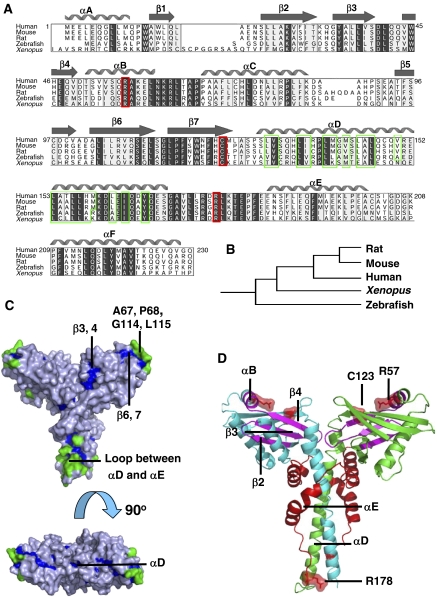
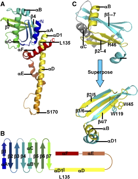
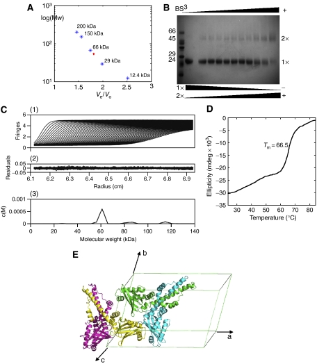
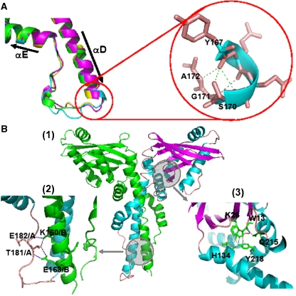
The recently characterised 299-residue human XLF/Cernunnos protein plays a crucial role in DNA repair by non-homologous end joining (NHEJ) and interacts with the XRCC4-DNA Ligase IV complex. Here, we report the crystal structure of the XLF (1-233) homodimer at 2.3 A resolution, confirming the predicted structural similarity to XRCC4. The XLF coiled-coil, however, is shorter than that of XRCC4 and undergoes an unexpected reverse in direction giving rise to a short distorted four helical bundle and a C-terminal helical structure wedged between the coiled-coil and head domain. The existence of a dimer as the major species is confirmed by size-exclusion chromatography, analytical ultracentrifugation, small-angle X-ray scattering and other biophysical methods. We show that the XLF structure is not easily compatible with a proposed XRCC4:XLF heterodimer. However, we demonstrate interactions between dimers of XLF and XRCC4 by surface plasmon resonance and analyse these in terms of surface properties, amino-acid conservation and mutations in immunodeficient patients. Our data are most consistent with head-to-head interactions in a 2:2:1 XRCC4:XLF:Ligase IV complex.


Figures

Figure 1
Figure 1
Evolutionary analysis of XLF. (A) Multiple sequence alignment of XLF orthologues. Strongly conserved residues are highlighted in light grey, identical residues are in dark grey, hydrophobic residues in αD are in green frames, the reported human disease mutation sites are in red frames and secondary structure elements of human XLF are shown above the alignment. (B) Clustering of XLF orthologues generated by Evolutionary Trace Server (TraceSuite II) (Innis et al, 2000). (C) Evolutionarily conserved residues mapped onto XLF homodimer structure. Residues conserved but inaccessible to solvent are shown in blue, while those conserved and exposed to solvent are green. (D) Mapping onto XLF structure of the cancer-related mutations found in clinical cases. XLF homodimer is coloured by chain in green and cyan. Disease single-point mutation sites R57 and C123; the mutation of the polypeptide after R178 is indicated in red on both chains. Regions spanning from A25, β2 to R57, αB are magenta.
Figure 2
Figure 2
The XLF crystal structure. (A) The structure of the XLF protomer. The secondary structure is coloured in rainbow, including an N-terminal globular head and C-terminal α-helices. The protein starts at the navy-blue α-helix and ends at the red α-helix. (B) Topology diagram of XLF protomer, secondary structure elements are in the same colour as (A). (C) Superposition of β2, β3, β4, αB (yellow) to β5, β6, β7, αD1 (cyan). The two motifs were picked up from the XLF head domain and rotated to superpose. β strands overlap well, and α-helices are in similar orientations. W45 and W119 are found at the topologically equivalent positions.
Figure 3
Figure 3
Evidence for the XLF dimer. (A) Superdex-200 calibration curves were used to estimate the molecular weight of the XLF multimer. Proteins used for calibration are shown in blue stars, and the red diamond indicates XLF elution. (B) Crosslinking with BS3 indicated the existence of an XLF dimer, the amount of which was enhanced by increasing BS3, while the monomer decreased at the same time. (C) Sedimentation velocity profiles of XLF (1.8 mg/ml) centrifuged at 20°C and a rotor speed of 55 000 r.p.m. (1) and the residuals obtained after data fitting (2). The peak at 60 kDa (3) corresponds to the XLF dimer, which takes a relevant concentration of 92%. Data were analysed using SEDFIT program (Schuck, 2000). (D) Thermal denaturation experiment performed by CD. Tm is measured as 66.5°C. (E) XLF forms a homodimer with a two-fold axis relating protomers, and two XLF homodimers are packed in one asymmetric unit of the C2 cell.
Figure 4
Figure 4
Conserved structural motifs in the C-terminal residues of XLF. (A) Loop region between αD and αE. The colour keys are set according to chains, the same as in Figure 3. Left panel: superposition of loop regions in the four chains; right panel: hydrogen bonds between the C-terminal region of αD to the following loop (Y167, Q168, S170, G171 and A172). (B) (1) Overall view of the XLF homodimer, chain A is in green, and chain B is coloured by chain in cyan (helix), magenta (sheet) and wheat (loop). Circled areas are shown in greater detail in (2) and (3). (2) Inter-chain hydrogen bonds connecting the loop of chain A and αD of chain B. (3) Hydrogen bonds between head domain residues and residues of αF.
Figure 5
Figure 5
Superposition of XLF and XRCC4 structures. XLF red and XRCC4 green. (A) Head domains of XLF and XRCC4 superpose, especially in the antiparallel β-sheets and in the helix-turn-helix motif in the middle. XLF differs from XRCC4 in the coiled-coil region. The angle between the head domain and the coiled-coil is larger in XLF than in XRCC4 because of the insertion of αF and αA. (B) The coiled-coil in XLF is much shorter than that in XRCC4, and does not contain an equivalent region to the XRCC4–Ligase IV-binding site. DNA Ligase IV fragment bound to XRCC4 is in magenta.
Figure 6
Figure 6
(A) XLF–XRCC4 interactions evaluated by BIAcore 2000. Sensorgrams obtained from the injections of XRCC4 over the immobilised XLF surface at concentrations of 50, 25, 12.5, 10, 5 μM. (B, C) Prediction of regions that will favour protein–protein interactions in XRCC4 and XLF structures. The darker the blue colour of a region of the dimer, the greater the probability that it acts as a binding region as indicated by ODA (Fernandez-Recio et al, 2005). XRCC4 probably interacts with other molecules through the head domains and the coiled-coil region. XLF head domains are likely to interact with other factors but also a region surrounding the conserved, K160 residue, is highlighted. (D) Possible modes of interaction between XRCC4, XLF and Ligase IV. XRCC4 molecules are shown in green, XLF is in red and Ligase IV BRCR-linker region is in magenta. (1) Linker region between Ligase IV BRCT domains binds to XRCC4's coiled-coil, folded XLF/Cernunnos contacts XRCC4 via the head domains. (2) The C termini of XLF molecules are unfolded and bind to Ligase IV in a similar way to XRCC4. Thus, there are two Ligase IV molecules in this large complex. (3) XLF and XRCC4 form a heterodimer and bind to Ligase IV in the composite coiled-coil region.

References

    1. Ahnesorg P, Smith P, Jackson SP (2006) XLF interacts with the XRCC4–DNA ligase IV complex to promote DNA nonhomologous end-joining. Cell 124: 301–313 - PubMed
    1. Berman H, Henrick K, Nakamura H, Markley JL (2007) The worldwide Protein Data Bank (wwPDB): ensuring a single, uniform archive of PDB data. Nucleic Acids Res 35: D301–D303 - PMC - PubMed
    1. Buck D, Malivert L, de Chasseval R, Barraud A, Fondaneche MC, Sanal O, Plebani A, Stephan JL, Hufnagel M, le Deist F, Fischer A, Durandy A, de Villartay JP, Revy P (2006) Cernunnos, a novel nonhomologous end-joining factor, is mutated in human immunodeficiency with microcephaly. Cell 124: 287–299 - PubMed
    1. Callebaut I, Malivert L, Fischer A, Mornon JP, Revy P, de Villartay JP (2006) Cernunnos interacts with the XRCC4 x DNA–ligase IV complex and is homologous to the yeast nonhomologous end-joining factor Nej1. J Biol Chem 281: 13857–13860 - PubMed
    1. Critchlow SE, Bowater RP, Jackson SP (1997) Mammalian DNA double-strand break repair protein XRCC4 interacts with DNA ligase IV. Curr Biol 7: 588–598 - PubMed

Publication types

Associated data