Comparative Study
. 2008 Jan 9;27(1):234-43.
doi: 10.1038/sj.emboj.7601940. Epub 2007 Nov 29.

Structure-guided identification of a new catalytic motif of oligosaccharyltransferase

Affiliations
Comparative Study

Structure-guided identification of a new catalytic motif of oligosaccharyltransferase

Mayumi Igura et al. EMBO J. .

Abstract

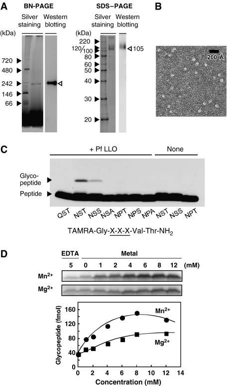
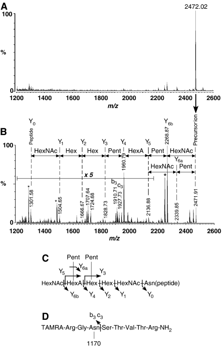
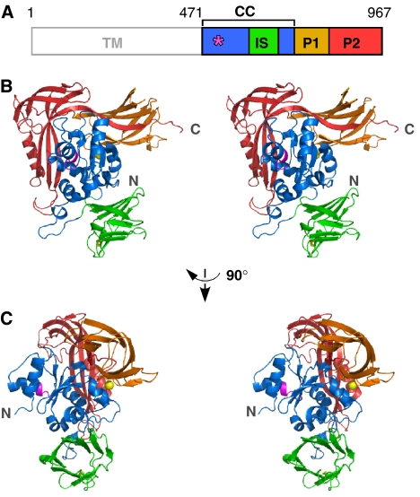
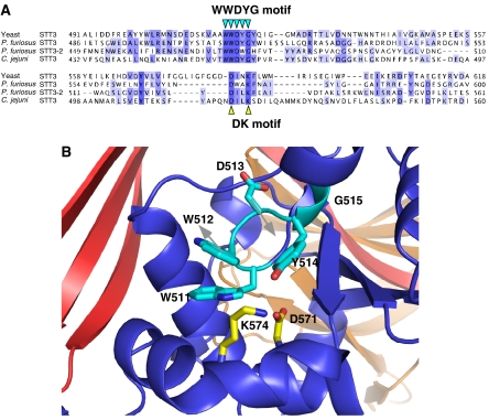
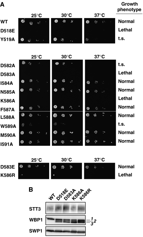
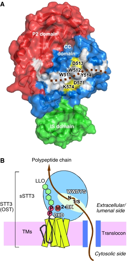
Asn-glycosylation is widespread not only in eukaryotes but also in archaea and some eubacteria. Oligosaccharyltransferase (OST) catalyzes the co-translational transfer of an oligosaccharide from a lipid donor to an asparagine residue in nascent polypeptide chains. Here, we report that a thermophilic archaeon, Pyrococcus furiosus OST is composed of the STT3 protein alone, and catalyzes the transfer of a heptasaccharide, containing one hexouronate and two pentose residues, onto peptides in an Asn-X-Thr/Ser-motif-dependent manner. We also determined the 2.7-A resolution crystal structure of the C-terminal soluble domain of Pyrococcus STT3. The structure-based multiple sequence alignment revealed a new motif, DxxK, which is adjacent to the well-conserved WWDYG motif in the tertiary structure. The mutagenesis of the DK motif residues in yeast STT3 revealed the essential role of the motif in the catalytic activity. The function of this motif may be related to the binding of the pyrophosphate group of lipid-linked oligosaccharide donors through a transiently bound cation. Our structure provides the first structural insights into the formation of the oligosaccharide-asparagine bond.


Figures

Figure 1
Figure 1
Oligosaccharyltransferase purified from Pyrococcus furiosus cells. (A) BN-PAGE and SDS–PAGE analyses of OST purified from P. furiosus cells by immunoaffinity, using an anti-P. furiosus sSTT3 antibody. Proteins on the gels were visualized by silver staining and western blotting, using an anti-P. furiosus sSTT3 antibody as the primary antibody. (B) Electron micrograph of the OST enzyme embedded in uranyl acetate stain. The purified STT3 protein in Triton X-100 micelles appeared as individual particles. Scale bar, 200 Å. (C) Effects of the amino-acid substitutions of the N-glycosylation sequon on the N-oligosaccharyltransferase reaction. The reaction mixtures were incubated for 16 h at 65°C in the presence and absence of P. furiosus LLO. (D) Metal ion dependence of the reaction and plot of the glycopeptide formation as a function of the final concentration of ions. The quantity of the glycopeptide was estimated using a TAMRA-labeled 22-residue peptide as an external standard, as described in Kohda et al (2007). The reaction mixtures were incubated for 1 h at 65°C.
Figure 2
Figure 2
MS/MS analysis of P. furiosus N-glycan structure. (A) MALDI-QIT-TOF MS spectrum of the OST reaction product. (B) MALDI-QIT-TOF MS/MS spectrum of the precursor ion at m/z 2472. The fragment ions originating from the sequential loss of oligosaccharide residues are indicated in the spectrum. Asterisks indicate other prominent peaks generated by dehydration (m/z 2250.9 and 1486.6) and the loss of ammonium (m/z 1284.6). (C) Estimated P. furiosus N-glycan structure, and its fragmentation pattern. (D) The observation of b3 and c3 fragment ions confirmed that the glycopeptide product is Asn-linked. TAMRA denotes carboxytetramethylrhodamine. ‘1170' represents the molecular mass of the attached oligosaccharide moiety.
Figure 3
Figure 3
Crystal structure of the C-terminal soluble domain of STT3. (A) Domain structure of P. furiosus STT3. TM, transmembrane domain; CC, central core domain, residues 471–600+683–725; IS, insertion domain, residues 601–682; P1, peripheral domain 1, residues 726–821; P2, peripheral domain 2, residues 822–967. The position of the WWDYG motif is indicated by an asterisk. (B) Stereoview of the overall structure of the C-terminal soluble domain of STT3 (residues 471–967). The WWDYG motif is shown in magenta. The disulfide bond between C638 and C658 is shown as yellow sticks. A bound metal cation is shown as a yellow sphere. (C) Different view from (B).
Figure 4
Figure 4
Putative active site of oligosaccharyltransferase comprising the two conserved motifs. (A) Sequence alignment of the region containing the known WWDYG motif and the newly found DK motif. An initial alignment was obtained with the Mafft algorithm (Katoh et al, 2005) in the program Jalview, and then was edited manually. (B) Close-up view of the putative active site, with the WWDYG motif in cyan (W511, W512, D513, Y514, and G515), and the DK motif in yellow (D571 and K574). Alternative possible side-chain directions of W512 and D513 in the absence of crystal packing effects are indicated by gray arrows.
Figure 5
Figure 5
Spotting growth assay of yeast STT3 point mutations. (A) Growth phenotype of yeast strains carrying point mutations in yeast STT3. Cells carrying wild type (WT) and mutations were spotted on −His+FOA plates. The growth of the colonies at three different temperatures was compared after 2 days. ‘t.s.' stands for temperature sensitive. Upper panel, WT and two mutations in the WWDYG motif; middle panel, alanine-scanning mutations in and near the DK motif; lower panel, two non-alanine mutations in the DK motif. Note that there was a single large colony at both 25 and 30°C for the K586R mutant. We confirmed that they were revertants, and thus K586R is regarded as a lethal phenotype. (B) The incorporation of mutated STT3 into the yeast OST complex. HA-tagged STT3 mutants in a yeast cell lysate containing 0.15% digitonin were immunoprecipitated under non-denaturing conditions, using an anti-HA antibody. The absorbed proteins were resolved by SDS–PAGE, followed by western blotting using anti-yeast STT3, anti-yeast WBP1, and anti-yeast SWP1 antibodies. Note that the underglycosylation of WBP1 resulted in three bands (labeled as 1, 2, and 3), whereas SWP1 lacks N-glycosylation sequons. STT3 migrated diffusely probably due to its many TM segments, irrespective of glycosylation.
Figure 6
Figure 6
Large hydrophobic surface and schematic of the overall structure of STT3. (A) Surface representation of the C-terminal soluble domain of P. furiosus STT3, colored according to the domain structure in Figure 3. The P1 domain is behind the molecule, and is not visible. A continuous, hydrophobic surface (white) consists of residues W491, W511, W512, Y514, G515, Y516, W517, I518, L522, L523, Y568, A577, I578, Y580, L581, and Y590 in the CC domain, and L872, F887, and I939 in the P2 domain. The brown dotted line indicates a hypothetical binding mode of a substrate polypeptide chain. Note that the putative catalytic amino-acid residues, D513 in the WWDYG motif, and D571 and K574 in the DK motif, shown in yellow, line both sides of the hydrophobic patch. (B) The overall structure of the full-length STT3, and the location of the three conserved motifs, WWDYG, DK, and DXD. A possible binding mode of a nascent polypeptide chain emerged from a translocon, and that of an oligosaccharide-PP-dolichol/undecaprenol are shown. The aspartate in the WWDYG motif functions as a catalytic base. The DK and DXD motifs are the binding sites for the pyrophosphate group of lipid-linked oligosaccharide donor, through a transiently bound Mn2+ or Mg2+, in a coordinated or sequential manner.

References

    1. Bause E, Breuer W, Peters S (1995) Investigation of the active site of oligosaccharyltransferase from pig liver using synthetic tripeptides as tools. Biochem J 312 (Pt 3): 979–985 - PMC - PubMed
    1. Bause E, Legler G (1981) The role of the hydroxy amino acid in the triplet sequence Asn-Xaa-Thr(Ser) for the N-glycosylation step during glycoprotein biosynthesis. Biochem J 195: 639–644 - PMC - PubMed
    1. Brünger AT, Adams PD, Clore GM, DeLano WL, Gros P, Grosse-Kunstleve RW, Jiang JS, Kuszewski J, Nilges M, Pannu NS, Read RJ, Rice LM, Simonson T, Warren GL (1998) Crystallography & NMR system: a new software suite for macromolecular structure determination. Acta Crystallogr D Biol Crystallogr 54: 905–921 - PubMed
    1. Burda P, Aebi M (1999) The dolichol pathway of N-linked glycosylation. Biochim Biophys Acta 1426: 239–257 - PubMed
    1. Chaban B, Voisin S, Kelly J, Logan SM, Jarrell KF (2006) Identification of genes involved in the biosynthesis and attachment of Methanococcus voltae N-linked glycans: insight into N-linked glycosylation pathways in Archaea. Mol Microbiol 61: 259–268 - PubMed

Publication types

MeSH terms

Substances