. 2008 Jan 15;56(2):233-40.
doi: 10.1002/glia.20608.

Testing NMDA receptor block as a therapeutic strategy for reducing ischaemic damage to CNS white matter

Affiliations

Testing NMDA receptor block as a therapeutic strategy for reducing ischaemic damage to CNS white matter

Yamina Bakiri et al. Glia. .

Abstract

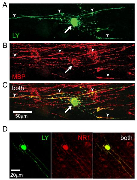
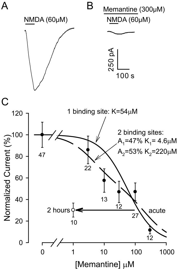
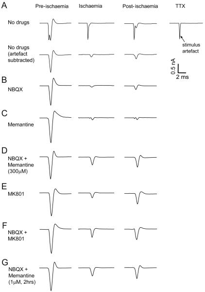
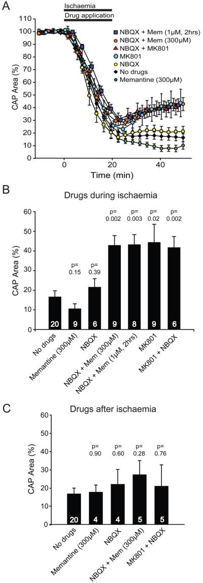
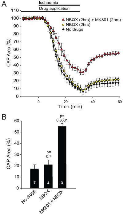
Damage to oligodendrocytes caused by glutamate release contributes to mental or physical handicap in periventricular leukomalacia, spinal cord injury, multiple sclerosis, and stroke, and has been attributed to activation of AMPA/kainate receptors. However, glutamate also activates unusual NMDA receptors in oligodendrocytes, which can generate an ion influx even at the resting potential in a physiological [Mg2+]. Here, we show that the clinically licensed NMDA receptor antagonist memantine blocks oligodendrocyte NMDA receptors at concentrations achieved therapeutically. Simulated ischaemia released glutamate which activated NMDA receptors, as well as AMPA/kainate receptors, on mature and precursor oligodendrocytes. Although blocking AMPA/kainate receptors alone during ischaemia had no effect, combining memantine with an AMPA/kainate receptor blocker, or applying the NMDA blocker MK-801 alone, improved recovery of the action potential in myelinated axons after the ischaemia. These data suggest NMDA receptor blockers as a potentially useful treatment for some white matter diseases and define conditions under which these blockers may be useful therapeutically. Our results highlight the importance of developing new antagonists selective for oligodendrocyte NMDA receptors based on their difference in subunit structure from most neuronal NMDA receptors.


Figures

Fig. 1
Fig. 1
Identification of corpus callosum oligodendrocytes expressing NMDA receptors. (A-C) Identification of corpus callosal oligodendrocyte. (A) Lucifer yellow (LY, green) fill of cell. (B) Antibody labelling (post-recording) for myelin basic protein (MBP, red). (C) Overlay of A and B (both). (D) Oligodendrocyte filled with Lucifer yellow, labelled post-recording for NR1 subunits of NMDA receptors.
Fig. 2
Fig. 2
Memantine blocks corpus callosum oligodendrocyte NMDA receptors. (A) Specimen current response to 60μM NMDA, at −70mV, of a mature oligodendrocyte. (B) NMDA-evoked current in another oligodendrocyte exposed to 300μM memantine for 1 min. (C) Dose-response curve for the effect of 1 min exposure to memantine (filled circles, ±s.e.m., number of cells shown under each point). The solid line fit shows a first order inhibition curve: current(current in absence of memantine)=IC50([memantine]+IC50) where the best fit (R2=0.81) concentration producing 50% block was IC50=54μM. The dashed line fit shows a second order inhibition curve: current(current in absence of memantine)=α.IC501([memantine]+IC501)+(100α).IC502([memantine]+IC502) where the best fit (R2=0.94) provided the following values: α = 51%, IC501=4.6μM and 100-α= 49%, IC502=220μM. Open circle shows effect of pre-incubating with 1μM memantine for 2 hours.
Fig. 3
Fig. 3
NMDA receptor mediated ischaemia-evoked inward current in corpus callosal oligodendrocytes. (A) Membrane current at −70mV in a mature oligodendrocyte during application of ischaemia solution. D-AP5 (200μM) blocks a component of the ischaemia-evoked current. (B) Mean current (±s.e.m.) blocked by D-AP5 (~6 mins after the start of ischaemia) in oligodendrocyte precursors (Prec) and mature myelinating oligodendrocytes (Mature). Numbers of cells shown on bars.
Fig. 4
Fig. 4
Ischaemia-evoked changes in optic nerve compound action potential (CAP). (A) Top row, traces from left are: CAP in control solution, after 20 mins ischaemia, 30 mins after returning to control solution, and the stimulus artefact recorded subsequently in 1μM TTX. Bottom row shows same traces with stimulus artefact trace subtracted. (B-G) Artefact-subtracted responses with 25μM NBQX (B), 300μM memantine (C), NBQX + memantine (D), 50μM MK-801 (E) or NBQX + MK-801 (F) present during ischaemia, and (G) NBQX present during ischaemia + 1 μM memantine pre-incubated 2 hours before ischemia and also present during ischaemia.
Fig. 5
Fig. 5
Effect of glutamate receptor blockers on ischaemia-evoked changes in optic nerve compound action potential (CAP). (A) Normalized area of rectified CAP before, during and after ischaemia (from 0-20 mins), using ischaemia solutions containing no glutamate receptor blockers (No drugs), 25μM NBQX, 300μM memantine (Mem), NBQX + Mem, 50μM MK-801, or NBQX + MK-801 (drugs were applied during ischaemia as shown by the bar), and also using solutions containing 1μM Mem from 2 hours before the ischaemia solution was applied (1μM Mem was present for the whole experiment, with NBQX present during ischaemia). (B) Normalised CAP area after 30 mins in normal solution, following 20 mins application of ischaemia solution containing different glutamate receptor blockers. Number of nerves studied is shown on each bar. (C) Normalised CAP area after 30 mins in normal solution containing the glutamate receptor blockers indicated, which was preceded by 20 mins of ischaemia solution lacking blockers. P values in (B) and (C) are for comparison with ‘No drugs’.
Fig. 6
Fig. 6
Effect of pre-incubation with glutamate receptor blockers on ischaemia-evoked changes in optic nerve compound action potential (CAP). (A) Normalized area of rectified CAP before, during and after ischaemia, using solutions containing no glutamate receptor blockers (No drugs), pre-incubation with 25μM NBQX from 2 hours before ischaemia until the end of ischaemia, or pre-incubation with 25μM NBQX and 50μM MK-801 from 2 hours before ischaemia until the end of ischaemia. These experiments used strong stimulation that evoked a near maximal response. (B) Normalised CAP area after 30 mins in normal solution, following the period of ischaemia. Number of nerves studied is shown on each bar. P values are compared with ‘No drugs’.

References

    1. Allen NJ, Káradóttir R, Attwell D. A preferential role for glycolysis in preventing the anoxic depolarization of rat hippocampal area CA1 pyramidal cells. J Neurosci. 2005;25:848–859. - PMC - PubMed
    1. Back SA, Craig A, Kayton RJ, Luo NL, Meshul CK, Allcock N, Fern R. Hypoxia-ischemia preferentially triggers glutamate depletion from oligodendroglia and axons in perinatal cerebral white matter. J Cereb Blood Flow Metab. 2007;27:334–347. - PubMed
    1. Blanpied TA, Boeckman FA, Aizenman E, Johnson JW. Trapping block of NMDA-activated responses by amantadine and memantine. J Neurophysiol. 1997;77:309–323. - PubMed
    1. Bolton S, Butt AM. The optic nerve: a model for axon-glia interactions. J Pharm Toxicol Meth. 2005;51:221–233. - PubMed
    1. Craig A, Ling Luo N, Beardsley DJ, Wingate-Pearse N, Walker DW, Hohimer AR, Back SA. Quantitative analysis of perinatal rodent oligodendrocyte lineage progression and its correlation with human. Exp Neurol. 2003;181:231–240. - PubMed

Publication types

MeSH terms