Review
. 2007 Nov 22;8 Suppl 1(Suppl 1):S11.
doi: 10.1186/1471-2091-8-S1-S11.

The role of the UPS in cystic fibrosis

Affiliations
Review

The role of the UPS in cystic fibrosis

Emma L Turnbull et al. BMC Biochem. .

Abstract

CF is an inherited autosomal recessive disease whose lethality arises from malfunction of CFTR, a single chloride (Cl-) ion channel protein. CF patients harbor mutations in the CFTR gene that lead to misfolding of the resulting CFTR protein, rendering it inactive and mislocalized. Hundreds of CF-related mutations have been identified, many of which abrogate CFTR folding in the endoplasmic reticulum (ER). More than 70% of patients harbor the DeltaF508 CFTR mutation that causes misfolding of the CFTR proteins. Consequently, mutant CFTR is unable to reach the apical plasma membrane of epithelial cells that line the lungs and gut, and is instead targeted for degradation by the UPS. Proteins located in both the cytoplasm and ER membrane are believed to identify misfolded CFTR for UPS-mediated degradation. The aberrantly folded CFTR protein then undergoes polyubiquitylation, carried out by an E1-E2-E3 ubiquitin ligase system, leading to degradation by the 26S proteasome. This ubiquitin-dependent loss of misfolded CFTR protein can be inhibited by the application of 'corrector' drugs that aid CFTR folding, shielding it from the UPS machinery. Corrector molecules elevate cellular CFTR protein levels by protecting the protein from degradation and aiding folding, promoting its maturation and localization to the apical plasma membrane. Combinatory application of corrector drugs with activator molecules that enhance CFTR Cl- ion channel activity offers significant potential for treatment of CF patients. Publication history: Republished from Current BioData's Targeted Proteins database (TPdb; http://www.targetedproteinsdb.com).


Figures

Figure 1
Figure 1
CFTR domain layout in the ER membrane lipid bilayer during ATP hydrolysis, depicting the membrane spanning domains (MSD), nucleotide binding domains (NBD) and regulatory domain (R). The membrane spanning domains are depicted in sepia and the cytosolic domains in aqua.
Figure 2
Figure 2
CFTR is co-translationally inserted into the ER membrane during ribosomal translation of CFTR mRNA from the nucleus. If CFTR is misfolded in the ER it is ubiquitylated and retrotranslocated to the cytosol, where it is degraded by the 26S proteasome. Upon inhibition of the proteasome, ubiquitylated CFTR is localized to a pericentriolar aggresome structure. Correctly folded CFTR proteins are transferred to the Golgi apparatus for glycolytic maturation via the coat complex II (COPII) machinery. Mature CFTR is exported to the plasma membrane to function as a chloride ion channel. CFTR protein levels at the plasma membrane are regulated by sub-apical vesicles delivering CFTR protein for either lysosomal degradation or recycling. For simplicity, we have represented CFTR with a single membrane spanning sepia symbol. Readers are invited to refer to figures 1 and 3 for the full domain architecture of the protein.
Figure 3
Figure 3
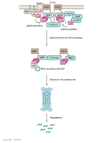
The UPS complexes located in the ER membrane (Derlin-1, RMA1, Ubc6e and p97) and cytoplasm (Hsp70 and CHIP) are shown. The figure demonstrates the polyubiquitylation of CFTR due to membrane-bound and cytosolic E3 ubiquitin ligase complexes. While the ubiquitylation likely occurs on the cytoplasmic domains of CFTR, the exact lysine residues conjugated with ubiquitin are unknown. p97 and possibly other associated factors are thought to participate in the extraction and delivery of CFTR from the ER membrane to the cytosolic proteasome. However, it is unknown whether the CFTR protein is extracted from the membrane in one piece, or degraded into smaller domains before retrotranslocation. Membrane spanning domains are depicted in sepia and the cytosolic domains in aqua.

References

    1. Rommens J.M., et al. Identification of the cystic fibrosis gene: chromosome walking and jumping. Science. 1989;245:1059–65. doi: 10.1126/science.2772657. - DOI - PubMed
    1. Riordan J.R., et al. Identification of the cystic fibrosis gene: cloning and characterization of complementary DNA. Science. 1989;245:1066–73. doi: 10.1126/science.2475911. - DOI - PubMed
    1. Sheppard D.N., et al. The amino-terminal portion of CFTR forms a regulated Cl- channel. Cell. 1994;76:1091–8. doi: 10.1016/0092-8674(94)90385-9. - DOI - PubMed
    1. Kreda S.M., et al. Characterization of wild-type and deltaF508 cystic fibrosis transmembrane regulator in human respiratory epithelia. Mol Biol Cell. 2005;16:2154–67. doi: 10.1091/mbc.E04-11-1010. - DOI - PMC - PubMed
    1. Roomans G.M. Pharmacological approaches to correcting the ion transport defect in cystic fibrosis. Am J Respir Med. 2003;2:413–31. - PubMed

Publication types

MeSH terms

Substances