. 2008 Feb 1;366(1):1-7.
doi: 10.1016/j.bbrc.2007.11.077. Epub 2007 Nov 29.

High definition profiling of autoantibodies to glutamic acid decarboxylases GAD65/GAD67 in stiff-person syndrome

Affiliations

High definition profiling of autoantibodies to glutamic acid decarboxylases GAD65/GAD67 in stiff-person syndrome

Peter D Burbelo et al. Biochem Biophys Res Commun. .

Abstract

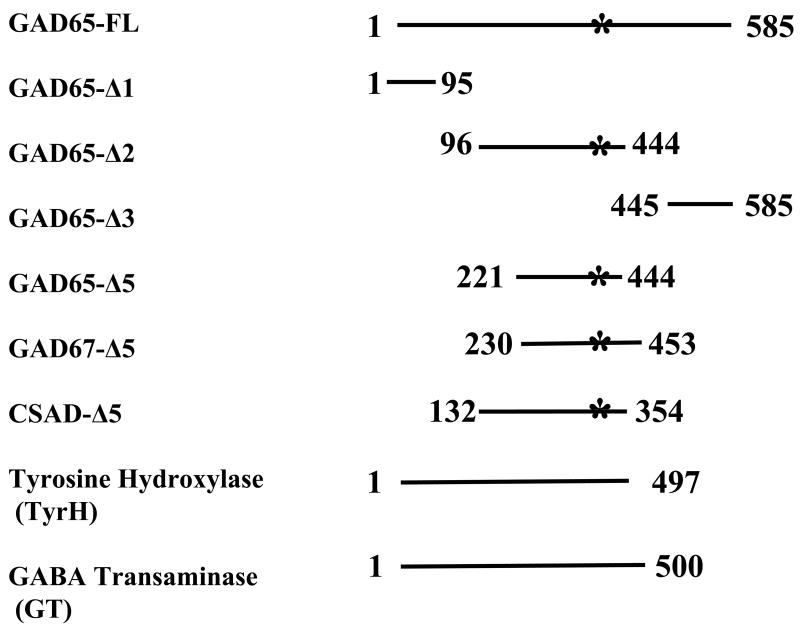
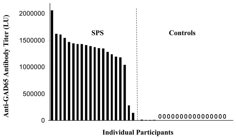
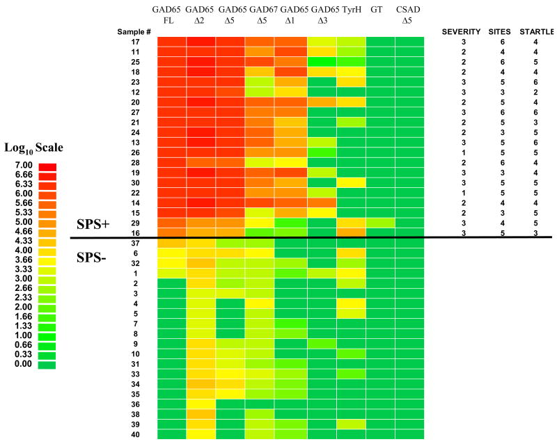
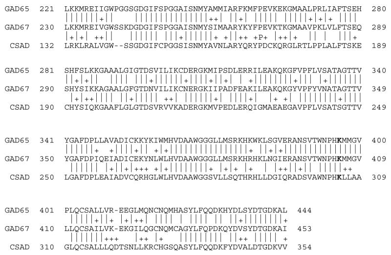
Highly reliable biomarkers for the diagnosis of neurological diseases are not widely available. Here we evaluated a luciferase immunoprecipitation technology (LIPS) for the diagnosis of a CNS autoimmune disorder, stiff-person syndrome (SPS). Analysis by LIPS of 40 sera samples from SPS and control subjects for anti-GAD65 antibodies revealed dramatic titer differences allowing diagnosis of SPS with 100% sensitivity and 100% specificity. Anti-GAD65 antibody titers of SPS were segregated from controls with values greater than 23 standard deviations above the control subject mean. By analyzing patient antibody responses directly to GAD65 sub-fragments, the central region containing the decarboxylase catalytic domain was highly immunoreactive with all of the SPS sera, while the N- and C-terminal regions showed lower antibody titers and only reacted with subsets of SPS sera. Additional profiling revealed that some SPS patients showed autoantibodies against GAD67 and tyrosine hydroxylase, but no significant immunoreactivity was detected with cysteine sulfinic acid decarboxylase or GABA transaminase. This study validates LIPS as a robust method to interrogate autoantibodies for the diagnosis of SPS and potentially other neurological diseases.


Figures

Fig. 1
Fig. 1
Proteins and protein fragments used as Ruc-fusions for autoantibody detection in SPS. Various coding sequences were amplified using PCR adapter primers and cloned in the pREN2 vector. Numbers represent amino acid residues used for each construct. The asterisk denotes the location of the pyridoxal phosphate-binding sites within GAD65, GAD67 and CSAD. Two other neurotransmitter related enzymes, tyrosine hydroxylase (TyrH) and GABA transaminase (GT) were also constructed.
Fig. 2
Fig. 2
Detection of patient antibodies to GAD65. Forty blinded sera samples from controls and SPS patients were utilized to evaluate anti-GAD65 antibody titers. Sera were analyzed using LIPS in two independent assays, the values averaged, and samples rank ordered from high to low titers corresponding to left to right, respectively. By analyzing the anti-GAD65 antibody titers of the 40 coded sera, the samples were statistically segregated (P-value of 3.3 × E−16) into two groups: 20 sera with a high anti-GAD65 antibody titer (left panel) and 20 sera with a low anti-GAD65 antibody titer (right panel). Sixteen of the samples showed values of zero (0). For SPS disease status, samples on the left panel with high anti-GAD65 antibody titer were predicted as positive. Following unblinding, the exact disease status of the 40 sera samples was revealed, showing that the LIPS data predicted SPS status from controls with 100% sensitivity and 100% specificity.
Fig. 3
Fig. 3
Detection of antibodies against GAD65 and its sub-fragments in SPS and control sera. Antibody titers to Ruc-antigen fusions for full-length GAD65 (FL) and five GAD65 sub-fragments (Δ1, Δ2, Δ3, and Δ5), GAD67-Δ5, tyrosine hydroxylase (TyrH), GABA transaminase (GT) and CSAD-Δ5 were determined in duplicate from 40 sera samples (1–40) using LIPS. The average titer values for each serum were log10 transformed and the samples were rank ordered from highest to lowest based on the antibody titer to full-length GAD65. Titer levels were color-coded as indicated by the log10 scale on the left, in which signal intensities range from red to green indicating high and low titers, respectively. SPS positive samples are above the black line. At the right is the clinical assessment profile including overall severity scale (1–3; mild, moderate, severe), number of sites affected (1–6) and startle response (1–7). No correlation was found between any of the autoantibody titers and these measures of disease severity.
Fig. 4
Fig. 4
Amino acid sequence homology within the corresponding regions of the GAD65-Δ5, GAD67-Δ5 and CSAD-Δ5 deletion mutants. The corresponding amino acid residues within each protein are shown. Identical amino acids residues are denoted by a dash line and the + denotes conservative amino acid substitutions. The active site lysine involved in pyridoxal phosphate binding is shown in bold.

References

    1. Turck CW, Maccarrone G, Sayan-Ayata E, Jacob AM, Ditzen C, Kronsbein H, Birg I, Doertbudak CC, Haegler K, Lebar M, Teplytska L, Kolb N, Uwaje N, Zollinger R. The quest for brain disorder biomarkers. J Med Invest. 2005;52(Suppl):231–5. - PubMed
    1. Sharp V, Utz PJ. Technology insight: can autoantibody profiling improve clinical practice? Nat Clin Pract Rheumatol. 2007;3:96–103. - PubMed
    1. Sodoyez-Goffaux F, Koch M, Dozio N, Brandenburg D, Sodoyez JC. Advantages and pitfalls of radioimmune and enzyme linked immunosorbent assays of insulin antibodies. Diabetologia. 1988;31:694–702. - PubMed
    1. Levy LM, Dalakas MC, Floeter MK. The stiff-person syndrome: an autoimmune disorder affecting neurotransmission of gamma-aminobutyric acid. Ann Intern Med. 1999;131:522–30. - PubMed
    1. Solimena M, Folli F, Aparisi R, Pozza G, De Camilli P. Autoantibodies to GABA-ergic neurons and pancreatic beta cells in stiff-man syndrome. N Engl J Med. 1990;322:1555–60. - PubMed

Publication types

LinkOut - more resources