. 2007 Dec 4;104(49):19583-8.
doi: 10.1073/pnas.0709846104. Epub 2007 Nov 28.

A helix-breaking mutation in TRPML3 leads to constitutive activity underlying deafness in the varitint-waddler mouse

Affiliations

A helix-breaking mutation in TRPML3 leads to constitutive activity underlying deafness in the varitint-waddler mouse

Christian Grimm et al. Proc Natl Acad Sci U S A. .

Abstract

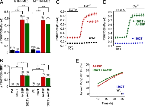
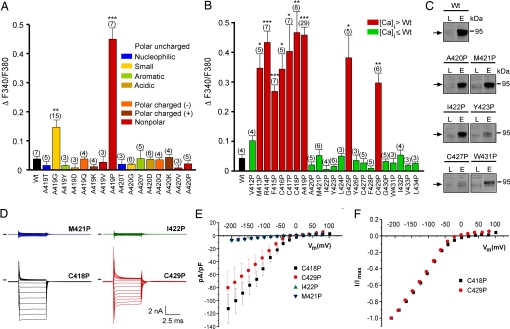
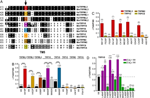
Homozygote varitint-waddler (Va) mice, expressing a mutant isoform (A419P) of TRPML3 (mucolipin 3), are profoundly deaf and display vestibular and pigmentation deficiencies, sterility, and perinatal lethality. Here we show that the varitint-waddler isoform of TRPML3 carrying an A419P mutation represents a constitutively active cation channel that can also be identified in native varitint-waddler hair cells as a distinct inwardly rectifying current. We hypothesize that the constitutive activation of TRPML3 occurs as a result of a helix-breaking proline substitution in transmembrane-spanning domain 5 (TM5). A proline substitution scan demonstrated that the inner third of TRPML3's TM5 is highly susceptible to proline-based kinks. Proline substitutions in TM5 of other TRP channels revealed that TRPML1, TRPML2, TRPV5, and TRPV6 display a similar susceptibility at comparable positions, whereas other TRP channels were not affected. We conclude that the molecular basis for deafness in the varitint-waddler mouse is the result of hair cell death caused by constitutive TRPML3 activity. To our knowledge, our study provides the first direct mechanistic link of a mutation in a TRP ion channel with mammalian hearing loss.


Conflict of interest statement

The authors declare no conflict of interest.

Figures

Fig. 1.
Fig. 1.
Elevated intracellular Ca2+ and Na+ levels and annexin V binding to HEK293 cells expressing varitint-waddler TRPML3 mutants. Ca2+ imaging (A) and Na+ imaging (B) results showing intracellular Ca2+ and Na+ levels of HEK293 cells expressing human (Hs) and murine (Mm) TRPML3 and the respective mutants. All experiments were performed 10–15 h after transfection (mean ± SEM; n is in parentheses. ***, P < 0.0001; **, P < 0.001 (Student's t test, unpaired, comparison with wild type). Transfected cells were identified by YFP fluorescence of the expressed proteins. (C and D) Ca2+ imaging experiments showing that the [Ca2+]i increase depends on extracellular Ca2+. After incubation in 2 mM EGTA for 5 min, the external buffer solution was switched to 2 mM calcium. (E) Quantification of the number of transfected HEK293 cells that bound Cy5-conjugated annexin V, an indication of early signs of apoptosis. Shown are time points after transfection with expression vectors for the mutants indicated. We found no annexin V-positive cells expressing wild-type TRPML3 or TRPML3(I362T) 25 h after transfection.
Fig. 2.
Fig. 2.
Constitutively active conductance elicited by varitint-waddler TRPML3 mutants in HEK293 cells and cochlear outer hair cells. (A) Currents elicited from wild type (black), A419P mutant (red), and I362/A419P mutant (green) in response to 5-ms voltage steps from a holding potential of −50 mV between −200 mV and +100 mV in 20-mV incremental steps. Black bars indicate zero current line. (B) Steady-state current–voltage plots from A normalized to cell capacitance. A two-tailed Student t test revealed no significant difference (P > 0.01) between the data points for any voltage measured (n = 9 for A419P, and n = 9 for I362T/A419P). (C) Same plot, but normalized to maximal current elicited at −200 mV to demonstrate similarity in responses. (D) Currents from a wild-type outer hair cell measured with Cs+-based intracellular solution. Membrane voltage range: −104 mV to +96 mV, in 20-mV increments, from a holding potential of −84 mV. (E) Currents recorded from a VaJ/VaJ outer hair cell under the same conditions as in D. (F) Comparison of the current–voltage curves for experiments similar to those shown in D and E, determined just after the start of the voltage steps. Conductance at −84 mV was 1.95 ± 0.26 nS for wild type (n = 4 OHCs, mean ± SEM) and 28.0 ± 7.4 nS for VaJ/VaJ (n = 7 OHCs) (P < 0.02, unpaired t test). (G) Normalized hair cell current from VaJ/VaJ mouse OHCs (n = 7) compared with expressed I362T/A419P (n = 9), normalized to −100 mV, to demonstrate similarity between responses. No statistical differences were found between the outer hair cell currents and the expressed currents at any voltage using the Student two-tailed t test (P > 0.05).
Fig. 3.
Fig. 3.
Effects of amino acid substitutions in TM5 of TRPML3. (A) Effect of the substitution of alanine at positions 419 and 420 in murine TRPML3 by various amino acids on [Ca2+]i. Note that only proline and glycine substitution at position 419 significantly led to an increase of [Ca2+]i. Shown are mean values ± SEM with n in parentheses. All experiments were performed 10–15 h after transfection. (B) Effect on [Ca2+]i of proline substitution of the residues predicted to constitute TM5 of murine TRPML3. Shown are mean values ± SEM with n in parentheses. Nine (red) of the 23 mutant isoforms showed significantly elevated basal intracellular calcium levels compared with wild-type TRPML3 (Wt). V412P is not part of the predicted TM5. ***, P < 0.0001; **, P < 0.001; *, P < 0.01 (Student's t test, unpaired, comparison with wild type). (C) Surface biotinylation analysis. TRPML3 wild-type protein (Wt) and the mutant isoforms are present in the plasma membrane. L indicates input load (1% of total), and E indicates elution from neutravidin beads of surface-biotinylated TRPML3 visualized by Western blot. (D) Voltage-clamp recording from mutants as listed, expressed in HEK293 cells. Voltage protocol consists of 20-mV incremental steps between −200 and +80 mV. Inward rectifier current is present in mutant TRPML3-expressing cells that showed elevated calcium and is not present in those where calcium was not elevated. Black bars indicate zero current line. (E) Steady-state current–voltage plots generated from data in D. (F) Current–voltage plots normalized to current at −200 mV to demonstrate similar relationships between mutants.
Fig. 4.
Fig. 4.
Susceptibility to proline substitutions in TM5 is a general feature of TRPML channels as well as TRPV5 and TRPV6. (A) Amino acid sequence comparison of TM5 of human (Hs) and murine (Mm) TRPML3 with TRPML1, TRPML2, TRPV5, TRPV6, TRPV2, TRPM2, and TRPC6. Positions equivalent to TRPML3(A419P) (red) are highlighted: yellow, TRPML1; dark brown, TRPML2; magenta, TRPV5; blue, TRPV6; orange, TRPV2; brown, TRPM2; olive, TRPC6. (B) Relative [Ca2+]i levels of HEK293 cells expressing mmTRPML3, hsTRPML1, mmTRPML2, mmTRPV5, mmTRPV6, mmTRPV2, hsTRPM2, and hsTRPC6 wild type and mutant isoforms as indicated in A. Shown are mean values ± SEM with the number of independent experiments with at least 5–10 cells each shown in parentheses. (C) Proline substitutions at additional positions in TM5 of TRPV2, TRPM2, and TRPC6. Proline substitutions of TRPML3 at equivalent positions resulted in elevated [Ca2+]i (red bars). None of the mutated TRPV2, TRPM2, and TRPC6 isoforms displayed elevated [Ca2+]i. (D) Effect on [Ca2+]i of proline substitution along TM5 of murine TRPV5. Shown are mean values ± SEM with n in parentheses. ***, P < 0.0001; **, P < 0.001; *, P < 0.05 (Student's t test, unpaired, comparison with wild type).

References

    1. Clapham DE. Nature. 2003;426:517–524. - PubMed
    1. Montell C. Sci STKE. 2005;2005:re3. - PubMed
    1. Walker RG, Willingham AT, Zuker CS. Science. 2000;287:2229–2234. - PubMed
    1. Kim J, Chung YD, Park DY, Choi S, Shin DW, Soh H, Lee HW, Son W, Yim J, Park CS, et al. Nature. 2003;424:81–84. - PubMed
    1. Gong Z, Son W, Chung YD, Kim J, Shin DW, McClung CA, Lee Y, Lee HW, Chang DJ, Kaang BK, et al. J Neurosci. 2004;24:9059–9066. - PMC - PubMed

Publication types