. 2007 Dec 25;46(51):15076-88.
doi: 10.1021/bi701732a. Epub 2007 Dec 1.

On the relationship between thermal stability and catalytic power of enzymes

Affiliations

On the relationship between thermal stability and catalytic power of enzymes

Maite Roca et al. Biochemistry. .

Abstract

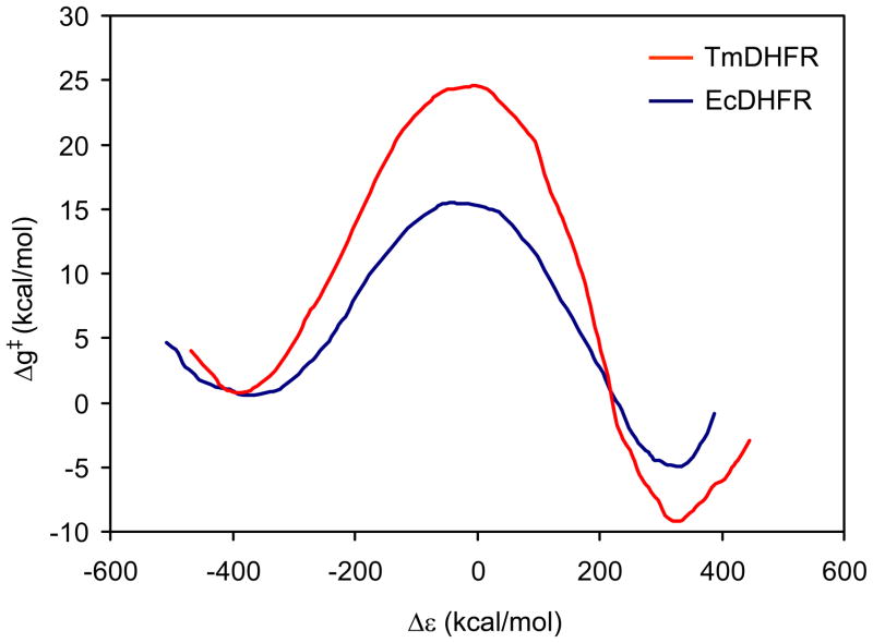
The possible relationship between the thermal stability and the catalytic power of enzymes is of great current interest. In particular, it has been suggested that thermophilic or hyperthermophilic (Tm) enzymes have lower catalytic power at a given temperature than the corresponding mesophilic (Ms) enzymes, because the thermophilic enzymes are less flexible (assuming that flexibility and catalysis are directly correlated). These suggestions presume that the reduced dynamics of the thermophilic enzymes is the reason for their reduced catalytic power. The present paper takes the specific case of dihydrofolate reductase (DHFR) and explores the validity of the above argument by simulation approaches. It is found that the Tm enzymes have restricted motions in the direction of the folding coordinate, but this is not relevant to the chemical process, since the motions along the reaction coordinate are perpendicular to the folding motions. Moreover, it is shown that the rate of the chemical reaction is determined by the activation barrier and the corresponding reorganization energy, rather than by dynamics or flexibility in the ground state. In fact, as far as flexibility is concerned, we conclude that the displacement along the reaction coordinate is larger in the Tm enzyme than in the Ms enzyme and that the general trend in enzyme catalysis is that the best catalyst involves less motion during the reaction than the less optimal catalyst. The relationship between thermal stability and catalysis appears to reflect the fact that to obtain small electrostatic reorganization energy it is necessary to invest some folding energy in the overall preorganization process. Thus, the optimized catalysts are less stable. This trend is clearly observed in the DHFR case.


Figures

Figure 1
Figure 1
A schematic representation of the reaction catalyzed by DHFR. This reaction involves a hydride transfer from the NADPH coenzyme to the 7,8-dihydrofolate substrate.
Figure 2
Figure 2
X-ray structures of the mesophilic Dihydrofolate Reductase (monomer) from Escherichia coli (EcDHFR) and the hyperthermophilic Dihydrofolate Reductase (dimer) from Thermotoga maritime (TmDHFR). The 7,8-dihydrofolate substrate (in orange) and NADPH coenzyme (in blue) are represented in sphere model.
Figure 3
Figure 3
(a) Free energy landscapes for the EcDHFR and TmDHFR. The free energy surfaces were obtained by the FEP/US method and are represented in terms of the radius of gyration (Rg) and the root mean square deviation (RMSD) from the most stable structure. Energies are expressed in kcal/mol and distances in Å. (b) Free energy surfaces for the EcDHFR and TmDHFR obtained by long molecular dynamics simulations without any constraint.
Figure 4
Figure 4
The EVB free energy profiles for the hydride transfer reaction of the EcDHFR (in blue) and the TmDHFR (in red). The profiles were obtained by averaging several EVB simulations as described elsewhere (44) and are given in terms of the EVB reaction coordinate (Δε =ε2−ε1 energy gap).
Figure 5
Figure 5
Representation of the total diabatic free energy functional (a), the solvent (b) and solute (c) contributions for the EcDHFR (in black) and the TmDHFR (in blue). The figures display the functional after shifting the minima to the same heights. The intersection region is magnified in each case. As discussed in ref (44) the larger the reorganization (λ) the higher the activation barrier.
Figure 5
Figure 5
Representation of the total diabatic free energy functional (a), the solvent (b) and solute (c) contributions for the EcDHFR (in black) and the TmDHFR (in blue). The figures display the functional after shifting the minima to the same heights. The intersection region is magnified in each case. As discussed in ref (44) the larger the reorganization (λ) the higher the activation barrier.
Figure 6
Figure 6
The free energy surface for EcDHFR (a) and TmDHFR (b). The surfaces are given in terms of the solute and solvent coordinates, which reflect the solute and solvent reorganization energies. The activation energies of the reaction are represented in EcDHFR and TmDHFR which are 15 kcal/mol and 23 kcal/mol, respectively. RS, TS and PS designate reactant state, transition state and product state, respectively.
Figure 7
Figure 7
The free energy surface as a function of the donor-acceptor distance (R(D-A)) and donor-hydrogen (R(D-H)) for the reaction catalyzed by the EcDHFR (a) and the TmDHFR (b). The contour lines are given in kcal/mol. RS, TS and PS designate reactant state, transition state and product state, respectively.
Figure 8
Figure 8
Dispersed polaron (DP) distribution analysis, taking into account only the electrostatic contribution of the solvent, for the EcDHFR (in blue) and TmDHFR (in red). This analysis gives the projection of the modes of the protein system along the reaction coordinate where the projection of each mode is represented by the corresponding reorganization energy λi=12ωiΔi2.
Figure 9
Figure 9
The relationship between the folding motions and the reactive modes in the reactant state of EcDHFR. In red arrows we represent the vector of the motion along the reaction coordinate for the residues near or at the active site while in black arrows are presented the direction of the folding coordinate. The figure considers (a) the direction of both the reaction coordinate and the folding coordinate in a partially unfolded structure. The two set of motions are almost orthogonal to each other (96° to each other) when we consider all the coordinates of the system. If we start the folding motion from a partially unfolded structure closer to the native structure they are close to be orthogonal (the angle between the two vectors is around 100°). (b) If we start from the occluded structure (49) the angle between both coordinates is around 93°.
Figure 9
Figure 9
The relationship between the folding motions and the reactive modes in the reactant state of EcDHFR. In red arrows we represent the vector of the motion along the reaction coordinate for the residues near or at the active site while in black arrows are presented the direction of the folding coordinate. The figure considers (a) the direction of both the reaction coordinate and the folding coordinate in a partially unfolded structure. The two set of motions are almost orthogonal to each other (96° to each other) when we consider all the coordinates of the system. If we start the folding motion from a partially unfolded structure closer to the native structure they are close to be orthogonal (the angle between the two vectors is around 100°). (b) If we start from the occluded structure (49) the angle between both coordinates is around 93°.
Figure 10
Figure 10
Representation of the donor-acceptor distance versus the acceptor-hydrogen distance along the reactive downhill trajectories in EcDHFR (a) in TmDHFR (b). The figures show that both systems oscillate in the RS for a very long time until it generates the rare reactive trajectory that goes to the TS.
Figure 11
Figure 11
Average projection of the downhill trajectories in EcDHFR on the folding coordinate (in blue) and on the reaction coordinate (in red). The negative time is taken relative to the start of the downhill trajectory and its time reversal corresponds to a reactive trajectory from the RS. The projection has been evaluated while considering the EVB reacting region and its surroundings up to 5 Å from the EVB atoms (see text).
Figure 12
Figure 12
Normalized autocorrelation of ΔrRC · δr(t)RC (in red) and ΔrFC · δr(t)RC (in blue) for the EcDHFR system. The fact that the autocorrelation decays in a very short time indicates that it is very unlikely that the fluctuation along the folding coordinate would transfer energy to the reaction coordinate.
Figure 13
Figure 13
A schematic representation of the folding and reaction processes in both systems the mesophile and the thermophile. Starting from a partially unfolded structure (a), the folding process takes place to reach the reactant state and folded structure (b). The rates in the figure correspond to the overall time until a given process occurs rather than to the time of a reactive trajectory. The rates for the motion along the folding coordinate are somewhat arbitrary. Note also that the actual time for a reactive trajectory is about 1ps. The figure illustrates schematically the fact that the enzyme spends most of the time at the reactant state in both systems and the motions are randomized by the thermal energy to lead to reactive trajectories only according to the Boltzmann probability of being in the TS (c).

References

    1. Marti S, Roca M, Andres J, Moliner V, Silla E, Tunon I, Bertran J. Theoretical insights in enzyme catalysis. Chemical Society Reviews. 2004;33:98–107. - PubMed
    1. Warshel A, Sharma PK, Kato M, Xiang Y, Liu HB, Olsson MHM. Electrostatic basis for enzyme catalysis. Chemical Reviews. 2006;106:3210–3235. - PubMed
    1. Epstein DM, Benkovic SJ, Wright PE. Dynamics of the Dihydrofolate-Reductase Folate Complex - Catalytic Sites and Regions Known to Undergo Conformational Change Exhibit Diverse Dynamical Features. Biochemistry. 1995;34:11037–11048. - PubMed
    1. Schnell JR, Dyson HJ, Wright PE. Structure, dynamics, and catalytic function of dihydrofolate reductase. Annual Review of Biophysics and Biomolecular Structure. 2004;33:119–140. - PubMed
    1. Hammes-Schiffer S. Quantum-classical simulation methods for hydrogen transfer in enzymes: a case study of dihydrofolate reductase. Current Opinion in Structural Biology. 2004;14:192–201. - PubMed

Publication types

Substances