. 2007 Dec 25;46(51):14878-88.
doi: 10.1021/bi701532r. Epub 2007 Dec 4.

Crystal structure of a type II dihydrofolate reductase catalytic ternary complex

Affiliations

Crystal structure of a type II dihydrofolate reductase catalytic ternary complex

Joseph M Krahn et al. Biochemistry. .

Abstract

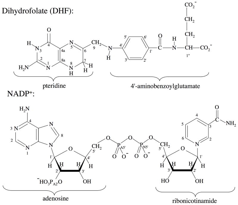
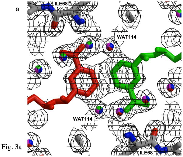
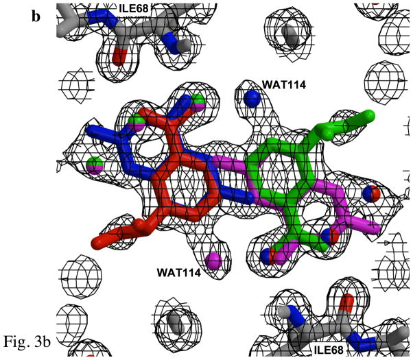
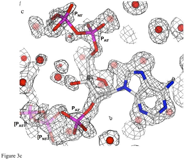
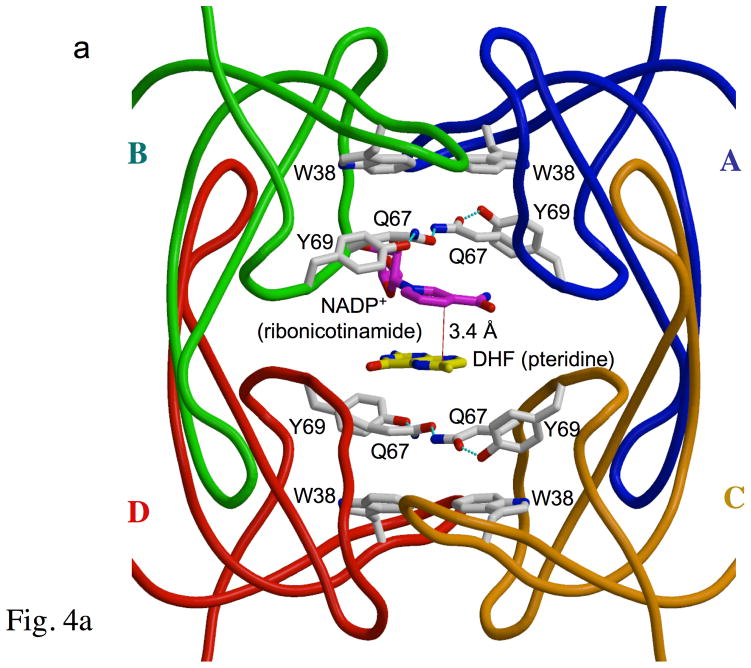
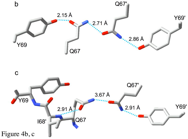
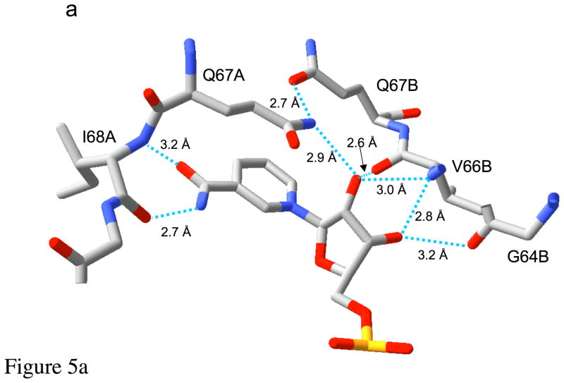
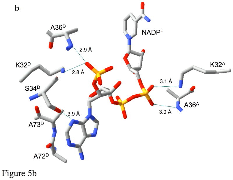
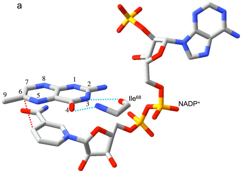
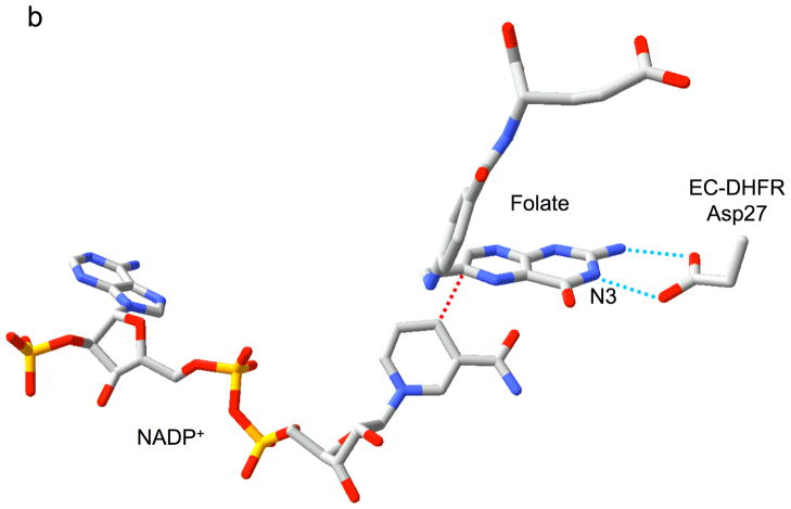
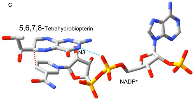
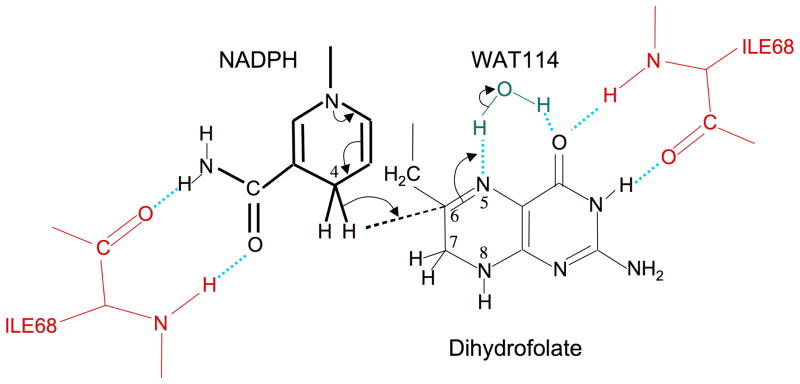
Type II dihydrofolate reductase (DHFR) is a plasmid-encoded enzyme that confers resistance to bacterial DHFR-targeted antifolate drugs. It forms a symmetric homotetramer with a central pore which functions as the active site. Its unusual structure, which results in a promiscuous binding surface that accommodates either the dihydrofolate (DHF) substrate or the NADPH cofactor, has constituted a significant limitation to efforts to understand its substrate specificity and reaction mechanism. We describe here the first structure of a ternary R67 DHFR.DHF.NADP+ catalytic complex, resolved to 1.26 A. This structure provides the first clear picture of how this enzyme, which lacks the active site carboxyl residue that is ubiquitous in Type I DHFRs, is able to function. In the catalytic complex, the polar backbone atoms of two symmetry-related I68 residues provide recognition motifs that interact with the carboxamide on the nicotinamide ring, and the N3-O4 amide function on the pteridine ring. This set of interactions orients the aromatic rings of substrate and cofactor in a relative endo geometry in which the reactive centers are held in close proximity. Additionally, a central, hydrogen-bonded network consisting of two pairs of Y69-Q67-Q67'-Y69' residues provides an unusually tight interface, which appears to serve as a "molecular clamp" holding the substrates in place in an orientation conducive to hydride transfer. In addition to providing the first clear insight regarding how this extremely unusual enzyme is able to function, the structure of the ternary complex provides general insights into how a mutationally challenged enzyme, i.e., an enzyme whose evolution is restricted to four-residues-at-a-time active site mutations, overcomes this fundamental limitation.


Figures

Figure 1
Figure 1
Structure and numbering of the dihydrofolate and NADP+ ligands present in the R67 DHFR•DHF•NADP+ ternary crystal structure.
Figure 2
Figure 2
The crystallographic problem posed by the DHFR•DHF•NADP+ ternary complex. A schematic, cross sectional view of R67 DHFR is shown in blue, with the pore shown in gray. The three perpendicular two-fold axes are indicated in red. On the left, a single pair of ligands is shown, while on the right, the four symmetry related pairs are indicated. In the crystal, each relative orientation is expected to have 25 % occupancy.
Figure 3
Figure 3
Fits of electron density. a) Fit of the electron density at the center of the pore for the binary complex, revealing the nicotinamide rings for two of the four symmetry-related NADP+ positions. Electron density represents 2Fobs-Fcalc, contoured at 0.5σ in order to illustrate density for ligands at 25% occupancy. Each color (red, blue, green, and magenta) identifies a unique set of NADP+ or water ligands among the four symmetry-related groups which are averaged in the crystal. Two symmetry-related nicotinamide rings are shown. Since only one NADP+ binds per tetramer (9), the ligand is present at 25% occupancy at each position. Symmetry-related water molecules bind to each of the sites not occupied by NADP+, resulting in 75% occupancy. For example, the green, magenta and blue water molecules describe water seen in three symmetry related sites when the red colored cofactor is bound. The protein side chains at the top and bottom of the figure correspond to the NH and O backbone atoms from symmetry related Ile68 residues, which interact with the amide of the nicotinamide. b) Electron density for one side of the pore in the ternary complex (contoured the same as a), showing overlapping nicotinamide and pteridine groups. Color-coding is as in panel a, however in this case, the two ligands exclude several water molecules from two of the four sites. Electron density for the pyridine ring of the nicotinamide overlaps closely with that arising from the pyrazine ring of DHF. Note that these figures illustrate electron density for symmetry-related molecules, rather than for an interacting substrate-cofactor pair, which is discussed below. WAT114, positioned near the pteridine O4, has only 25 % occupancy and appears to be associated with the pteridine ring. c) Electron density corresponding to the phospho-adenosine pyrophosphate group of NADP+. The bracketed PN5′ and PA5′ from a symmetry-related NADP+ molecule, shown in lighter shades of magenta and red, are also included. The density was taken from data for the ternary complex. Symmetry-related water molecules are also indicated by faded coloring.
Figure 3
Figure 3
Fits of electron density. a) Fit of the electron density at the center of the pore for the binary complex, revealing the nicotinamide rings for two of the four symmetry-related NADP+ positions. Electron density represents 2Fobs-Fcalc, contoured at 0.5σ in order to illustrate density for ligands at 25% occupancy. Each color (red, blue, green, and magenta) identifies a unique set of NADP+ or water ligands among the four symmetry-related groups which are averaged in the crystal. Two symmetry-related nicotinamide rings are shown. Since only one NADP+ binds per tetramer (9), the ligand is present at 25% occupancy at each position. Symmetry-related water molecules bind to each of the sites not occupied by NADP+, resulting in 75% occupancy. For example, the green, magenta and blue water molecules describe water seen in three symmetry related sites when the red colored cofactor is bound. The protein side chains at the top and bottom of the figure correspond to the NH and O backbone atoms from symmetry related Ile68 residues, which interact with the amide of the nicotinamide. b) Electron density for one side of the pore in the ternary complex (contoured the same as a), showing overlapping nicotinamide and pteridine groups. Color-coding is as in panel a, however in this case, the two ligands exclude several water molecules from two of the four sites. Electron density for the pyridine ring of the nicotinamide overlaps closely with that arising from the pyrazine ring of DHF. Note that these figures illustrate electron density for symmetry-related molecules, rather than for an interacting substrate-cofactor pair, which is discussed below. WAT114, positioned near the pteridine O4, has only 25 % occupancy and appears to be associated with the pteridine ring. c) Electron density corresponding to the phospho-adenosine pyrophosphate group of NADP+. The bracketed PN5′ and PA5′ from a symmetry-related NADP+ molecule, shown in lighter shades of magenta and red, are also included. The density was taken from data for the ternary complex. Symmetry-related water molecules are also indicated by faded coloring.
Figure 4
Figure 4
The “club sandwich motif” of R67 DHFR. Panel a shows a crystallographic tetramer with a single (asymmetric) pair of ligands. Each monomer is color-coded and labeled A-D. The active site pore faces the viewer. The four Trp38 residues, which contribute to the multi-tiered stack, occur on the outer layers of the “sandwich”. Moving inwards, the next layers are the symmetry-related Y69-Q67-Q67′-Y69′ residues, which “clamp” the pteridine and nicotinamide rings at the center. These residues sandwich the ligands, DHF (yellow) and NADP+ (magenta; only the ribonicotinamide ring is shown for simplicity). A red line shows the proximity of the C4 (nicotinamide) and C6 (pteridine) atoms, which are involved in hydride transfer. Panels b and c show details of the R67 DHFR “clamp”. In panel b, the structure of one of the two hydrogen bonding networks formed from Q67 and Y69 residues on chains A and B is shown. This panel corresponds to the top of the pore in panel a, rotated 90° about the horizontal axis. In panel c, the structure of the Y69-Q67-Q67′-Y69′ group in the apo enzyme is shown. In this case, one of the Gln67 residues tends to adopt an alternate conformation, which relieves the congestion and allows the sidechain to form an alternate hydrogen bond with Ile68 NH instead of with Tyr69 OH.
Figure 4
Figure 4
The “club sandwich motif” of R67 DHFR. Panel a shows a crystallographic tetramer with a single (asymmetric) pair of ligands. Each monomer is color-coded and labeled A-D. The active site pore faces the viewer. The four Trp38 residues, which contribute to the multi-tiered stack, occur on the outer layers of the “sandwich”. Moving inwards, the next layers are the symmetry-related Y69-Q67-Q67′-Y69′ residues, which “clamp” the pteridine and nicotinamide rings at the center. These residues sandwich the ligands, DHF (yellow) and NADP+ (magenta; only the ribonicotinamide ring is shown for simplicity). A red line shows the proximity of the C4 (nicotinamide) and C6 (pteridine) atoms, which are involved in hydride transfer. Panels b and c show details of the R67 DHFR “clamp”. In panel b, the structure of one of the two hydrogen bonding networks formed from Q67 and Y69 residues on chains A and B is shown. This panel corresponds to the top of the pore in panel a, rotated 90° about the horizontal axis. In panel c, the structure of the Y69-Q67-Q67′-Y69′ group in the apo enzyme is shown. In this case, one of the Gln67 residues tends to adopt an alternate conformation, which relieves the congestion and allows the sidechain to form an alternate hydrogen bond with Ile68 NH instead of with Tyr69 OH.
Figure 5
Figure 5
Recognition of NADP+. Panel a shows the interactions of the ribonicotinamide-ribose moiety of bound NADP+ with the protein. The extensive hydrogen bond network to the ribose helps to position the reactive centers and also tilts the rings into a more reactive geometry. Panel b illustrates many of the remaining interactions of the R67 DHFR with the cofactor. Ionic interactions between symmetry related Lys32 residues and two of the phosphate groups as well as several H-bonds are involved in binding. The ionic interactions have been experimentally monitored by ionic strength effects on binding (33). The terminal subscript indicates the chain identity of the residue.
Figure 5
Figure 5
Recognition of NADP+. Panel a shows the interactions of the ribonicotinamide-ribose moiety of bound NADP+ with the protein. The extensive hydrogen bond network to the ribose helps to position the reactive centers and also tilts the rings into a more reactive geometry. Panel b illustrates many of the remaining interactions of the R67 DHFR with the cofactor. Ionic interactions between symmetry related Lys32 residues and two of the phosphate groups as well as several H-bonds are involved in binding. The ionic interactions have been experimentally monitored by ionic strength effects on binding (33). The terminal subscript indicates the chain identity of the residue.
Figure 6
Figure 6
Relative substrate orientation. a) Relative orientation of NADP+ and the pteridine ring of DHF determined here for the R67 DHFR•DHF•NADP+ ternary complex. The backbone of Ile68 on chain D, which interacts with the N3-O4 amide of DHF, is also shown. The nicotinamide and pteridine ring systems adopt an endo conformation in which the closest approach corresponds to the reactive nicotinamide C4 and pteridine C6 carbons. The NADP+ is wrapped around the pteridine ring, so that the phosphates are positioned near the 2-amino group: distances: Ad-5′-P---N = 5.4 Å; Nic-5′-P---N = 6.1 Å; Ad-2′-P---N = 6.8 Å. b) Relative substrate orientation in a ternary DHFR•folate•NADP+ complex corresponding to the E. coli (Type I) enzyme (pdb entry 1RX2; (27)). The Asp-27 sidechain from the E. coli enzyme, which binds to folate N2 and N3, is also shown. The relative exo orientation contrasts with that observed for the Type II enzyme. c) Relative substrate orientation in a ternary PTR•tetrahydrobiopterin•NADP+ complex corresponding to the leishmania pteridine reductase (pdb entry 2BFP (67)). The relative endo orientation is analogous to that observed for R67 DHFR, however the enzyme catalyzes a B-side hydride transfer, so that the orientaton of the nicotinamide ring is flipped. Hydrogen bonding/salt bridge interactions with N3 are shown as blue dotted lines, and the reactive centers on the substrates are connected with red dotted lines. In order to facilitate comparison, the orientation of the pteridine ring system is similarly oriented in each frame.
Figure 6
Figure 6
Relative substrate orientation. a) Relative orientation of NADP+ and the pteridine ring of DHF determined here for the R67 DHFR•DHF•NADP+ ternary complex. The backbone of Ile68 on chain D, which interacts with the N3-O4 amide of DHF, is also shown. The nicotinamide and pteridine ring systems adopt an endo conformation in which the closest approach corresponds to the reactive nicotinamide C4 and pteridine C6 carbons. The NADP+ is wrapped around the pteridine ring, so that the phosphates are positioned near the 2-amino group: distances: Ad-5′-P---N = 5.4 Å; Nic-5′-P---N = 6.1 Å; Ad-2′-P---N = 6.8 Å. b) Relative substrate orientation in a ternary DHFR•folate•NADP+ complex corresponding to the E. coli (Type I) enzyme (pdb entry 1RX2; (27)). The Asp-27 sidechain from the E. coli enzyme, which binds to folate N2 and N3, is also shown. The relative exo orientation contrasts with that observed for the Type II enzyme. c) Relative substrate orientation in a ternary PTR•tetrahydrobiopterin•NADP+ complex corresponding to the leishmania pteridine reductase (pdb entry 2BFP (67)). The relative endo orientation is analogous to that observed for R67 DHFR, however the enzyme catalyzes a B-side hydride transfer, so that the orientaton of the nicotinamide ring is flipped. Hydrogen bonding/salt bridge interactions with N3 are shown as blue dotted lines, and the reactive centers on the substrates are connected with red dotted lines. In order to facilitate comparison, the orientation of the pteridine ring system is similarly oriented in each frame.
Figure 7
Figure 7
Enzyme-substrate interactions and catalytic mechanism. The enzyme exploits a subtle symmetry between the NADP+ cofactor and the DHF substrate by interacting similarly with the nicotinamide amide group in the first case, and the N3-O4 amide group of the pteridine. In both cases, a pair of hydrogen bonds is formed with the backbone carbonyl and amide groups of the Ile68 residues on chains A and D. As a result of these and other interactions described in the text, the relative positions of two ring systems are optimized for hydride transfer to C6. This transfer follows or is concerted with N5 protonation, presumably from WAT114. WAT114 is within hydrogen bonding distance of DHF O4, but is solvent accessible and does not otherwise appear to be specifically activated.

Similar articles

Cited by

References

    1. Fleming MP, Datta N, Gruneberg RN. Trimethoprim resistance determined by R factors. Br Med J. 1972;1:726–8. - PMC - PubMed
    1. Pattishall KH, Acar J, Burchall JJ, Goldstein FW, Harvey RJ. Two distinct types of trimethoprim-resistant dihydrofolate reductase specified by R-plasmids of different compatibility groups. J Biol Chem. 1977;252:2319–23. - PubMed
    1. Stone D, Smith SL. The amino acid sequence of the trimethoprim- resistant dihydrofolate reductase specified in Escherichia coli by R-plasmid R67. J Biol Chem. 1979;254:10857–61. - PubMed
    1. Smith SL, Stone D, Novak P, Baccanari DP, Burchall JJ. R plasmid dihydrofolate reductase with subunit structure. J Biol Chem. 1979;254:6222–5. - PubMed
    1. Narayana N, Matthews DA, Howell EE, Nguyen-huu X. A plasmid-encoded dihydrofolate reductase from trimethoprim-resistant bacteria has a novel D2-symmetric active site. Nat Struct Biol. 1995;2:1018–1025. - PubMed

Publication types