. 2008 Apr;10(4):994-1007.
doi: 10.1111/j.1462-5822.2007.01102.x. Epub 2007 Dec 4.

The Pseudomonas aeruginosa type III secreted toxin ExoT is necessary and sufficient to induce apoptosis in epithelial cells

Affiliations

The Pseudomonas aeruginosa type III secreted toxin ExoT is necessary and sufficient to induce apoptosis in epithelial cells

Sasha H Shafikhani et al. Cell Microbiol. 2008 Apr.

Abstract

Type III secreted (T3SS) effectors are important virulence factors in acute infections caused by Pseudomonas aeruginosa. PA103, a well-studied human lung isolate, encodes and secretes two effectors, ExoU and ExoT. ExoU is a potent cytotoxin that causes necrotic cell death. In addition, PA103 can induce cell death in macrophages in an ExoU-independent but T3SS-dependent manner. We now demonstrate that ExoT is both necessary and sufficient to cause apoptosis in HeLa cells and that it activates the mitochondrial/cytochrome c-dependent apoptotic pathway. We further show that ExoT induction of cell death is primarily dependent on its ADP ribosyltransferase domain activity. Our data also indicate that the T3SS apparatus can cause necrotic cell death, which is effectively blocked by ExoT, suggesting that P. aeruginosa may have evolved strategies to prevent T3SS-induced necrosis.


Figures

Fig. 1.
Fig. 1.
PA103ΔU induces cell death by at least two distinct pathways in HeLa cells. HeLa cells were infected in the absence (A) or presence (B) of ZVAD-fmk (60 mM) with PA103ΔU, PA103ΔUΔT, PA103pscJ at an moi of ca. 10 for the indicated time. Host cell death was assessed by uptake of PI by simultaneous time-lapse phase and fluorescent videomicroscopy. Note that both PA103ΔU and PA103ΔUΔT cause cell death whereas the T3SS mutant strain does not. ZVAD inhibits cell death induced by the ExoT-producing strain PA103ΔU but fails to prevent cell death induced by PA103ΔUΔT.
Fig. 2.
Fig. 2.
ExoT-expressing bacteria induce apoptosis in HeLa cells. HeLa cells were infected with the indicated strains for ~5 h at an moi of ca. 10 in the absence (A) or presence (B) of ZVAD. Bacteria were then removed and fresh medium containing antibiotics was added to kill remaining bacteria. Cell death was analysed by flow cytometry (A–C) or time-lapse videomicroscopy (D). In (A)– (C), cells were collected 20 h post infection and analysed for DNA content by flow cytometry. Each experiment was repeated in triplicate and the average ± SEM (*P < 0.001 and **P < 0.01 is graphed in (C). In (D) cell morphology was analysed by time-lapse videomicroscopy after removal of bacteria (T = 0). Note that ExoT-producing PA103ΔU strain increases the fraction of sub-G1 cells (A–C) in a ZVAD-sensitive manner and causes membrane blebbing and apoptotic body formation (D).
Fig. 3.
Fig. 3.
ExoT activates the mitochondrial/cytochrome c-dependent apoptosis. A. HeLa cells were infected with the indicated strains at an moi of ca. 10 for 5 h, bacteria were removed and medium containing MitoCapture stain was added to stain cells. Cells were imaged by IF microscopy without fixation. Punctate mitochondrial staining is apparent in uninfected, PA103pscJ-infected and PA103ΔUΔT-infected cells. In contrast, PA103ΔU-infected or camptothecin-treated cells exhibit diffuse cytoplasmic staining, consistent with disruption of the mitochondrial membrane potential. B. The fraction of cells with diffuse staining from six random fields for each sample is tabulated and shown as the mean ± SEM (*P < 0.001). C. HeLa cells were infected with the indicated strains in the presence or absence of ZVAD as described in Fig. 2. Bacteria were removed 5 h post infection, cells were harvested at 20 h post infection, and triplicate samples were analysed for activated caspase-3 by flow cytometry using APO ACTIVE 3. The mean ± SEM is shown (*P < 0.001).
Fig. 4.
Fig. 4.
ExoT is sufficient to induce cell death in HeLa cells. A and B. HeLa cells were transiently transfected in the absence (A) or presence (B) of ZVAD with a mammalian expression vector expressing either wild-type ExoT or ExoT(G−A−) fused to GFP at the C-termini. Cell death was analysed by time-lapse videomicroscopy in the presence of PI. Video images were captured every 15 min. The arrows show representative transfected cells. C. The tabulated results, collected from multiple movies, are shown. Expression of ExoT induces nearly 100% cell death whereas the ExoT double mutant or vector alone results in much less cell death. ZVAD significantly inhibits ExoT-induced cell death. The numbers above each column indicates the total number of transfected cells that were scored (*P < 0.001; **P < 0.01, χ2 test).
Fig. 5.
Fig. 5.
Transient transfection of ExoT is sufficient to induce apoptosis in HeLa cells. A and B. HeLa cells were transiently transfected in the absence (A) or presence (B) of ZVAD with a mammalian expression vector expressing wild-type ExoT or ExoT(G−A−) fused to GFP at the C-termini, or control vector. At 15–20 h post transfection, cells were fixed and stained with DAPI (blue). Transfected cells (green) were assessed for nuclear condensation and nuclear fragmentation, indicators of apoptosis. The arrow points to a representative ExoT transfected cell, showing apoptotic nuclear condensation. C. The tabulated results, collected from multiple transfection events are shown. The numbers above each column indicates the total number of transfected cells that were scored (*P < 0.001; χ2 test).
Fig. 6.
Fig. 6.
Transient transfection of ExoT is sufficient to activate caspase-3 in HeLa cells. HeLa cells, transiently transfected with the indicated constructs fused to GFP, were fixed and stained with DAPI (nucleus, blue) and an antibody to activated caspase-3 (red). Representative ExoT-transfected cells are indicated with an arrow. ExoT-transfected cells exhibit caspase-3 activation, whereas vector-transfected or ExoT(G−A−) cells do not. Camptothecin-treated cells serve as a positive control.
Fig. 7.
Fig. 7.
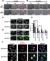
ExoT induction of cell death is primarily due to its ADPRT activity. A and B. HeLa cells were infected in the absence (A) or presence (B) of ZVAD (60 mM) with PA103ΔU/T(G−A+) or PA103ΔU/T(G+A−) at an moi of ca. 10 for the indicated time. Host cell death was assessed by uptake of PI (red) by simultaneous time-lapse phase and fluorescent videomicroscopy. Note that while infection with PA103ΔU/T(G−A+) results in rapid, ZVAD-sensitive cell death, in the presence of PA103ΔU/T(G+A−) cell death is delayed and is partially ZVAD-sensitive. C–E. HeLa cells were transiently transfected in the absence (C) or presence (D) of ZVAD with a mammalian expression vector expressing either ExoT(G+A−) or ExoT(G−A+) fused to GFP at the C-termini. Cell death was analysed by time-lapse videomicroscopy in the presence of PI. Video images were captured every 15 min. The arrows show representative transfected cells. The tabulated results, collected from multiple movies, are shown in (E). The numbers above each column in (E) indicates the total number of transfected cells that were scored (*P < 0.001; **P < 0.01, χ2 test). F. Nuclear condensation or caspase-3 activation of pExoT(G−A+)- or pExoT(G+A−)-transfected cells are assessed by IF microscopy at 1000× magnification. Note that expression of mutant GAP but wild-type ADPRT, ExoT(G−A+), results in nearly 100% cell death whereas the ExoT(G+A−) with wild-type GAP and mutant ADPRT induces only 48% cell death. ZVAD significantly inhibits both the GAP- and the ADPRT-induced cell death. Also worth noting is that both GAP and ADPRT expression results in nuclear condensation and caspase-3 activation.

References

    1. Alaoui-El-Azher M, Jia J, Lian W, and Jin S (2006) ExoS of Pseudomonas aeruginosa induces apoptosis through a Fas receptor/caspase 8-independent pathway in HeLa cells. Cell Microbiol 8: 326–338. - PubMed
    1. Balachandran P, Dragone L, Garrity-Ryan L, Lemus A, Weiss A, and Engel J (2007) The ubiquitin ligase Cbl-b limits Pseudomonas aeruginosa Exotoxin T-mediated virulence. J Clin Invest 117: 419–427. - PMC - PubMed
    1. Barbieri JT, and Sun J (2004) Pseudomonas aeruginosa ExoS and ExoT. Rev Physiol Biochem Pharmacol 152: 79–92. - PubMed
    1. Callus BA, and Vaux DL (2007) Caspase inhibitors: viral, cellular and chemical. Cell Death Differ 14: 73–78. - PubMed
    1. Cho SY, and Klemke RL (2000) Extracellular-regulated kinase activation and CAS/Crk coupling regulate cell migration and suppress apoptosis during invasion of the extracellular matrix. J Cell Biol 149: 223–236. - PMC - PubMed

Publication types

MeSH terms

Substances

LinkOut - more resources