

Pleomorphism of the nuclear envelope in breast cancer: a new approach to an old problem
- PMID: 18053086
- PMCID: PMC3823482
- DOI: 10.1111/j.1582-4934.2007.00176.x
Pleomorphism of the nuclear envelope in breast cancer: a new approach to an old problem
Abstract
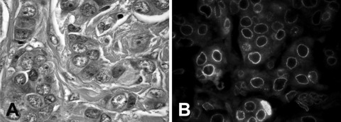
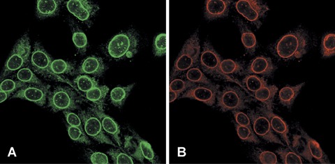
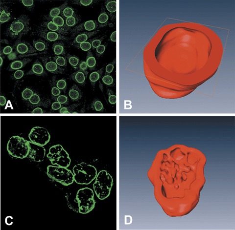
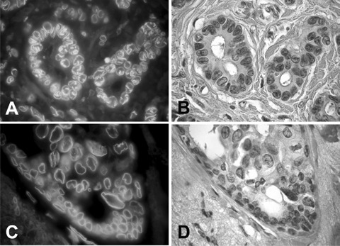
In routine practice, nuclear pleomorphism of tumours is assessed by haematoxylin staining of the membrane-bound heterochromatin. However, decoration of the nuclear envelope (NE) through the immunofluorescence staining of NE proteins such as lamin B and emerin can provide a more objective appreciation of the nuclear shape. In breast cancer, nuclear pleomorphism is one of the least reproducible parameters to score histological grade, thus we sought to use NE proteins to improve the reproducibility of nuclear grading. First, immuno-fluorescence staining of NE as well as confocal microscopy and three-dimensional reconstruction of nuclei in cultured cells showed a smooth and uniform NE of normal breast epithelium in contrast to an irregular foldings of the membrane and the presence of deep invaginations leading to the formation of an intranuclear scaffold of NE-bound tubules in breast cancer cells. Following the above methods and criteria, we recorded the degree of NE pleomorphism (NEP) in a series of 273 invasive breast cancers tested by immunofluorescence. A uniform nuclear shape with few irregularities (low NEP) was observed in 135 cases or, alternatively, marked folds of the NE and an intranuclear tubular scaffold (high NEP cases) were observed in 138 cases. The latter features were significantly correlated (P-value <0.002) with lymph node metastases in 54 histological grade 1 and in 173 cancers with low mitotic count. Decoration of the NE might thus be regarded as a novel diagnostic parameter to define the grade of malignancy, which parallels and enhances that provided by routine histological procedures.
Figures
References
-
- Elston CW, Ellis IO. Pathological prognostic factors in breast cancer. I. The value of histological grade in breast cancer: experience from a large study with long-term follow-up. Histopathology. 1991;19:403–10. - PubMed
-
- Volpi A, Bacci F, Paradiso A, Saragoni L, Scarpi E, Ricci M, Aldi M, Bianchi S, Muretto P, Nuzzo F, Simone G, Mangia A, Schittulli F, Amadori D. Prognostic relevance of histological grade and its component in node-negative breast cancer patients. Mod Pathol. 2004;17:1038–44. - PubMed
-
- Sloane JP, Amendoeira I, Apostolikas N, Bellocq JP, Bianchi S, Boecker W, Bussolati G, Coleman D, Connolly CE, Dervan P, Eusebi V, De Miguel C, Drijkoningen M, Elston CW, Faverley D, Gad A, Jacquemier J, Lacerda M, Martinez-Penuela J, Munt C, Peterse JL, Rank F, Sylvan M, Tsakraklides V, Zafrani B. Consistency achieved by 23 European pathologists from 12 countries in diagnosing breast disease and reporting prognostic features of carcinomas. European Commission Working Group on Breast Screening Pathology. Virchows Arch. 1999;434:3–10. - PubMed
-
- Meyer JS, Alvarez C, Milikowski C, Olson N, Russo I, Russo J, Glass A, Zehnbauer BA, Lister K, Parwaresch R. Cooperative Breast Cancer Tissue Resource. Breast carcinoma malignancy grading by Bloom-Richardson system vs proliferation index:reproducibility of grade and advantages of proliferation index. Mod Pathol. 2005;18:1067–78. - PubMed
-
- Ghadially FN. Ultrastructural pathology of the cell and matrix: a text and atlas of physiological and pathological alterations in the fine structure of cellular and extracellular components. 3. London: Butterworths; 1988.
