. 2007;9(6):R125.
doi: 10.1186/ar2336.

The proinflammatory cytokines IL-2, IL-15 and IL-21 modulate the repertoire of mature human natural killer cell receptors

Affiliations

The proinflammatory cytokines IL-2, IL-15 and IL-21 modulate the repertoire of mature human natural killer cell receptors

Casimir de Rham et al. Arthritis Res Ther. 2007.

Abstract

Natural killer (NK) cells play a crucial role in the immune response to micro-organisms and tumours. Recent evidence suggests that NK cells also regulate the adaptive T-cell response and that it might be possible to exploit this ability to eliminate autoreactive T cells in autoimmune disease and alloreactive T cells in transplantation. Mature NK cells consist of a highly diverse population of cells that expresses different receptors to facilitate recognition of diseased cells and possibly pathogens themselves. Ex vivo culture of NK cells with cytokines such as IL-2 and IL-15 is an approach that permits significant expansion of the NK cell subpopulations, which are likely to have potent antitumour, antiviral, or immunomodulatory effects in autoimmunity. Our data indicate that the addition of IL-21 has a synergistic effect by increasing the numbers of NK cells on a large scale. IL-2 and IL-15 may induce the expression of killer cell immunoglobulin-like receptors (KIRs) in KIR-negative populations, the c-lectin receptor NKG2D and the natural cytotoxic receptor NKp44. The addition of IL-21 to IL-15 or IL-2 can modify the pattern of the KIR receptors and inhibit NKp44 expression by reducing the expression of the adaptor DAP-12. IL-21 also preserved the production of interferon-gamma and enhanced the cytotoxic properties of NK cells. Our findings indicate that the proinflammatory cytokines IL-2, IL-15 and IL-21 can modify the peripheral repertoire of NK cells. These properties may be used to endow subpopulations of NK cells with specific phenotypes, which may be used in ex vivo cellular immunotherapy strategies.


Figures
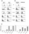
Figure 1
Figure 1
Ex vivo NK proliferation with IL-2 or IL-15 in the presence or absence of IL-21. (a) Proliferation of bulk natural killer (NK) cells. NK cells stained with CFSE (5-carboxyfluorescein diacetate succinimidyl ester) were gated onto the CD3-negative fraction from peripheral blood mononuclear cells and cultured for seven days with IL-2 or IL-15 in the presence or absence of IL-21. The number of cells undergoing division was analyzed at days 5 and 7. Proliferating NK cells are expressed in percentages. This experiment is representative of three individual experiments performed. (b) Proliferation of NK cell subpopulations. NK cells stained with CFSE were purified by magnetic beads, and CD56dim and CD56bright subpopulations were isolated by fluorescence-activated cell sorting. The two populations were cultured for 7 days with IL-2 or IL-15 in the presence or absence of IL-21 and analyzed at day 7. The percentage indicates proliferating NK cells. This experiment is representative of three individual experiments performed. (c) Amplification of CD56dim and CD56bright subpopulations. Sorted 200,000 CD56dim and CD56bright NK cells were plated at day 0 and cultured for 7 days with IL-2 or IL-15 in the presence or absence of IL-21. At day 7 NK cells were stained for their purity (data not shown) and counted. The results are expressed as fold increase as compared to day 0.
Figure 2
Figure 2
Expression of KIR repertoire after culture ex vivo of NK cells with IL-2 or IL-15 with/without IL-21. (a) Expression of different killer cell immunoglobulin-like receptor (KIRs) on the CD56dim natural killer (NK) population. NK cells were purified by magnetic beads, and the CD56dim subpopulations were isolated by fluorescence-activated cell sorting (FACS) and cultured for seven days with IL-2 or IL-15 in the presence or absence of IL-21. Each bar represents different culture conditions, and within the bar are shown the percentages of expression of KIR combinations: no KIR, and one, two, three, or four KIR combinations. KIR combination signifies KIR recognized by a single antibody. Anti-CD158a recognizes KIR2DL1 (CD158a) and KIR2DS1 (CD158h). Anti-CD158b recognizes KIR2DL2 (CD158b1), KIR2DL3 (CD158b2) and KIR2DS2 (CD158j). Anti-NKB1 is specific for KIR3DL1 (CD158e1), and anti-KARp50.3 (CD158i) recognizes KIR2DS4. This experiment is representative of five individual experiments performed. (b) Expression of the different KIR receptors on the CD56bright NK population. NK cells were purified by magnetic beads and the CD56bright subpopulations were isolated by FACS sorting and cultured for 7 days with IL-2 or IL-15 in the presence or absence of IL-21. Each bar represents different culture conditions, and within the bar are shown the percentages of expression KIR combinations: no KIR, and one, two, three, or four KIR combinations. KIR combination signifies KIR recognized by a single antibody. Anti-CD158a recognizes KIR2DL1 (CD158a) and KIR2DS1 (CD158h). Anti-CD158b recognizes KIR2DL2 (CD158b1), KIR2DL3 (CD158b2) and KIR2DS2 (CD158j). Anti-NKB1 is specific for KIR3DL1 (CD158e1), and anti-KARp50.3 (CD158i) recognizes KIR2DS4. This experiment is representative of five individual experiments performed.
Figure 3
Figure 3
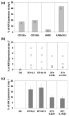
KIR expression in a population of KIR-negative NK cells. (a) Killer cell immunoglobulin-like receptor (KIR) repertoire of a prototypical blood donor at day 0. The KIR repertoire was assessed on the natural killer (NK) bulk population at day 0 before the sorting of the KIR-negative fraction. Anti-CD158a recognizes KIR2DL1 (CD158a) and KIR2DS1 (CD158h). Anti-CD158b recognizes KIR2DL2 (CD158b1), KIR2DL3 (CD158b2) and KIR2DS2 (CD158j). Anti-NKB1 is specific for KIR3DL1 (CD158e1), and anti-KARp50.3 (CD158i) recognizes KIR2DS4. This experiment is representative of three individual experiments performed. (b) KIR repertoire of the KIR-negative population after 7 days of culture. The KIR-negative population of the same donor (panel a) was selected by fluorescence-activated cell sorting (FACS; left column), and cultured for 7 days with IL-2 or IL-15 in the presence or absence of IL-21 (the four columns to the right). The KIR repertoire was assessed after 7 days of culture. Anti-CD158a recognizes KIR2DL1 (CD158a) and KIR2DS1 (CD158h). Anti-CD158b recognizes KIR2DL2 (CD158b1), KIR2DL3 (CD158b2) and KIR2DS2 (CD158j). Anti-NKB1 is specific for KIR3DL1 (CD158e1), and anti-KARp50.3 (CD158i) recognizes KIR2DS4. This experiment is representative of three individual experiments performed. The symbols used are defined in panel a. (c) Effect of IL-2 and IL-15 and/or IL-21 on the KIR repertoire of several donors. The KIR-negative population was selected by FACS sorting (left column) and cultured for 7 days with IL-2 or IL-15 in the presence or absence of IL-21. This experiment represents the fraction of KIR-negative sorted cells from five normal donors who expressed KIRs after 7 days.
Figure 4
Figure 4
Proliferation of KIR-negative and KIR-positive NK cells. Peripheral blood mononuclear cells were stained with CFSE (5-carboxyfluorescein diacetate succinimidyl ester) and natural killer (NK) cells were purified by magnetic beads. The CFSE-positive, killer cell immunoglobulin-like receptor (KIR)-negative fraction was purified by fluorescence-activated cell sorting before culture for 7 days with IL-2 or IL-15 in the presence or absence of IL-21. The grey dots represent the KIR-negative fraction and the black dots represent the KIR-positive fraction after 7 days of culture (left panels). The percentage indicates the fraction KIR-positive and KIR-negative NK cells after 7 days of culture. The number of cells undergoing division in the KIR negative (in grey) and the KIR-positive (in black) fraction were analyzed after 7 days of culture on CD56+/KIR- (in grey) or CD56+/KIR+ (black) gated cells (right panel).
Figure 5
Figure 5
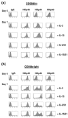
NCR repertoire expressed after culture ex vivo of NK cells with IL-2 or IL-15 with-without IL-21. (a) Expression of the natural cytotoxicity receptor (NCR) family on the CD56dim population. Natural killer (NK) cells were purified by magnetic beads, and the CD56dim subpopulation was isolated by fluorescence-activated cell sorting (FACS) and cultured for 7 days with IL-2 or IL-15 in the presence or absence of IL-21. NKp46, NKp44 and NKp30 were assessed on the CD56dim population at days 0 and 7. One of three similar experiments is shown. (b) Expression of the NCR receptor family, NKp46, NKp44 and NKp30 on the CD56bright population. NK cells were purified by magnetic beads and the CD56bright subpopulations were isolated by FACS sorting and cultured for 7 days with IL-2 or IL-15 in the presence or absence of IL-21. NKp46, NKp44 and NKp30 were assessed on the CD56bright population at days 0 and 7. One of three similar experiments is shown.
Figure 6
Figure 6
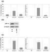
IL-21 downregulated cell surface expression of NKp44 and NKG2D and expression of DAP10 and DAP12. (a) IL-21 downregulated NKp44 and DNAX-activating protein of 12 kDa (DAP12). The percentage of natural killer (NK) cells expressing NKp44 at the cell surface after addition of IL-21 to IL-15 (left) was diminished. Simultaneously, transcripts of the adaptor DAP12 were quantified by real-time PCR after 7 days of culture with IL-15 and compared with the same culture conditions when IL-21 was added (right). The results indicate a reduction in DAP12 transcripts after addition of IL-21 compared with day 0. The expression of DAP12 cDNA was normalized against the housekeeping gene 18S. Mean ± standard deviation of three experiments is shown. (b) IL-21 downregulated DAP12 at the protein level. NK cells were purified by magnetic beads and cultured with IL-15 in presence or absence of IL-21. After 7 days of culture, the adaptor DAP12 protein was quantified by Western blot. The blots were probed with anti-DAP12 and anti-β-tubulin antibodies. (c) IL-21 downregulated NKG2D and DAP10. The percentage of NK cells expressing NKG2D at the cell surface after addition of IL-21 to IL-15 (left) is shown. Simultaneously, the adaptor DAP10 transcripts were quantified by real-time PCR after 7 days of culture with IL-15 and compared with the same culture condition when IL-21 was added (right). As previously shown by Burgess and coworkers [24], in the presence of IL-2, IL-21 down-regulated the expression of C-lectin receptor NKG2D at the cell surface and the transcript of the adaptor DAP10 after 7 days of culture with IL-15. The expression of DAP10 cDNA was normalized against the housekeeping gene 18S. Mean ± standard deviation of three experiments is shown.
Figure 7
Figure 7
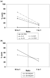
Cytotoxicity of NK cells after ex vivo proliferation in the presence of cytokines. (a) Cytotoxicity of natural killer (NK) cells to K562 cells. Human primary NK cells were purified by magnetic beads and cultured for 7 days with IL-2 or IL-15 in the presence or absence of IL-21. Both populations were analyzed for cytoxicity to K562 target cells at ratios of 10:1 and 1:1. One of three similar experiments is shown. (b) Cytotoxicity of NKp44-positive and NKp44-negative NK cells. Human primary NK cells were purified by magnetic beads (bulk) and cultured for 7 days with IL-15 and IL-21. The NKp44-positive and NKp44-negative subpopulations were sorted by fluorescence-activated cell sorting and were analyzed for their cytotoxicity to K562 targets cells at ratios of 10:1 and 1:1. One of three similar experiments is shown.
Figure 8
Figure 8
Expression of IFN-γ on NK cells after their proliferation ex vivo in presence of cytokines. IFN-γ was analyzed by capture assay on natural killer (NK) cells at day 0 without cytokines, and after 7 days of culture with IL-2 or IL-15 in the presence or absence of IL-21. To determine the capacity of NK cells to produce IFN-γ, cells were subjected to stimulation by IL-12 for 12 hours under all conditions. One of three similar experiments is shown.

References

    1. Parham P. MHC class I molecules and KIRs in human history, health and survival. Nat Rev Immunol. 2005;5:201–214. doi: 10.1038/nri1570. - DOI - PubMed
    1. Shi FD, Van Kaer L. Reciprocal regulation between natural killer cells and autoreactive T cells. Nat Rev Immunol. 2006;6:751–760. doi: 10.1038/nri1935. - DOI - PubMed
    1. Lanier LL. NK cell recognition. Annu Rev Immunol. 2005;23:225–274. doi: 10.1146/annurev.immunol.23.021704.115526. - DOI - PubMed
    1. Uhrberg M, Valiante NM, Shum BP, Shilling HG, Lienert-Weidenbach K, Corliss B, et al. Human diversity in killer cell inhibitory receptor genes. Immunity. 1997;7:753–763. doi: 10.1016/S1074-7613(00)80394-5. - DOI - PubMed
    1. Hsu KC, Chida S, Geraghty DE, Dupont B. The killer cell immunoglobulin-like receptor (KIR) genomic region: gene-order, haplotypes and allelic polymorphism. Immunol Rev. 2002;190:40–52. doi: 10.1034/j.1600-065X.2002.19004.x. - DOI - PubMed

Publication types

MeSH terms

LinkOut - more resources