. 2007 Dec 5:7:135.
doi: 10.1186/1471-213X-7-135.

Expression and function of nr4a2, lmx1b, and pitx3 in zebrafish dopaminergic and noradrenergic neuronal development

Affiliations

Expression and function of nr4a2, lmx1b, and pitx3 in zebrafish dopaminergic and noradrenergic neuronal development

Alida Filippi et al. BMC Dev Biol. .

Abstract

Background: Dopaminergic neurons form in diverse areas of the vertebrate di- and mesencephalon to constitute several major neuromodulatory systems. While much is known about mammalian mesencephalic dopaminergic neuron development, little is known about the specification of the diencephalic dopaminergic groups. The transcription factors Pitx3 and Lmx1b play an important role in mammalian mesencephalic dopaminergic specification, and Nurr1/Nr4a2 has been shown to contribute to specification of the dopaminergic neurotransmitter phenotype. We use zebrafish to analyze potentially evolutionarily conserved roles of these transcription factors in a vertebrate brain that lacks a mesencephalic dopaminergic system, but has an ascending dopaminergic system in the ventral diencephalon.

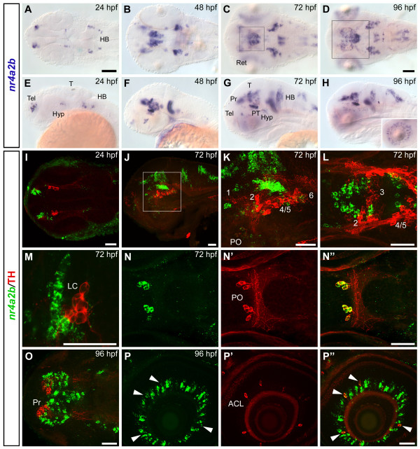
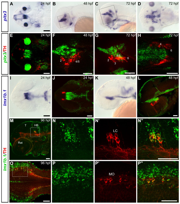
Results: We use a combination of fluorescent in situ hybridization and immunohistochemistry to determine whether nr4a2, lmx1b, and pitx3 genes are expressed in mature dopaminergic neurons or in potential precursor populations. We identify a second nr4a2 paralogue, nr4a2a, and find it co-expressed with Tyrosine hydroxylase in preoptic, pretectal and retinal amacrine dopaminergic neurons, while nr4a2b is only expressed in preoptic and retinal dopaminergic neurons. Both zebrafish nr4a2 paralogues are not expressed in ventral diencephalic dopaminergic neurons with ascending projections. Combined morpholino antisense oligo mediated knock-down of both nr4a2a and nr4a2b transcripts reveals that all zebrafish dopaminergic neurons expressing nr4a2a depend on Nr4a2 activity for tyrosine hydroxylase and dopamine transporter expression. Zebrafish lmx1b.1 is expressed in noradrenergic neurons of the locus coeruleus and medulla oblongata, but knock-down reveals that it is specifically required for tyrosine hydroxylase expression only in the medulla oblongata area postrema noradrenergic neurons. Both lmx1b genes and pitx3 are not expressed in dopaminergic neurons, but in a diencephalic territory that might contain precursor cells for ventral diencephalic dopaminergic neurons. Upon morpholino knock-down of both lmx1b paralogues, the number of neurons in diencephalic dopaminergic clusters with ascending projections appears specifically reduced. Thus lmx1b paralogues may contribute to the generation of diencephalic dopaminergic precursors. Conversely, knock-down of pitx3 does not specifically affect any diencephalic DA cluster.

Conclusion: Our data indicate a conserved evolutionary role of Nr4a2 proteins in specification of the neurotransmitter phenotype, albeit it appears to be only one of several regulatory modules of dopaminergic differentiation, as most ventral diencephalic dopaminergic neurons do not express nr4a2 genes in zebrafish. For zebrafish lmx1b genes, which are not expressed in mature dopaminergic neurons, our data suggest a role in diencephalic precursor populations contributing to the ascending dopaminergic systems. A di-mesencephalic longitudinal domain of lmx1b expression may be the basis for the expansion and posterior shift of ventral di-/mesencephalic dopaminergic populations with ascending projections during evolution.


Figures

Figure 1
Figure 1
nr4a2a is co-expressed with TH in DA neurons of the pretectum, the preoptic area and in amacrine cells of the retina. (A-H) Whole mount in situ hybridization showing nr4a2a expression pattern at 24 hpf (A, E), 48 hpf (B, F), 72 hpf (C, G) and 96 hpf (D, H). Dorsal (A-D) and lateral (E-H) views of the head, anterior is to the left. (I-P") The spatial relationship between nr4a2a-expressing cells and CA neurons in different areas of the brain was analyzed by whole mount FISH to nr4a2a (green) and anti-TH immunohistochemistry (red). Expression was documented by confocal stacks of images, and information for regions corresponding to specific CA neuronal groups was summarized by generation of z-projections from subsets of focal planes of these stacks. (I) Dorsal overview of a 24 hpf embryo (35 μm projection, the approximate head region framed in A is shown): the THir domain is located anterior to the diencephalic nr4a2a domain but there is no co-expression. (J) High magnification of the diencephalic DA clusters at 48 hpf (18 μm projection, approximate area framed in F). (K) High magnification of the diencephalic DA clusters at 72 hpf (15 μm projection, approximate area delimited by the big frame in G). (L) High magnification of the region of the locus coeruleus at 72 hpf (6 μm projection, approximate area delimited by the small frame in G). (M-M") Lateral view of a 96 hpf embryo showing the brain region framed in H (23 μm projection): nr4a2a (green channel, M) and TH (red channel, M'); co-expression is detectable in the pretectum (Pr, arrowhead in M') and in the preoptic area (PO, arrow in M') (merged channels: M"). High magnification dorsal views of the pretectal area and the preoptic dopaminergic neurons are shown respectively in N-N" (6 μm projection, approximate area framed in D) and in O-O" (11 μm projection, arrowheads). (P-P") nr4a2a is expressed in numerous cells of the inner nuclear layer of the retina (P and inset in H), and in all THir amacrine cells (P'-P", arrowheads). In this and in the following figures the DA groups in the ventral diencephalon are numbered from 1 to 6, according to [28]. I, N-N", O-O", dorsal views; J, K, L, M-M", P-P", lateral views; anterior is to the left. Scale bar in A for A-C, E-G and in D for D, H: 100 μm; scale bars in I-P": 50 μm. Abbreviations: ACL, amacrine cell layer; HB, hindbrain; Hyp, hypothalamus; LC, locus coeruleus; PO, preoptic area; Pr, pretectum; T, tectum; Tel, telencephalon; Ret, retina.
Figure 2
Figure 2
nr4a2b is co-expressed with TH in the DA neurons of the preoptic area and in the amacrine cells of the retina. (A-H) Whole mount in situ hybridization showing nr4a2b expression pattern at 24 hpf (A, E), 48 hpf (B, F), 72 hpf (C, G) and 96 hpf (D, H). Dorsal (A-D) and lateral (E-H) views of the head are represented, anterior is to the left. (I-P") The spatial relationship between nr4a2b-expressing cells and CA neurons in different areas of the brain was analyzed by whole mount FISH to nr4a2b (green) and anti-TH immunohistochemistry (red). (I) Dorsal view (56 μm projection) of the head at 24 hpf. (J) Lateral overview (21 μm projection through the diencephalic DA groups) of a 72 hpf embryo. Scattered cells among THir neurons express nr4a2b but double labeled cells are not detectable in this region. A higher magnification of the framed area in J is showed in K (15 μm projection), and a dorsal view of the diencephalic clusters at the same developmental stage is presented in L (9 μm projection). (M) Single confocal plane showing the close proximity of nr4a2b-expressing cells to the THir NA neurons of the locus coeruleus at 72 hpf. Similar to nr4a2a, nr4a2b is co-expressed with TH in the preoptic area (N-N", 12 μm projection, 72 hpf) and in the amacrine cells of the retina (arrowheads in P-P", 7 μm projection, 96 hpf), but no co-expression is detectable in the pretectum at 96 hpf (O, 4 μm projection, approximate area framed in D). I, L, M, N-N", O, dorsal views; J, K, P-P", lateral views; anterior is to the left. Scale bar in A for A-C, E-G and in D for D, H: 100 μm; scale bars in I-P": 50 μm. Abbreviations: ACL, amacrine cell layer; HB, hindbrain; Hyp, hypothalamus; LC, locus coeruleus; PO, preoptic area; Pr, pretectum; PT, posterior tuberculum; T, tectum; Tel, telencephalon; Ret, retina.
Figure 3
Figure 3
Areas of co-expression between nr4a2a/b, pitx3 and lmx1b.1 in the posterior tuberculum. (A-D") Confocal z-projections of double whole mount FISH at 48 hpf show overlapping expression domains for nr4a2a/b, pitx3 and lmx1b.1. The projections encompass the focal planes through the ventral diencephalon. The antisense probes used for the FISH are indicated on the top of each panel. (A-A") 28 μm projection; (B-B") 19 μm projection; (C-C") 16 μm projection; (D-D") 13 μm projection. Anterior is to the left. Scale bars: 50 μm. Abbreviation: PT, posterior tuberculum.
Figure 4
Figure 4
Expression domains of pitx3 and lmx1b.1 genes in relation to catecholaminergic groups. (A-D) Whole mount in situ hybridization showing pitx3 expression pattern at 24 hpf (A, dorsal view), 48 hpf (B, lateral view) and 72 hpf (C, lateral view, and D, dorsal view). (E-H) Confocal z-projections of whole mount FISH to pitx3 (green) and anti-TH immunohistochemistry (red) revealing the spatial relationship between pitx3-expressing and THir cells. (E) Dorsal overview of the head at 24 hpf (35 μm projection). (F) Single plane confocal image of a 48 hpf embryo, the approximate diencephalic area framed in B is showed. (G) Lateral view (38 μm projection) of a 72 hpf embryo (area framed in C). (H) Single dorsal plane showing the diencephalic area of a 72 hpf embryo: pitx3 expression is mainly detectable in medial and dorsal position with respect to the THir neurons of the posterior tuberculum, and no co-localization can be detected with any DA group (see also additional file 5). (I-L) Expression profile of lmx1b.1 at 24 hpf (I-J) and 48 hpf (K-L) analyzed by traditional WISH (I, K) or by whole mount FISH (J, L, green) and anti-TH immunohistochemistry (J, L, red). Dorsal (I-J) and lateral (K-L) views are represented. J and L are 35 μm and 17 μm confocal projections respectively. No co-expression of lmx1b.1 and TH is detected in the DA neurons of the posterior tuberculum. Double labelling is instead observed in the NA neurons of the locus coeruleus (LC) (M-N") and medulla oblongata (MO) (O-P"). (M) Single confocal image at the level of the locus coeruleus of a 96 hpf embryo. High magnification of the framed area is showed in N-N" (23 μm projection). (O) Dorsal overview of the medulla oblongata in a 96 hpf embryo (4 μm projection). A high magnification of the framed area is showed in P-P" (single plane) (see also additional files 6 and 7). Anterior is always to the left. Scale bars in A and I (for A-D and I, K): 100 μm; all the other scale bars: 50 μm. Abbreviations: HB, hindbrain; LC, locus coeruleus; MO, medulla oblongata; T, tectum; Ret, retina.
Figure 5
Figure 5
Nr4a2a is required for formation of DA neurons in pretectum, preoptic area and retina. Morphants were analyzed by WISH to detect th expression at 96hpf. (A) Embryos injected with control morpholino show normal formation of CA groups, including DA nuclei in the pretectum (arrowhead) and the preoptic region (arrow). (B) Injection of 2ng MOnr4a2, which targets both nr4a2 genes, leads to a strong reduction of DA neurons in the pretectum (arrowhead) and the preoptic region (arrow). (C) Control morphants form DA amacrine cells in the retina (arrow), which are absent or strongly reduced in embryos injected with MOnr4a2 (D). All the ventral diencephalic DA groups develop in the nr4a2 morphant embryos (F), including group 3 (arrow), although the spatial organization of the neurons appears altered when compared to control embryos (E). (G-H) When dat expression was analyzed (G, H), nr4a2 morphants showed lack of DA neurons in the pretectum (H, arrowhead), in the retina (inset in H) and the preoptic area (not shown). A-B, G-H: lateral views; C-F: dorsal views. Anterior is to the left. Scale bars in A for A-B, in E for E-F and in G for G-H: 100 μm. Abbreviations: LC, locus coeruleus; MO, medulla oblongata; OB, olfactory bulb; PO, preoptic area; Pr, pretectum; SP, subpallium.
Figure 6
Figure 6
Loss of lmx1b.1 and lmx1b.2 changes the spatial organization of CA cell bodies in the vDC and the hindbrain. (A) Embryos injected with control morpholino display a normal development of DA neurons in the vDC. (B) Embryos injected with both MOlmx1b1 and MOlmx1b2 similarly generate th expressing cells in the vDC, but they are dispersed towards lateral positions. (C, D) Confocal analysis of anti-TH immunostaining shows that vDC group 3 neurons develop normally in morphant embryos (D, arrow) compared to controls (C), while group 1 neurons are somewhat reduced (D, arrowhead). Confocal z-projections of 80 μm (C) and 74 μm (D) are reported. (E) Control morphant embryos form noradrenergic neurons in the area postrema (arrow), while (F) in the same area lmx1b.1 and lmx1b.2 double morphants show a gap of th expression (white arrows). Surprisingly, double morphants frequently exhibit ectopic th-expressing cells in the hindbrain (F, black arrow). (G-H) Analysis of pitx3 expression in control (G) and lmx1b.1/2 morphant embryo (H) at 28 hpf. The diencephalic expression domain of pitx3 is strongly reduced in MOlmx1b1/2 injected embryos. In addition, the most ventral diencephalic domain of nr4a2a expression is reduced in 48hpf morphant embryos (J, compare to I; arrows). A-H: dorsal views; I-J: lateral views. Anterior is to the left. Scale bars in A are for A-B, E-F, in G for G-H and in I are for I-J: 100 μm; scale bar in C is for C-D: 50 μm.
Figure 7
Figure 7
nr4a2a is expressed in a population of differentiating neurons. (A-H") Dorsal views of 48 hpf embryos hybridized with lmx1b.1 and sox2 (A-B"), nr4a2a and sox2 (C-D"), nr4a2a and gfap (E-F"), or nr4a2a and elavl3 (G-H") probes show that nr4a2a is not expressed in progenitor cells, but in early differentiating cells. A, C, E and G show dorsal overviews of the head, at the level of the ventral diencephalon. High magnifications of the posterior tubercular area are reported on the right side of each overview. All the images are single confocal planes, except G, which is a 22 μm confocal projection. Anterior is to the left. Scale bars: 50 μm.

Similar articles

Cited by

References

    1. Dauer W, Przedborski S. Parkinson's disease: mechanisms and models. Neuron. 2003;39:889–909. doi: 10.1016/S0896-6273(03)00568-3. - DOI - PubMed
    1. Marin F, Herrero MT, Vyas S, Puelles L. Ontogeny of tyrosine hydroxylase mRNA expression in mid- and forebrain: neuromeric pattern and novel positive regions. Dev Dyn. 2005;234:709–17. doi: 10.1002/dvdy.20467. - DOI - PubMed
    1. Puelles L, Verney C. Early neuromeric distribution of tyrosine-hydroxylase-immunoreactive neurons in human embryos. J Comp Neurol. 1998;394:283–308. doi: 10.1002/(SICI)1096-9861(19980511)394:3<283::AID-CNE2>3.0.CO;2-Y. - DOI - PubMed
    1. Björklund A, Lindvall O. Dopamine containing systems in the CNS. In: Björklund A, Höckfelt T, editor. Handbook of chemical neuroanatomy: Classical Transmitters in the CNS. Vol. 2. Amsterdam: Elsevier; 1984. pp. 55–121.
    1. Prakash N, Wurst W. Development of dopaminergic neurons in the mammalian brain. Cell Mol Life Sci. 2006;63:187–206. doi: 10.1007/s00018-005-5387-6. - DOI - PMC - PubMed

Publication types

MeSH terms