. 2008 Feb;44(2):429-34.
doi: 10.1016/j.yjmcc.2007.10.016. Epub 2007 Dec 3.

Regulation of Akt/PKB activity by P21-activated kinase in cardiomyocytes

Affiliations

Regulation of Akt/PKB activity by P21-activated kinase in cardiomyocytes

Kai Mao et al. J Mol Cell Cardiol. 2008 Feb.

Abstract

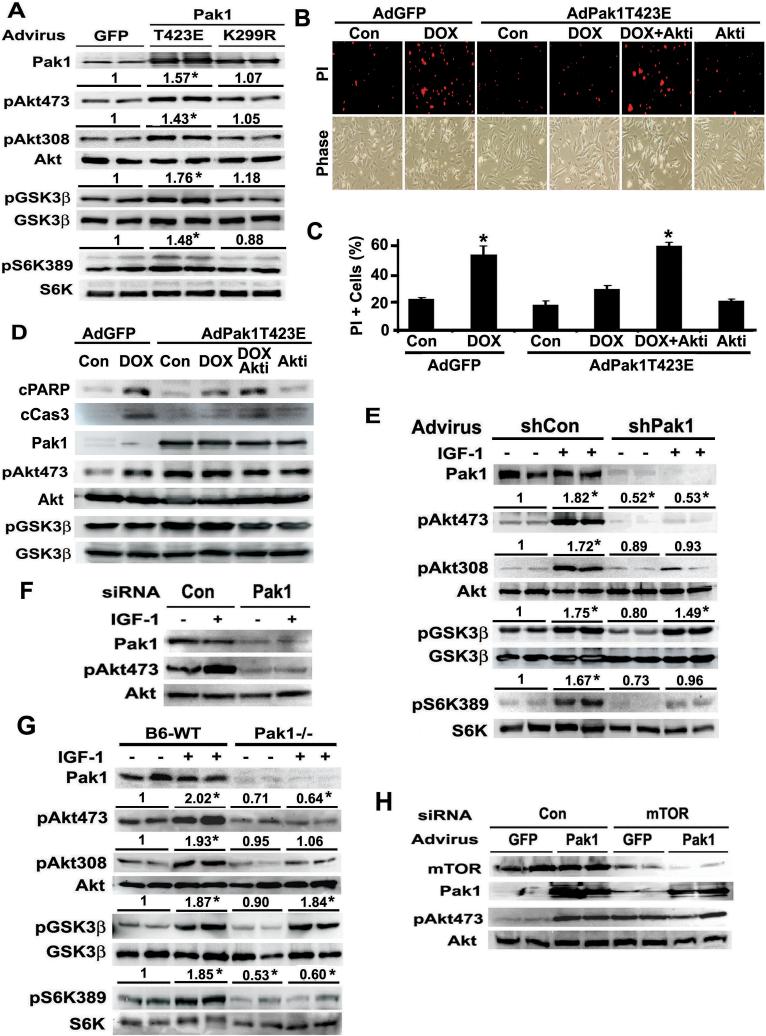
Akt/PKB is a critical regulator of cardiac function and morphology, and its activity is governed by dual phosphorylation at active loop (Thr308) by phosphoinositide-dependent protein kinase-1 (PDK1) and at carboxyl-terminal hydrophobic motif (Ser473) by a putative PDK2. P21-activated kinase-1 (Pak1) is a serine/threonine protein kinase implicated in the regulation of cardiac hypertrophy and contractility and was shown previously to activate Akt through an undefined mechanism. Here we report Pak1 as a potential PDK2 that is essential for Akt activity in cardiomyocytes. Both Pak1 and Akt can be activated by multiple hypertrophic stimuli or growth factors in a phosphatidylinositol-3-kinase (PI3K)-dependent manner. Pak1 overexpression induces Akt phosphorylation at both Ser473 and Thr308 in cardiomyocytes. Conversely, silencing or inactivating Pak1 gene diminishes Akt phosphorylation in vitro and in vivo. Purified Pak1 can directly phosphorylate Akt only at Ser473, suggesting that Pak1 may be a relevant PDK2 responsible for AKT Ser473 phosphorylation in cardiomyocytes. In addition, Pak1 protects cardiomyocytes from cell death, which is blocked by Akt inhibition. Our results connect two important regulators of cellular physiological functions and provide a potential mechanism for Pak1 signaling in cardiomyocytes.


Figures

Figure-1
Figure-1
A. Pak1 is activated by multiple hypertrophic stimuli and growth factors in NRVC. NRVC were cultured under serum-free condition and stimulated with indicated agonists for 5 to 15 minutes. Pak1 activity was determined by immuno-complex kinase assay using MBP as the substrate in the presence of 10 μCi [γ-32P] ATP. 32P-labeled MBP was detected by autoradiography. The numbers above each immuno-blot are fold changes relative to control vehicle-treated cells as determined by densitometry. Results are means (n=4, SE not shown). * indicates p<0.05 compared with control (Con). B. Phosphorylation of Pak1-T423, Akt-S473 or T308, GSK-3β-S9 and S6K-T389 was determined by Western blot analysis. C. IGF-1 and insulin (INS)-induced Pak1 kinase activity was attenuated by PI3K inhibitor wortmanin (WT) or LY294002 (LY). Shown are autoradiographs of p32-labeled MBP. D. IGF-1 and INS-induced phosphorylation of Pak1 and Akt was blocked by LY as shown with western blot analysis. E. Pak1 activity was increased in pressure overloaded mouse hearts. FVB/N or C57B/6 mice underwent transverse aortic constriction (TAC), and Pak1 activity was determined by immuno-complex kinase assay 1 or 3 days after TAC. Results are means (n=3, SE not shown). * indicates p<0.05 compared with sham-operated mice. F. Cardiac Akt activity was increased by TAC as shown by Western blot analysis for pAkt473. G. Endogenous Pak1 is physically associated with Akt in cardiomyocytes. Protein lysates from either NRVC (left half of the image) or FVB/N mouse heart (right half) were incubated with Pak1 or Akt antibodies (Ab), and precipitated with protein A/G agarose beads (IP), which was followed by Western blot analysis (WB) with Pak1 or Akt Ab. The input is one tenth of the amount of protein lysates used for IP. Note: Akt Ab is efficient to pull down Akt and Pak1 proteins from NRVC but not from heart lysates. H. Purified Pak1 can phosphorylate a wild type Akt but not a mutant Akt with serine converted to alanine at the amino acid residue 473. The in vitro phosphorylation of GST-Akt-S fusion protein by purified Pak1 was performed in a cell-free system. Akt phosphorylation was determined by Western blot analysis with phospho-specific antibodies. I. Purified Pak1 can directly phosphorylate Akt-S473 but not T308 in GST-Akt-F fusion protein that contain the full length Akt.
Figure-2
Figure-2
A. Adenovirus-mediated gene transfer of active Pak1-T423E, but not kinase-dead Pak1-K299R, was sufficient to activate Akt signaling in cultured cardiomyocytes. NRVC were infected with indicated adenoviruses, and the phosphorylation of Akt, GSK-3β and S6K was determined by Western blot analysis 24 hours after infection. The numbers above each immuno-blot are fold changes relative to AdGFP-infected cells as determined by densitometry. Results are mean (n=4, SE not shown). * indicates p<0.05 compared with GFP. B. Overexpression of Pak1 in NRVCs protects from anticancer drug doxorubicin (DOX)-induced myocyte death. NRVCs were infected with AdGFP or AdPak1-T423E, and exposed to DOX (1 μmol/L) for 16 hours with or without the Akt inhibitor X (Akti, 2 μmol/L). Myocyte death was determined by propidium iodide (PI, red) staining with fluorescent microscopy. C. PI positive NRVCs were expressed as the percentage of total myocytes counted under phase contrast. Shown are means ± SE from 5 independent experiments. *p<0.01 vs control. D. Pak1-T423E inhibits DOX-induced apoptosis as shown by reduced cleavage of PARP and caspase3. NRVC were infected with indicated adenovirus and treated with indicated drugs as in B and C. Cleaved PARP (cPARP) and caspase3 (cCas3) were determined by Western blot analysis. E. IGF-1-induced Akt phosphorylation was dramatically diminished by shRNA-mediated Pak1 silencing in cultured cardiomyocytes. NRVC were infected with adenovirus expressing shPak1 or shCon, and Akt activation by IGF-1 was determined 72 hours after viral infection. The numbers above each immuno-blot are fold changes relative to shCon-expressing cells in the absence of IGF-1 as determined by densitometry. Results are means (n=4, SE not shown). * indicates p<0.05 compared with shCon. F. Synthetic siRNA-mediated Pak1 Gene silencing diminished IGF-1-induced Akt phosphorylation. NRVC were transfected with 50 nmol/L of siRNA targeting Pak1, and the ability of IGF-1 to induce Akt phosphorylation was determined 60-hour later by Western blot analysis. G. Targeted deletion of Pak1 gene markedly diminished Akt activity in mouse hearts. Wild type control mice (B6-WT) and Pak1−/− mice were injected i.p. with IGF-1 (4 mg/kg), and Akt activity was determined by Western blot analysis 1 hour after injection. The numbers above each immuno-blot are fold changes relative to B6-WT in the absence of IGF-1 as determined by densitometry. Results are means (n=4, SE not shown). * indicates p<0.05 compared with B6-WT. H. SiRNA-mediated knockdown of mTOR protein did not prevent Pak1-induced Akt phsophorylation at S473. NRVC were transfected with a synthetic siRNA targeting rat mTOR, and the ability of Pak1-T423E to induce Akt phosphorylation was determined by Western blot analysis.
Figure-2
Figure-2
A. Adenovirus-mediated gene transfer of active Pak1-T423E, but not kinase-dead Pak1-K299R, was sufficient to activate Akt signaling in cultured cardiomyocytes. NRVC were infected with indicated adenoviruses, and the phosphorylation of Akt, GSK-3β and S6K was determined by Western blot analysis 24 hours after infection. The numbers above each immuno-blot are fold changes relative to AdGFP-infected cells as determined by densitometry. Results are mean (n=4, SE not shown). * indicates p<0.05 compared with GFP. B. Overexpression of Pak1 in NRVCs protects from anticancer drug doxorubicin (DOX)-induced myocyte death. NRVCs were infected with AdGFP or AdPak1-T423E, and exposed to DOX (1 μmol/L) for 16 hours with or without the Akt inhibitor X (Akti, 2 μmol/L). Myocyte death was determined by propidium iodide (PI, red) staining with fluorescent microscopy. C. PI positive NRVCs were expressed as the percentage of total myocytes counted under phase contrast. Shown are means ± SE from 5 independent experiments. *p<0.01 vs control. D. Pak1-T423E inhibits DOX-induced apoptosis as shown by reduced cleavage of PARP and caspase3. NRVC were infected with indicated adenovirus and treated with indicated drugs as in B and C. Cleaved PARP (cPARP) and caspase3 (cCas3) were determined by Western blot analysis. E. IGF-1-induced Akt phosphorylation was dramatically diminished by shRNA-mediated Pak1 silencing in cultured cardiomyocytes. NRVC were infected with adenovirus expressing shPak1 or shCon, and Akt activation by IGF-1 was determined 72 hours after viral infection. The numbers above each immuno-blot are fold changes relative to shCon-expressing cells in the absence of IGF-1 as determined by densitometry. Results are means (n=4, SE not shown). * indicates p<0.05 compared with shCon. F. Synthetic siRNA-mediated Pak1 Gene silencing diminished IGF-1-induced Akt phosphorylation. NRVC were transfected with 50 nmol/L of siRNA targeting Pak1, and the ability of IGF-1 to induce Akt phosphorylation was determined 60-hour later by Western blot analysis. G. Targeted deletion of Pak1 gene markedly diminished Akt activity in mouse hearts. Wild type control mice (B6-WT) and Pak1−/− mice were injected i.p. with IGF-1 (4 mg/kg), and Akt activity was determined by Western blot analysis 1 hour after injection. The numbers above each immuno-blot are fold changes relative to B6-WT in the absence of IGF-1 as determined by densitometry. Results are means (n=4, SE not shown). * indicates p<0.05 compared with B6-WT. H. SiRNA-mediated knockdown of mTOR protein did not prevent Pak1-induced Akt phsophorylation at S473. NRVC were transfected with a synthetic siRNA targeting rat mTOR, and the ability of Pak1-T423E to induce Akt phosphorylation was determined by Western blot analysis.

Similar articles

Cited by

References

    1. Shiojima I, Walsh K. Regulation of cardiac growth and coronary angiogenesis by the Akt/PKB signaling pathway. Genes Dev. 2006;20:3347–3365. - PubMed
    1. Alessi DR, James SR, Downes CP, Holmes AB, Gaffney PR, Reese CB, Cohen P. Characterization of a 3-phosphoinositide-dependent protein kinase which phosphorylates and activates protein kinase Balpha. Curr Biol. 1997;7:261–269. - PubMed
    1. Dong LQ, Liu F. PDK2: the missing piece in the receptor tyrosine kinase signaling pathway puzzle. Am J Physiol Endocrinol Metab. 2005;289:E187–196. - PubMed
    1. Guertin DA, Stevens DM, Thoreen CC, Burds AA, Kalaany NY, Moffat J, Brown M, Fitzgerald KJ, Sabatini DM. Ablation in mice of the mTORC components raptor, rictor, or mLST8 reveals that mTORC2 is required for signaling to Akt-FOXO and PKCalpha, but not S6K1. Dev Cell. 2006;11:859–871. - PubMed
    1. Bokoch GM. Biology of the p21-activated kinases. Annu Rev Biochem. 2003;72:743–781. - PubMed

Publication types

MeSH terms