. 2007 Dec;133(6):1869-81.
doi: 10.1053/j.gastro.2007.09.008. Epub 2007 Sep 14.

Toll-like receptor-4 promotes the development of colitis-associated colorectal tumors

Affiliations

Toll-like receptor-4 promotes the development of colitis-associated colorectal tumors

Masayuki Fukata et al. Gastroenterology. 2007 Dec.

Abstract

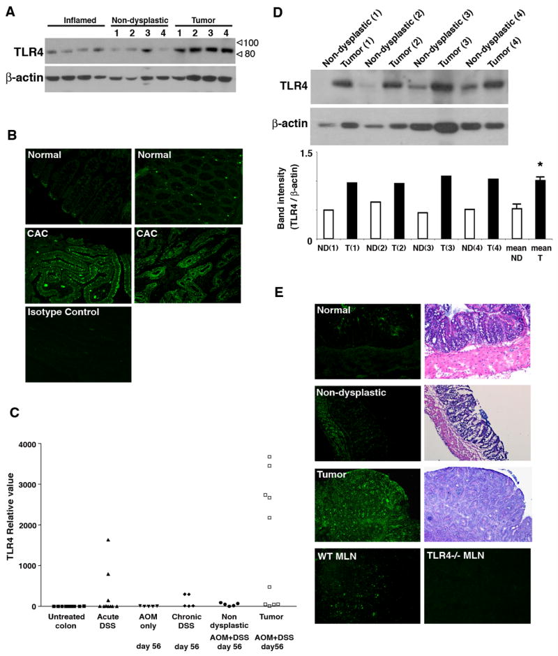
Background & aims: Chronic inflammation is a risk factor for colon cancer in patients with ulcerative colitis (UC). The molecular mechanisms linking inflammation and colon carcinogenesis are incompletely understood. We tested the hypothesis that Toll-like receptor 4 (TLR4) is involved in tumorigenesis in the setting of chronic inflammation.

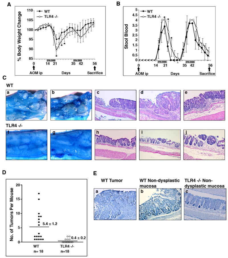
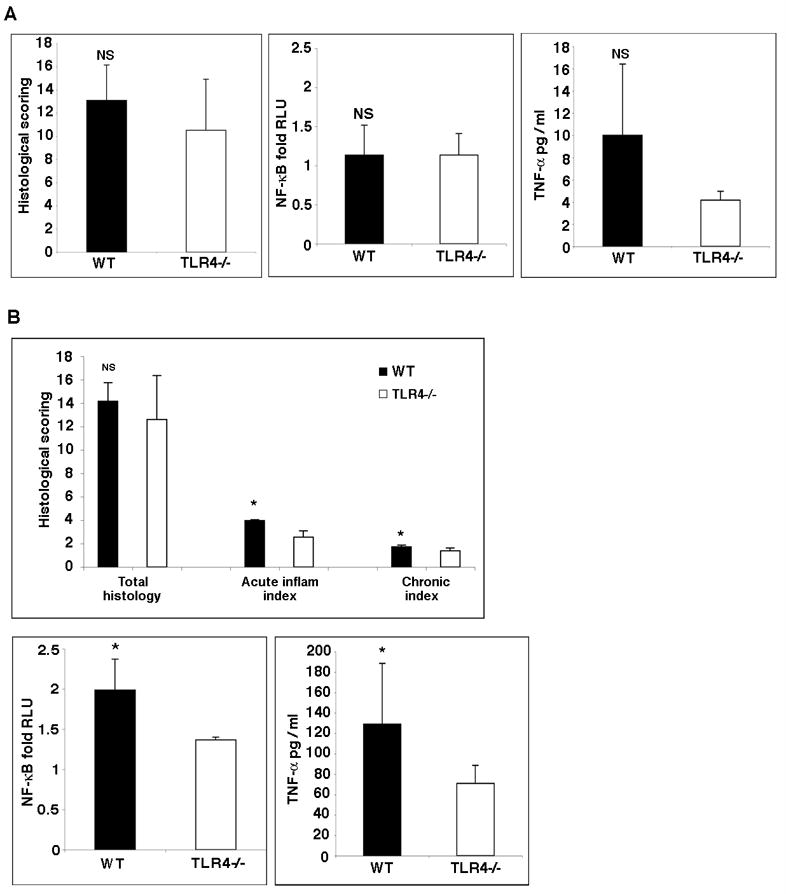
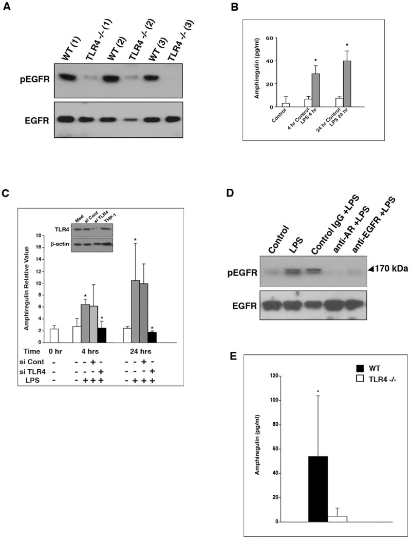
Methods: Tissues from UC patients with cancer were examined for TLR4 expression. Colitis-associated neoplasia was induced using azoxymethane injection followed by dextran sodium sulfate treatment in TLR4-deficient or wild-type mice. Inflammation, polyps, and microscopic dysplasia were scored. Cyclooxygenase (Cox)-2 and prostaglandin E(2) production were analyzed by real-time polymerase chain reaction, immunohistochemistry, or enzyme immunoassay. Epidermal growth factor receptor (EGFR) phosphorylation and amphiregulin production were examined by Western blot analysis and enzyme-linked immunosorbent assay, respectively.

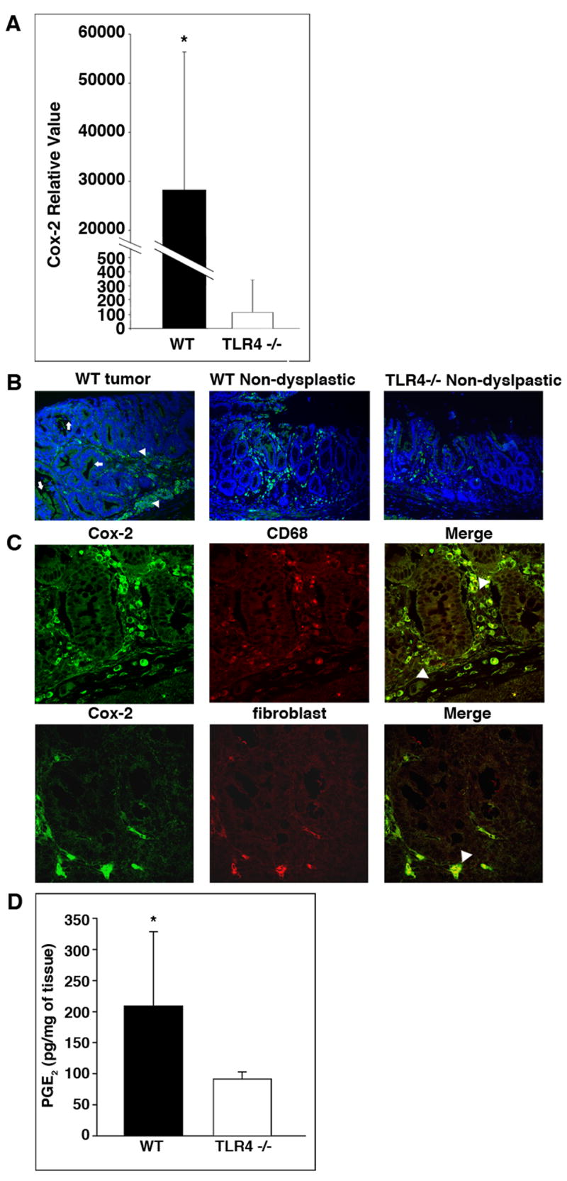
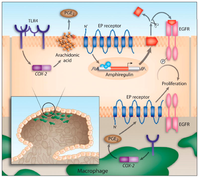
Results: We show that TLR4 is overexpressed in human and murine inflammation-associated colorectal neoplasia. TLR4-deficient mice were protected markedly from colon carcinogenesis. Mechanistically, we show that TLR4 is responsible for induction of Cox-2, increased prostaglandin E(2) production, and activation of EGFR signaling in chronic colitis. Amphiregulin, an EGFR ligand, was induced in a TLR4, Cox-2-dependent fashion and contributes to activation of EGFR phosphorylation in colonic epithelial cells.

Conclusions: TLR4 signaling is critical for colon carcinogenesis in chronic colitis. TLR4 activation appears to promote the development of colitis-associated cancer by mechanisms including enhanced Cox-2 expression and increased EGFR signaling. Inhibiting TLR4 signaling may be useful in the prevention or treatment of colitis-associated cancer.


Conflict of interest statement

The authors have no conflicts of interest to disclose.

Figures

Figure 1
Figure 1. TLR4 expression is up-regulated in colitis-associated tumors
A. Expression of TLR4 in normal mucosa and tumors from patients with UC. Samples of tumor tissue (n=4) and corresponding surrounding non-dysplastic colon (n=4) were examined for TLR4 protein by Western blot analysis. Colonic tissues from patients with active ulcerative colitis were also examined (Inflamed) (n=4). 25 μg/lane of protein was loaded per lane. β-actin staining is used as a loading control. B. Immunofluorescent staining for TLR4 in human colitis-associated cancer. Normal human colon tissue (2 representative examples of 11 patients) (Top panels) and colon cancer in patients with ulcerative colitis (2 representative examples of 15 patients) (middle panels) were stained for TLR4. C. TLR4 mRNA expression in the AOM-DSS model. Normal colon (n=10), acute phase after 7 days of DSS (n=10), day 56 after injection of AOM (n=5), day 56 of chronic DSS colitis after two cycles of DSS (n=5), non-dysplastic mucosa in AOM+DSS treated mice at day 56 (n=5), and tumor tissues taken from AOM+DSS treated mice at day 56 were analyzed for TLR4 mRNA expression by real-time PCR. P <0.05 for tumor tissue versus non-dysplastic mucosa, non-dysplastic mucosa versus normal colon, and chronic DSS versus normal mucosa. D. Western blot analysis for TLR4 in murine colon samples. Immunoblots of tissue lysates (25 μg/lane) prepared from colonic samples of tumor versus corresponding surrounding non-dysplastic mucosa (n=4). Graph shows ratios of TLR4 band intensity normalized with β-actin band (ND= Non-dysplastic mucosa, T= Tumor). Mean ratios of non-dysplastic mucosa and tumor are shown at the end (*P<0.0001). E. Immunofluorescent staining for TLR4 in the mouse CAC model. Serial sections of tumors or surrounding mucosa are stained with H&E for histology and immunofluorescent staining for TLR4. Mesenteric lymph node (MLN) is used as a positive control; mesenteric lymph node from a TLR4−/− mouse is used as a negative control.
Figure 2
Figure 2. TLR4−/− mice are protected from colonic tumors in the setting of inflammation
A, B. AOM and DSS were administered as shown. Weight change (A) and stool blood (B) were examined during the study period. The data represent the average (± SD) of four independent experiments with a total of 36 mice (TLR4−/− (n=18), and WT controls (n=18)) (*p<0.05). C. Tumors in the CAC model. Colons were cut open longitudinally and the mucosal surface was stained with 1% Alcian blue. All WT mice showed multiple polypoid lesions (a, b) but not the TLR4−/− mice (f, g). Microscopically, two examples of WT dysplasias are shown at lower (c) and higher (d) magnifications. Non-dysplastic mucosa from a WT mouse is shown in e. Most TLR4−/− mucosa following AOM-DSS appeared as in panel h. An example of a dysplastic lesion in a TLR4−/− mouse is shown at lower (i) and higher (j) magnifications. D. Incidence of dysplasia. The number of dysplastic lesions was counted per mouse under the microscope. Data are expressed as mean ± SEM (P < 0.0001). E. Proliferation in the CAC model. BrDU incorporation was assessed using anti-BrDU Ab staining. Representative pictures are taken from a WT dysplastic lesion (a), and WT non-dysplastic mucosa (b). Mucosa of AOM-DSS treated TLR4−/− mice (c) is also shown.
Figure 3
Figure 3. Comparison of inflammatory activity in TLR4−/− versus WT mice
A. Histology scores in WT versus TLR4−/− mice at day 56 following AOM+DSS (left panel). Table 1 demonstrates the criteria used for scoring. The total histology score was similar in WT and TLR4−/− mice. NF-κB activation state (middle panel) and TNF-α secretion (right panel) in WT versus TLR4−/− mice at day 56 following AOM+DSS. There are no significant differences between WT and TLR4−/− mice in NF-κB activation or TNF-α production in the intestine. B. Histology scores in WT versus TLR4−/− mice at day 7 following DSS. The acute and chronic sub-scores for the indices are shown. NF-κB activation state and TNF-α secretion in WT versus TLR4−/− mice after seven days of DSS treatment. TLR4−/− mice have significantly decreased NF-κB activation and TNF-α secretion.
Figure 4
Figure 4. Mucosal Cox-2 expression is decreased in TLR4−/− mice in the CAC model
A. TaqMan real-time PCR was used to compare mucosal expression of Cox-2 mRNA in WT and TLR4−/− mice in the CAC model (n=6 each). Data are represented as mean ± SD of relative values of expression (*P < 0.05). B. Immunofluorescent staining for Cox-2 in the colon. Representative pictures show Cox-2 positive cells (green - FITC) in lamina propria cells (arrowhead) and IEC (arrow) in WT mice and TLR4−/− mice. C. Double immunostaining for Cox-2 (green - FITC) and the macrophage marker CD68 (red - TRITC) in WT mucosa (top panel) or anti-fibroblast antibodies (red-TRITC) (bottom panel). Arrows indicate double positive cells (right most panels). D. PGE2 production by colonic tissue from TLR4−/− or WT mice in the CAC model. Colonic tissues from TLR4−/− (n=9) and WT mice (n=10). Data are shown as mean ± SD. (*P < 0.05).
Figure 5
Figure 5. TLR4 signal regulates EGFR tyrosine phosphorylation and amphiregulin expression in colonocytes
A. Western blot analysis of phosphorylated EGFR and EGFR in the colon. Results from three representative samples obtained from WT or TLR4−/− mice are shown. 25μg/lane of protein was loaded per lane. The membrane was sequentially probed for phospho-EGFR and EGFR. Beta-actin was used to demonstrate equal protein loading. B. LPS induces the release of amphiregulin protein. SW480 cells were treated with vehicle (control) or LPS (2 μg/ml) for the indicated periods. Amphiregulin concentration in supernatants was measured by ELISA. Data are expressed as mean ± SD of relative values of expression in three individual experiments with triplicate samples (*P < 0.05). C. LPS-mediated induction of amphiregulin mRNA is TLR4-dependent. SW480 cells were transiently transfected with control siRNA or siRNA against TLR4 and then stimulated with LPS (2μg/ml) for the indicated periods of time. Untransfected control samples were not LPS treated. Levels of amphiregulin mRNA were determined by real time PCR. The data are represented as mean ± SD of relative values of expression in three individual experiments with triplicate samples (*P < 0.05). Inset: Western blot shows TLR4 expression with medium alone, control siRNA, and TLR4 siRNA in SW480 cells. The last bar is THP-1 cells as a positive control. D. LPS-mediated activation of EGFR is ligand-dependent. SW480 cells were pre-treated with antibodies to the ligand-binding site of EGFR, anti-amphiregulin antibody or control IgG for two hours. Subsequently, cells were stimulated with LPS (2μg/ml) for 30 minutes. Blots of whole cell lysates (25 μg/lane) were sequentially probed for phospho-EGFR and EGFR. The data are one representative experiment of three with similar results. β-actin was used as an internal control for protein loading. E. TLR4 deficiency is associated with reduced colonic mucosal production of amphiregulin. The production of amphiregulin was measured in colonic mucosa from WT (n=10) and TLR4−/− (n=8) mice. Data are expressed as mean ± SD (*P < 0.05).
Figure 6
Figure 6. Model of TLR4-mediated colon carcinogenesis
TLR4 expression is increased in chronic intestinal inflammation. TLR4 signaling in response to LPS induces Cox-2 expression and PGE2 production. PGE2 through its receptors (EP) can act in a paracrine or autocrine fashion on colonocytes to stimulate the expression and release of amphiregulin, an EGFR ligand. EGFR signaling is associated with increased proliferation of colonocytes. Likewise, TLR4 expression in tumor-associated macrophages may also respond to LPS by inducing Cox-2 and PGE2, which may then act on the epithelium to stimulate proliferation of colonocytes.

Similar articles

Cited by

References

    1. Sin DD, Man SF, McWilliams A, Lam S. Progression of airway dysplasia and C-reactive protein in smokers at high risk of lung cancer. Am J Respir Crit Care Med. 2006;173:535–9. - PMC - PubMed
    1. Rao VP, Poutahidis T, Ge Z, Nambiar PR, Boussahmain C, Wang YY, Horwitz BH, Fox JG, Erdman SE. Innate immune inflammatory response against enteric bacteria Helicobacter hepaticus induces mammary adenocarcinoma in mice. Cancer Res. 2006;66:7395–400. - PubMed
    1. Gunter MJ, Stolzenberg-Solomon R, Cross AJ, Leitzmann MF, Weinstein S, Wood RJ, Virtamo J, Taylor PR, Albanes D, Sinha R. A prospective study of serum C-reactive protein and colorectal cancer risk in men. Cancer Res. 2006;66:2483–7. - PubMed
    1. Surveillance, Epidemiology, and End Results (SEER) Program and the National Center for Health Statistics. http://seer.cancer.gov/
    1. Itzkowitz SH, Yio X. Inflammation and cancer IV. Colorectal cancer in inflammatory bowel disease: the role of inflammation. Am J Physiol Gastrointest Liver Physiol. 2004;287:G7–17. - PubMed

Publication types