. 2008 Jan 25;375(4):1152-64.
doi: 10.1016/j.jmb.2007.11.006. Epub 2007 Nov 13.

Bloom's syndrome helicase and Mus81 are required to induce transient double-strand DNA breaks in response to DNA replication stress

Affiliations

Bloom's syndrome helicase and Mus81 are required to induce transient double-strand DNA breaks in response to DNA replication stress

Tsutomu Shimura et al. J Mol Biol. .

Abstract

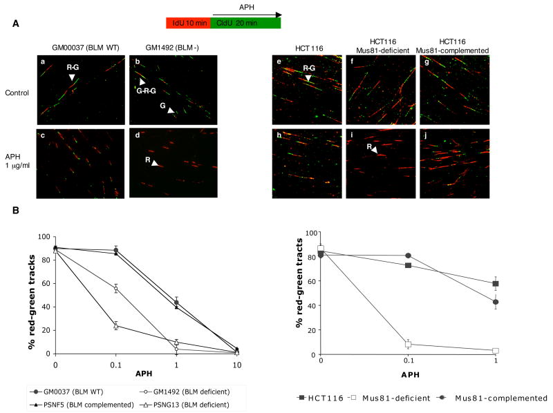
Perturbed DNA replication either activates a cell cycle checkpoint, which halts DNA replication, or decreases the rate of DNA synthesis without activating a checkpoint. Here we report that at low doses, replication inhibitors did not activate a cell cycle checkpoint, but they did activate a process that required functional Bloom's syndrome-associated (BLM) helicase, Mus81 nuclease and ataxia telangiectasia mutated and Rad3-related (ATR) kinase to induce transient double-stranded DNA breaks. The induction of transient DNA breaks was accompanied by dissociation of proliferating cell nuclear antigen (PCNA) and DNA polymerase alpha from replication forks. In cells with functional BLM, Mus81 and ATR, the transient breaks were promptly repaired and DNA continued to replicate at a slow pace in the presence of replication inhibitors. In cells that lacked BLM, Mus81, or ATR, transient breaks did not form, DNA replication did not resume, and exposure to low doses of replication inhibitors was toxic. These observations suggest that BLM helicase, ATR kinase, and Mus81 nuclease are required to convert perturbed replication forks to DNA breaks when cells encounter conditions that decelerate DNA replication, thereby leading to the rapid repair of those breaks and resumption of DNA replication without incurring DNA damage and without activating a cell cycle checkpoint.


Figures

Figure 1
Figure 1
DSBs formed after APH treatment evaluated by neutral comet assay. (A) Neutral comet assays were performed using cells exposed to H202 (500 μM) for 15 minutes or APH (1 μg/mL) for 10 minutes as indicated. (B) Average tail length was quantified as described in Methods. BLM-complemented PSNF5 or BLM-deficient PSNG13 cells were used, as indicated. For each data point, 50 nuclei were scored, using data from 2 independent experiments.
Figure 2
Figure 2
BLM-dependent γ–H2AX foci. Cells were treated with APH and then immunostained with PCNA and γ–H2AX. (A) Cells were treated with 1 μg/mL APH for the indicated amount of time and stained for PCNA (green) and γ–H2AX (red) as described in Methods. Inserts show images at higher magnification. (B) and (C). The number of γ–H2AX-positive PCNA-positive PSNF5 (closed circles) and PSNG13 cells (open circles) per 100 cells was counted. APH treatment was at the concentration and for the amount of time indicated. (D) The number of γ–H2AX-positive PCNA-positive GM0037 and GM1492 cells per 100 cells was counted. Cells were treated with 1 μg/mL APH for the indicated amount of time. Experiments were repeated 3 times with independent samples. Error bars represent standard deviations.
Figure 3
Figure 3
Mus81-dependent γ–H2AX foci. Cells were treated with APH and then immunostained with PCNA and γ–H2AX. (A) HCT116 Mus81−/−, or HCT116 Mus81−/− complemented cells were treated with 0.5 μg/ml APH for the indicated amount of time and immunostained for PCNA (green) and γ–H2AX (red), as described in Methods. Inserts show images at higher magnification. (B) and (C). The number of γ–H2AX–positive PCNA-positive nuclei per 100 cells was counted. Parental unmodified HCT116 (circles), HCT116 Mus81−/− cells (triangles), and HCT116 Mus81−/− +Mus81 cells (squares) were treated with APH (B) or HU (C). Each experiment was repeated 3 times with independent samples. Error bars represent standard deviations. (D) Western blot analysis of Phosphorylated Chk1 expression in HCT116 derived cell lines. When treated with 0.5 μg/ml of APH for 30 minutes, only Mus 81-defiecient cells exhibited staining for p-Chk-1. PCNA was used as a loading control (lower panel).
Figure 4
Figure 4
Phosphorylation of BLM and formation of DSBs after treatment with low levels of APH. (A) and (B). Cells were treated with 1 μg/mL of APH for 10 minutes and immunostained for PCNA (red) and p-BLM (green). Inserts show images at higher magnification. (C) ATR is required for formation of DSBs. The extent of DSB formation was measured by comet assay in cells that contain an active ATR kinase and in cells in which the ATR kinase was inactivated (ATR K/D). Comet assays were performed as described in the legend to Figure 1.
Figure 4
Figure 4
Phosphorylation of BLM and formation of DSBs after treatment with low levels of APH. (A) and (B). Cells were treated with 1 μg/mL of APH for 10 minutes and immunostained for PCNA (red) and p-BLM (green). Inserts show images at higher magnification. (C) ATR is required for formation of DSBs. The extent of DSB formation was measured by comet assay in cells that contain an active ATR kinase and in cells in which the ATR kinase was inactivated (ATR K/D). Comet assays were performed as described in the legend to Figure 1.
Figure 5
Figure 5
Stalled replication forks in cells treated with APH. Replication fork progression was assessed in PSNF5, PSNG13, GM00037, and GM01492 cells. Cell labeling protocol is shown schematically above Panel (A). Cells were labeled with IdU for 10 min. IdU was then washed out and the cells were exposed to APH and CldU for 20 min. IdU was immunodetected by Cy-3–labeled antibodies (red color). CldU was detected by Alexa488-labeled antibodies (green color). (A) Representative images of labeled cells are shown. Red-green tracks (R-G), red-only tracks (R), green-only tracks (G), and green-red-green tracks (G-R-G) are indicated by arrowheads. (B) Abundance of R-G (elongation), G (initiation) and R (termination/stalling) in GM00037, GM01492, PSNF5, and PSNG13 cells.(C) Abundance of R-G (elongation), G (initiation) and R (termination/stalling) in HCT116, Mus81 deficient and Mus81 complemented HCT116. Experiments were repeated at least 3 times with independent samples. Standard deviations are in parenthesis. . Experiments were repeated at least 3 times with independent samples.
Figure 6
Figure 6
PCNA levels and distribution in response to inhibition of DNA replication. (A) Abundance of PCNA in soluble and insoluble fraction in BLM-deficient and BLM-complemented cells treated with APH. LaminB is for loading control. Cells were treated with 1 μg/ml APH for 1 h and then immunoblotted with PCNA and LaminB as described in Methods. (B) Immunostaining for PCNA (green) and RPA (red); (C) Immunostaining for PCNA (red) and pol α (green). (D) Immunostaining for PCNA (green) and pol ε (red). Cells were treated with 1 μg/ml of APH as indicated. Inserts show images at higher magnification.
Figure 6
Figure 6
PCNA levels and distribution in response to inhibition of DNA replication. (A) Abundance of PCNA in soluble and insoluble fraction in BLM-deficient and BLM-complemented cells treated with APH. LaminB is for loading control. Cells were treated with 1 μg/ml APH for 1 h and then immunoblotted with PCNA and LaminB as described in Methods. (B) Immunostaining for PCNA (green) and RPA (red); (C) Immunostaining for PCNA (red) and pol α (green). (D) Immunostaining for PCNA (green) and pol ε (red). Cells were treated with 1 μg/ml of APH as indicated. Inserts show images at higher magnification.
Figure 7
Figure 7
Schematic representation of the proposed events triggered by exposure to replication inhibitors such as APH. Low doses of inhibitors cause temporary inhibition of replication forks that is resolved by conversion of those forks to transient DNA breaks by the action of BLM helicase and Mus81 nuclease. This activity requires phosphorylation of BLM by ATR. The transient breaks are rapidly repaired by the non-homologous end-joining pathway (NHEJ); repair lead to recovery of replication at a slow rate even in the presence of the drug. In the absence of components of the NHEJ pathway such as DNA-PK or XRCC4, the breaks persist and lead to the activation of the S-phase checkpoint. High doses of inhibitors, BLM, Mus81 or ATR deficiencies may also lead to persistent breaks and checkpoint activation.

Similar articles

Cited by

References

    1. Feijoo C, Hall-Jackson C, Wu R, Jenkins D, Leitch J, Gilbert DM, Smythe C. Activation of mammalian Chk1 during DNA replication arrest: a role for Chk1 in the intra-S phase checkpoint monitoring replication origin firing. J Cell Biol. 2001;154:913–23. - PMC - PubMed
    1. Hickson ID. RecQ helicases: caretakers of the genome. Nat Rev Cancer. 2003;3:169–78. - PubMed
    1. Bachrati CZ, Hickson ID. RecQ helicases: suppressors of tumorigenesis and premature aging. Biochem J. 2003;374:577–606. - PMC - PubMed
    1. Chaganti RS, Schonberg S, German J. A manyfold increase in sister chromatid exchanges in Bloom’s syndrome lymphocytes. Proc Natl Acad Sci U S A. 1974;71:4508–12. - PMC - PubMed
    1. Bischof O, Kim SH, Irving J, Beresten S, Ellis NA, Campisi J. Regulation and localization of the Bloom syndrome protein in response to DNA damage. J Cell Biol. 2001;153:367–80. - PMC - PubMed

Publication types

MeSH terms