. 2008 Jan 1;313(1):408-19.
doi: 10.1016/j.ydbio.2007.10.035. Epub 2007 Nov 1.

Dally regulates Dpp morphogen gradient formation by stabilizing Dpp on the cell surface

Affiliations

Dally regulates Dpp morphogen gradient formation by stabilizing Dpp on the cell surface

Takuya Akiyama et al. Dev Biol. .

Abstract

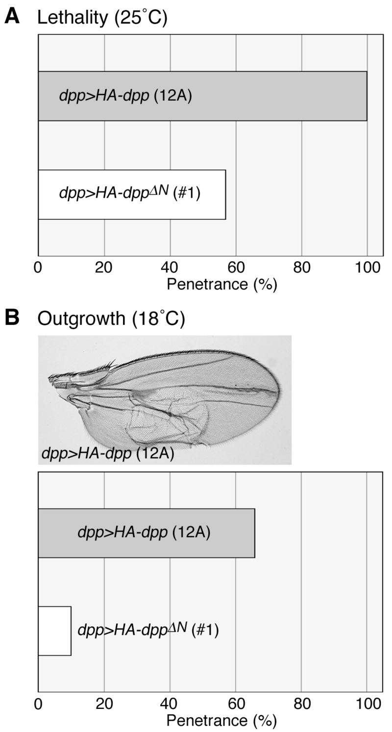
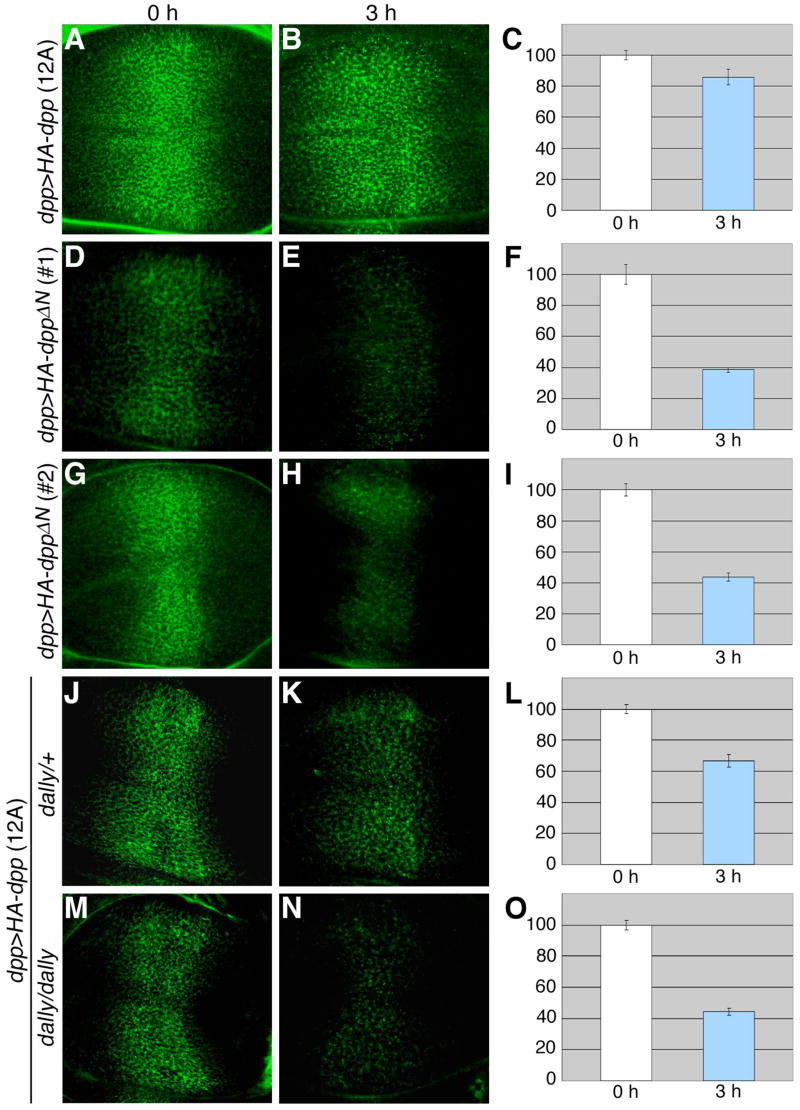
Decapentaplegic (Dpp), a Drosophila homologue of bone morphogenetic proteins, acts as a morphogen to regulate patterning along the anterior-posterior axis of the developing wing. Previous studies showed that Dally, a heparan sulfate proteoglycan, regulates both the distribution of Dpp morphogen and cellular responses to Dpp. However, the molecular mechanism by which Dally affects the Dpp morphogen gradient remains to be elucidated. Here, we characterized activity, stability, and gradient formation of a truncated form of Dpp (Dpp(Delta N)), which lacks a short domain at the N-terminus essential for its interaction with Dally. Dpp(Delta N) shows the same signaling activity and protein stability as wild-type Dpp in vitro but has a shorter half-life in vivo, suggesting that Dally stabilizes Dpp in the extracellular matrix. Furthermore, genetic interaction experiments revealed that Dally antagonizes the effect of Thickveins (Tkv; a Dpp type I receptor) on Dpp signaling. Given that Tkv can downregulate Dpp signaling by receptor-mediated endocytosis of Dpp, the ability of dally to antagonize tkv suggests that Dally inhibits this process. Based on these observations, we propose a model in which Dally regulates Dpp distribution and signaling by disrupting receptor-mediated internalization and degradation of the Dpp-receptor complex.


Figures

Figure 1
Figure 1. N-terminal basic domain of Dpp is essential for binding with Dally
(A) N-terminal amino acid sequence of vertebrate BMP4s, Drosophila Dpp, and a truncated form of Dpp which lacks seven amino acid residues at the N-terminus (DppΔN). Basic amino acid residues are represented in bold. (B) Binding of Dpp and DppΔN to heparin. S2 cells were transfected with HA-dpp or HA-dppΔN cDNA, and the conditioned media containing these proteins were subjected to heparin column chromatography. HA-tagged molecules in the conditioned media (T), in the unbound (U) and bound (B) fractions were detected by western blot analysis. (C) Binding of Dally to Dpp and DppΔN. Myc-Dally and HA-Dpp proteins expressed in S2 cells were subjected to coimmunoprecipitation using anti-HA antibody as described in Materials and Methods. The precipitates were analyzed by western blot analysis using anti-Myc antibody. When Myc-Dally and HA-Dpp were coexpressed in S2 cells, Myc-Dally was recovered from the cell lysate. No binding was detected between Dally and DppΔN. S2 cells that expressed only Myc-Dally or HA-tagged proteins were used as negative controls.
Figure 2
Figure 2. Secretion and in vitro signaling activity of DppΔN
(A) Secretion of Dpp and DppΔN. After transfection with HA-dpp or HA-dppΔN cDNA, S2 cells were incubated for 72 hours at 25°C. HA-tagged proteins in the cell (C) and medium (M) fractions were detected by western blot analysis using anti-HA antibody. Untransfected S2 cells serve as a negative control (NC). (B) In vitro signaling activity of Dpp and DppΔN. Mad-expressing S2 cells were treated with Dpp or DppΔN for 4 hours at 25°C. Signaling activity of Dpp and DppΔN was assayed by western blot analysis of the cell lysate using anti-pMad antibody. Middle panel shows western blotting of the same samples using anti-Flag antibody to show total Mad protein levels. Lower panel is a control blot stained with anti-HA antibody showing the same amount of Dpp and DppΔN was used in this assay.
Figure 3
Figure 3. Comparison of in vivo activities of Dpp and DppΔN
(A and B) Phenotypes induced by overexpression of dpp and dppΔN. Expression of UAS-HA-dpp (12A) or UAS-HA-dppΔN (#1) was induced by dpp-GAL4. Bar graphs show lethality (A) and penetrance of the wing outgrowth phenotype (B) of dpp (grey bars)- and dppΔN (white bars)-expressing animals. dpp>HA-dpp adult wing is shown in B.
Figure 4
Figure 4. Distribution of Dpp and DppΔN in the wing discs
(A and B) Synthesis of Dpp and DppΔN. Wing discs from dpp>HA-dpp (12A) and dpp>HA-dppΔN (#1) third instar larvae were stained with anti-HA antibody by the conventional staining protocol. Note that comparable levels of Dpp and DppΔN were detected in the dpp-expressing cells. (C–E) Extracellular distribution of Dpp and DppΔN. HA-tagged proteins in dpp>HA-dpp (12A) (C) and dpp>HA-dppΔN (#1) (D) wing discs were labeled with anti-HA antibody by the extracellular staining protocol. Intensity profiles for extracellular signals of Dpp (black) and DppΔN (red) generated by NIH Image are shown in E.
Figure 5
Figure 5. Stability of Dpp and DppΔN in S2 cells
(A) Stability of Dpp and DppΔN in a cell-free system. HA-Dpp and HA-DppΔN were incubated in M3 medium in the absence of S2 cells at 25°C. After the indicated times, HA-Dpp and HA-DppΔN were detected by western blot analysis using anti-HA antibody. The signal intensity at each incubation time relative to that of time 0 (shown as 1) was measured using the LI-COR Odyssey Blot system (LI-COR Biosciences) and is indicated as graphs on the right side. Relative levels of Dpp and DppΔN are shown by solid and dotted lines, respectively. The averaged values of results from three independent experiments are presented. (B and C) Degradation of Dpp and DppΔN in cell culture. HA-Dpp and HA-DppΔN were allowed to bind to S2 cells (B) or S2 cells overexpressing Dally (C) for 30 minutes at 25°C. After washing away unbound Dpp proteins and incubating cells for the indicated times at 25°C, the remaining proteins were detected from cell lysates as described above.
Figure 6
Figure 6. Stability of Dpp and DppΔN in the developing wing
Wing discs from UAS-HA-dpp (12A)/+; dpp-GAL4/+ (A–C), UAS-HA-dppΔN (#1)/dpp-GAL4 (D–F), UAS-HA-dppΔN (#2)/dpp-GAL4 (G–I), UAS-HA-dpp (12A)/+; dpp-GAL4/dallygem (J–L), and UAS-HA-dpp (12A)/+; dallygem, dpp-GAL4/dallygem (M-O) were pulse-labeled with anti-HA antibody extracellularly, and incubated in M3 medium for 3 hours at 25°C. Images are shown for 0 (A, D, G, J, and M) and 3 (B, E, H, K, and N) hours after pulse-labeling. Total intensity of signals in the wing pouch area was measured by NIH Image. The relative intensity of signals at 3 hours compared to 0 hours (100%) is shown by bar graphs (C, F, I, L, and O). Each value was calculated from results of at least 5 discs.
Figure 7
Figure 7. The effect of Dally on Dpp protein stability in the wing disc
A hs-HA-dpp transgenic strain was used to monitor stability of Dpp protein in the wing disc. At 4 hours after a heat-shock treatment (37°C, 90 min), wing discs were dissected from hs-HA-dpp larvae and Dpp protein was detected by the conventional staining protocol using anti-HA antibody. (A) A negative control staining of hs-HA-dpp/+; hh-GAL4/+ wing disc without the heat-shock induction. (B) A control disc of the same genotype where HA-Dpp expression was induced by heat-shock. (C) The same treatment was performed in a disc where a dally RNAi construct was expressed in the posterior cells by hh-GAL4 driver (hs-HA-dpp/UAS-IR-dally; hh-GAL4/UAS-IR-dally). (D) Same as C except that dally was overexpressed in the posterior compartment (hs-HA-dpp/UAS-Myc-dally; hh-GAL4/+). White double arrow lines in C and D show regions of dally RNAi expression and dally overexpression, respectively.
Figure 8
Figure 8. dally suppresses the effect of tkv on the Dpp gradient formation
Anti-pMad antibody staining of wing discs for wild-type (A), tkva12 heterozygote (B), dallygem homozygote (C), tkva12/+; dallygem/dallygem (D), en>dally (E), en>tkv (F), and en>dally, tkv (G). White bars represent domains with high levels of pMad.

Similar articles

Cited by

References

    1. Belenkaya TY, Han C, Yan D, Opoka RJ, Khodoun M, Liu H, Lin X. Drosophila Dpp morphogen movement is independent of dynamin-mediated endocytosis but regulated by the glypican members of heparan sulfate proteoglycans. Cell. 2004;119:231–44. - PubMed
    1. Bhanot P, Brink M, Samos CH, Hsieh JC, Wang Y, Macke JP, Andrew D, Nathans J, Nusse R. A new member of the frizzled family from Drosophila functions as a Wingless receptor. Nature. 1996;382:225–30. - PubMed
    1. Brand AH, Perrimon N. Targeted gene expression as a means of altering cell fates and generating dominant phenotypes. Development. 1993;118:401–15. - PubMed
    1. Cherbas L, Moss R, Cherbas P. Transformation techniques for Drosophila cell lines. Methods Cell Biol. 1994;44:161–79. - PubMed
    1. Dubois L, Lecourtois M, Alexandre C, Hirst E, Vincent JP. Regulated endocytic routing modulates wingless signaling in Drosophila embryos. Cell. 2001;105:613–24. - PubMed

Publication types

MeSH terms

LinkOut - more resources