

Doxazosin induces apoptosis of cells expressing hERG K+ channels
- PMID: 18054910
- PMCID: PMC2239015
- DOI: 10.1016/j.ejphar.2007.10.051
Doxazosin induces apoptosis of cells expressing hERG K+ channels
Abstract
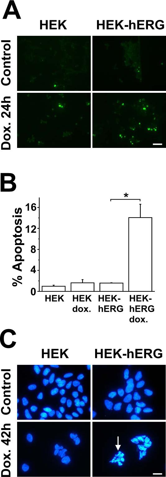
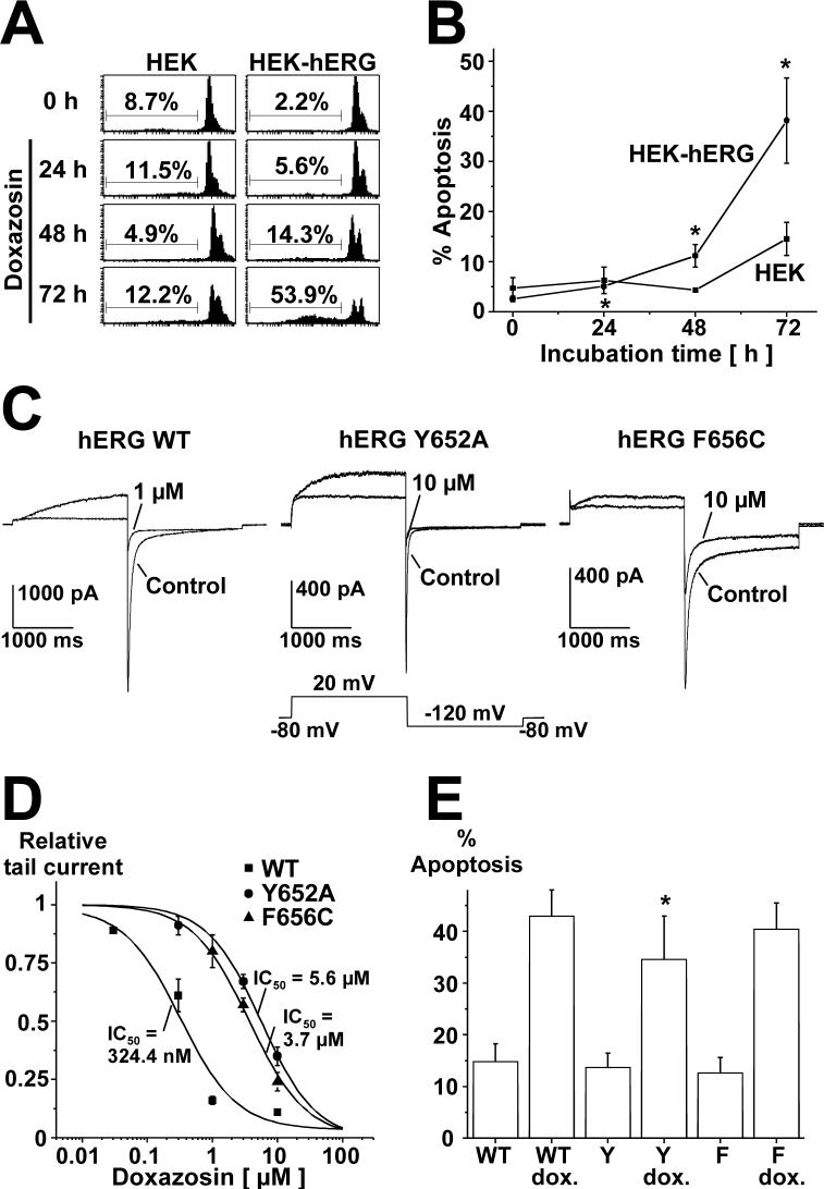
The antihypertensive drug doxazosin has been associated with an increased risk for congestive heart failure and cardiomyocyte apoptosis. Human ether-a-go-go-related gene (hERG) K(+) channels, previously shown to be blocked by doxazosin at therapeutically relevant concentrations, represent plasma membrane receptors for the antihypertensive drug. To elucidate the molecular basis for doxazosin-associated pro-apoptotic effects, cell death was studied in human embryonic kidney cells using three independent apoptosis assays. Doxazosin specifically induced apoptosis in hERG-expressing HEK cells, while untransfected control groups were insensitive to treatment with the antihypertensive agent. An unexpected biological mechanism has emerged: binding of doxazosin to its novel membrane receptor, hERG, triggers apoptosis, possibly representing a broader pathophysiological mechanism in drug-induced heart failure.
Figures
Similar articles
-
Novel roles for hERG K(+) channels in cell proliferation and apoptosis.Cell Death Dis. 2011 Aug 18;2(8):e193. doi: 10.1038/cddis.2011.77. Cell Death Dis. 2011. PMID: 21850047 Free PMC article. Review.
-
HERG K+ channel-dependent apoptosis and cell cycle arrest in human glioblastoma cells.PLoS One. 2014 Feb 6;9(2):e88164. doi: 10.1371/journal.pone.0088164. eCollection 2014. PLoS One. 2014. PMID: 24516604 Free PMC article.
-
Inhibition of human ether-a-go-go-related gene potassium channels by alpha 1-adrenoceptor antagonists prazosin, doxazosin, and terazosin.Naunyn Schmiedebergs Arch Pharmacol. 2004 May;369(5):462-72. doi: 10.1007/s00210-004-0931-8. Epub 2004 Apr 20. Naunyn Schmiedebergs Arch Pharmacol. 2004. PMID: 15098086
-
Doxazosin induces apoptosis in cardiomyocytes cultured in vitro by a mechanism that is independent of alpha1-adrenergic blockade.Circulation. 2003 Jan 7;107(1):127-31. doi: 10.1161/01.cir.0000043803.20822.d1. Circulation. 2003. PMID: 12515754
-
Apoptotic impact of alpha1-blockers on prostate cancer growth: a myth or an inviting reality?Prostate. 2004 Apr 1;59(1):91-100. doi: 10.1002/pros.10357. Prostate. 2004. PMID: 14991869 Review.
Cited by
-
Hsa_circ_0011385 accelerates the progression of thyroid cancer by targeting miR-361-3p.Cancer Cell Int. 2020 Feb 13;20:49. doi: 10.1186/s12935-020-1120-7. eCollection 2020. Cancer Cell Int. 2020. PMID: 32082079 Free PMC article.
-
Novel roles for hERG K(+) channels in cell proliferation and apoptosis.Cell Death Dis. 2011 Aug 18;2(8):e193. doi: 10.1038/cddis.2011.77. Cell Death Dis. 2011. PMID: 21850047 Free PMC article. Review.
-
Transforming growth factor-beta type I receptor/ALK5 contributes to doxazosin-induced apoptosis in H9C2 cells.Naunyn Schmiedebergs Arch Pharmacol. 2009 Dec;380(6):561-7. doi: 10.1007/s00210-009-0449-1. Epub 2009 Sep 2. Naunyn Schmiedebergs Arch Pharmacol. 2009. PMID: 19727674
-
HERG K+ channel-dependent apoptosis and cell cycle arrest in human glioblastoma cells.PLoS One. 2014 Feb 6;9(2):e88164. doi: 10.1371/journal.pone.0088164. eCollection 2014. PLoS One. 2014. PMID: 24516604 Free PMC article.
-
Multiple mechanisms of hERG liability: K+ current inhibition, disruption of protein trafficking, and apoptosis induced by amoxapine.Naunyn Schmiedebergs Arch Pharmacol. 2010 May;381(5):385-400. doi: 10.1007/s00210-010-0496-7. Epub 2010 Mar 13. Naunyn Schmiedebergs Arch Pharmacol. 2010. PMID: 20229012
References
-
- Eiras S, Fernandez P, Pineiro R, Iglesias MJ, Gonzalez-Juanatey JR, Lago F. Doxazosin induces activation of GADD153 and cleavage of focal adhesion kinase in cardiomyocytes en route to apoptosis. Cardiovasc. Res. 2006;71:118–128. - PubMed
-
- Gonzalez-Juanatey JR, Iglesias MJ, Alcaide C, Pineiro R, Lago F. Doxazosin induces apoptosis in cardiomyocytes cultured in vitro by a mechanism that is independent of α1-adrenergic blockade. Circulation. 2003;107:127–131. - PubMed
-
- Lang F, Foller M, Lang KS, Lang PA, Ritter M, Gulbins E, Vereninov A, Huber SM. Ion channels in cell proliferation and apoptotic cell death. J. Membrane Biol. 2005;205:147–157. - PubMed
-
- Latt SA, Stetten G. Spectral studies on 33258 Hoechst and related bisbenzimidazole dyes useful for fluorescent detection of deoxyribonucleic acid synthesis. J. Histochem. Cytochem. 1976;24:24–33. - PubMed
Publication types
MeSH terms
Substances
Grants and funding
LinkOut - more resources
Full Text Sources
Medical
Miscellaneous
