. 2008 Feb;29(2):336-53.
doi: 10.1016/j.nbd.2007.09.009. Epub 2007 Oct 16.

CD40L disruption enhances Abeta vaccine-mediated reduction of cerebral amyloidosis while minimizing cerebral amyloid angiopathy and inflammation

Affiliations

CD40L disruption enhances Abeta vaccine-mediated reduction of cerebral amyloidosis while minimizing cerebral amyloid angiopathy and inflammation

D Obregon et al. Neurobiol Dis. 2008 Feb.

Abstract

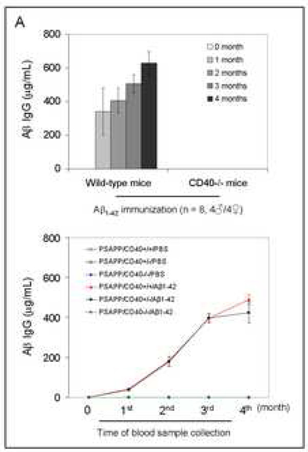
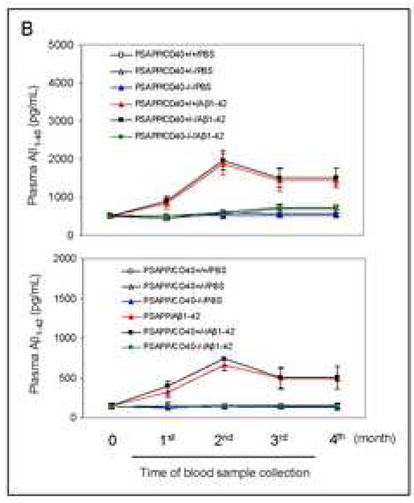
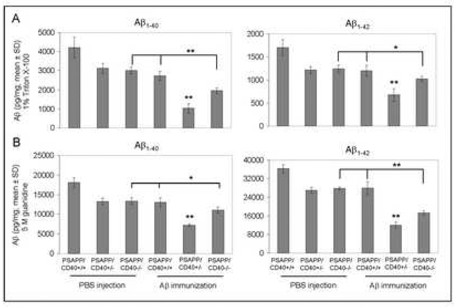
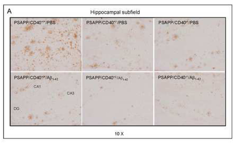
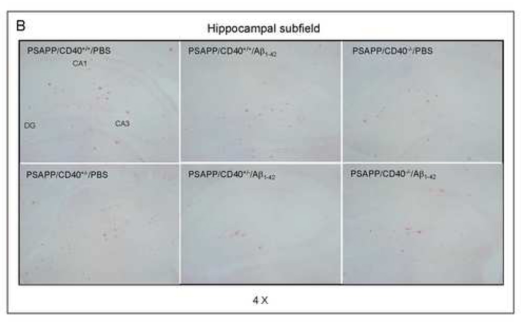
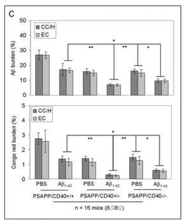
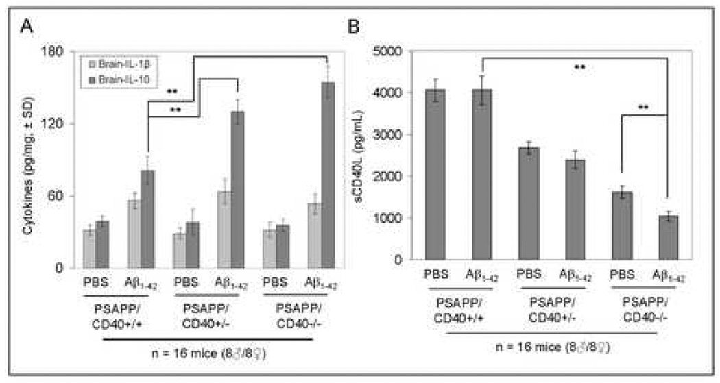
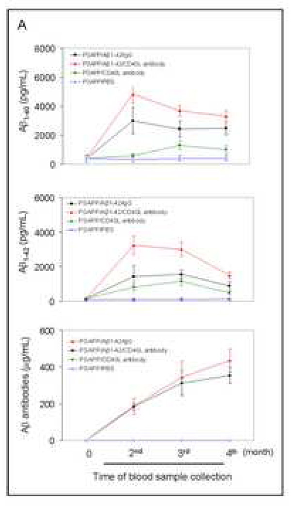
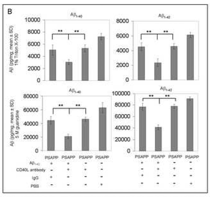
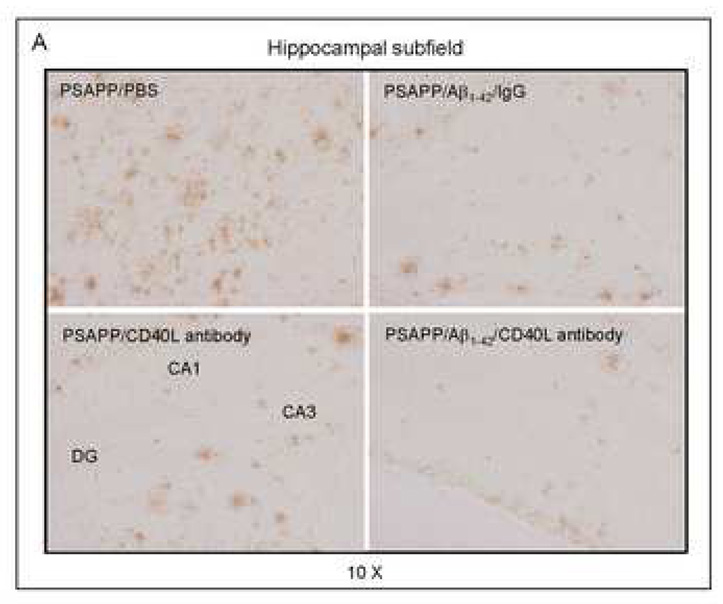
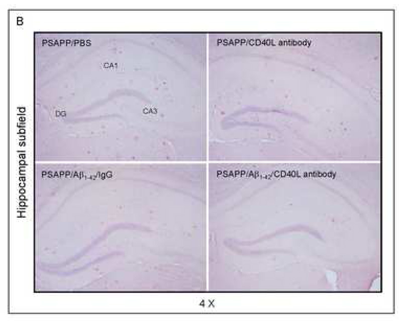
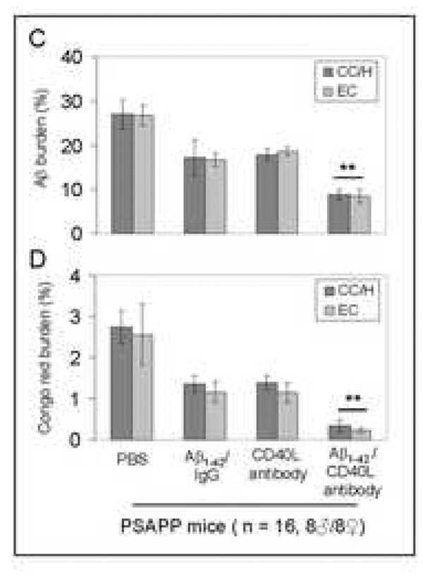
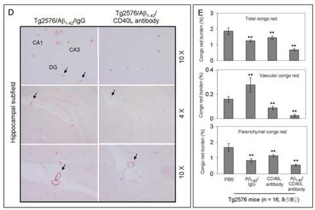
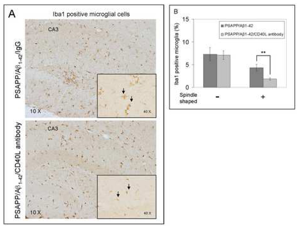
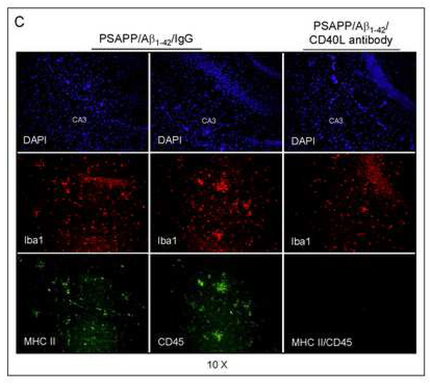
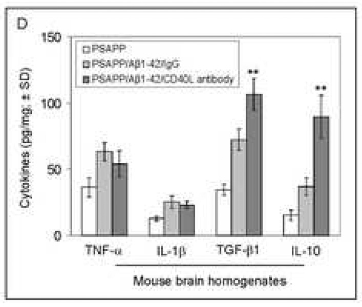
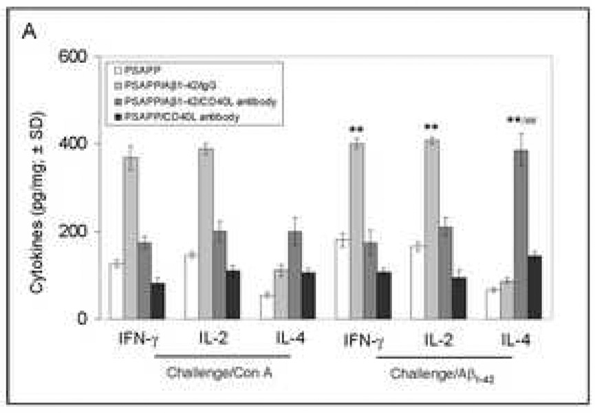
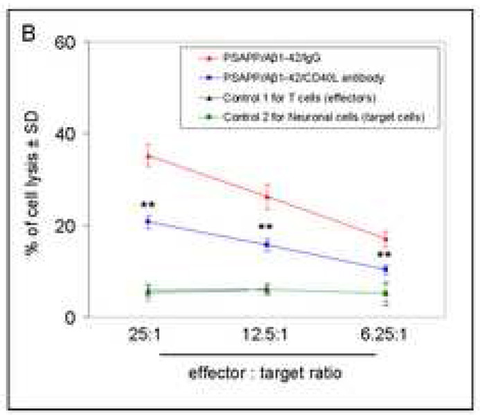
Amyloid-beta (Abeta) immunization efficiently reduces amyloid plaque load and memory impairment in transgenic mouse models of Alzheimer's disease (AD). Active Abeta immunization has also yielded favorable results in a subset of AD patients. However, a small percentage of patients developed severe aseptic meningoencephalitis associated with brain inflammation and infiltration of T-cells. We have shown that blocking the CD40-CD40 ligand (L) interaction mitigates Abeta-induced inflammatory responses and enhances Abeta clearance. Here, we utilized genetic and pharmacologic approaches to test whether CD40-CD40L blockade could enhance the efficacy of Abeta(1-42) immunization, while limiting potentially damaging inflammatory responses. We show that genetic or pharmacologic interruption of the CD40-CD40L interaction enhanced Abeta(1-42) immunization efficacy to reduce cerebral amyloidosis in the PSAPP and Tg2576 mouse models of AD. Potentially deleterious pro-inflammatory immune responses, cerebral amyloid angiopathy (CAA) and cerebral microhemorrhage were reduced or absent in these combined approaches. Pharmacologic blockade of CD40L decreased T-cell neurotoxicity to Abeta-producing neurons. Further reduction of cerebral amyloidosis in Abeta-immunized PSAPP mice completely deficient for CD40 occurred in the absence of Abeta immunoglobulin G (IgG) antibodies or efflux of Abeta from brain to blood, but was rather correlated with anti-inflammatory cytokine profiles and reduced plasma soluble CD40L. These results suggest CD40-CD40L blockade promotes anti-inflammatory cellular immune responses, likely resulting in promotion of microglial phagocytic activity and Abeta clearance without generation of neurotoxic Abeta-reactive T-cells. Thus, combined approaches of Abeta immunotherapy and CD40-CD40L blockade may provide for a safer and more effective Abeta vaccine.


Figures

Fig. 1
Fig. 1
Evaluation of the effects of CD40 deficiency on Aβ antibody generation and Aβ efflux in β1–42-immunized mice. Peripheral blood samples were collected monthly throughout the four-month Aβ immunization course. (A) The graph shows antibody levels for wild-type vs. CD40−/− mice (top panel) and PSAPP mice deficient for CD40 vs. appropriate controls as indicated (bottom panel) following Aβ1–42 vaccination. PSAPP/CD40+/+/Aβ1–42 and PSAPP/CD40+/−/Aβ1–42 mice produced similar elevations in Aβ IgG antibodies, in contrast to PSAPP/CD40−/−/Aβ1–42, PSAPP/CD40+/+/PBS, PSAPP/CD40+/−/PBS and PSAPP/CD40−/−/PBS mice that produced undetectable levels of Aβ IgG antibodies. Data are presented as mean ± SD of plasma Aβ antibodies (µg/mL). (B) Plasma Aβ1–40 and Aβ1–42 peptides were measured separately by ELISA. Data are represented as mean ± SD of Aβ1–40 (top panel) or Aβ1–42 (bottom panel). PSAPP/CD40+/+/Aβ1–42 and PSAPP/CD40+/−/Aβ1–42 mice produced similar elevations in plasma Aβ1–40 and Aβ1–42, in contrast to PSAPP/CD40−/−/Aβ1–42, PSAPP/CD40+/+/PBS, PSAPP/CD40+/−/PBS, and PSAPP/CD40−/−/PBS mice that produced minimal levels of plasma Aβ1–40 and Aβ1–42.
Fig. 1
Fig. 1
Evaluation of the effects of CD40 deficiency on Aβ antibody generation and Aβ efflux in β1–42-immunized mice. Peripheral blood samples were collected monthly throughout the four-month Aβ immunization course. (A) The graph shows antibody levels for wild-type vs. CD40−/− mice (top panel) and PSAPP mice deficient for CD40 vs. appropriate controls as indicated (bottom panel) following Aβ1–42 vaccination. PSAPP/CD40+/+/Aβ1–42 and PSAPP/CD40+/−/Aβ1–42 mice produced similar elevations in Aβ IgG antibodies, in contrast to PSAPP/CD40−/−/Aβ1–42, PSAPP/CD40+/+/PBS, PSAPP/CD40+/−/PBS and PSAPP/CD40−/−/PBS mice that produced undetectable levels of Aβ IgG antibodies. Data are presented as mean ± SD of plasma Aβ antibodies (µg/mL). (B) Plasma Aβ1–40 and Aβ1–42 peptides were measured separately by ELISA. Data are represented as mean ± SD of Aβ1–40 (top panel) or Aβ1–42 (bottom panel). PSAPP/CD40+/+/Aβ1–42 and PSAPP/CD40+/−/Aβ1–42 mice produced similar elevations in plasma Aβ1–40 and Aβ1–42, in contrast to PSAPP/CD40−/−/Aβ1–42, PSAPP/CD40+/+/PBS, PSAPP/CD40+/−/PBS, and PSAPP/CD40−/−/PBS mice that produced minimal levels of plasma Aβ1–40 and Aβ1–42.
Fig. 2
Fig. 2
Cerebral Aβ levels are significantly reduced in Aβ1–42-immunized PSAPP mice heterozygous for CD40. Detergent-soluble Aβ1–40 and Aβ1–42 (A) and insoluble (5M guanidine-soluble) Aβ1–40 and Aβ1–42 peptides (B) were measured separately in brain homogenates by ELISA. Data are presented as mean ± SD of Aβ1–40 or Aβ1–42 (pg/mg protein).
Fig. 3
Fig. 3
β-amyloid pathology is reduced in Aβ1–42-immunized PSAPP mice heterozygous for CD40. Mouse coronal brain sections were embedded in paraffin and stained with monoclonal human Aβ antibody (A), or were stained with congo red (B), and the hippocampus is shown. (C) Percentages [plaque area/total area; mean ± SD with n = 16 mice (8♂/8♀)] of Aβ antibody-immunoreactive Aβ plaques (top panel) and congo red-positive Aβ deposits (bottom panel) were calculated by quantitative image analysis for each brain region (CC/H: cingulate cortex and hippocampus; EC: entorhinal cortex) as indicated.
Fig. 3
Fig. 3
β-amyloid pathology is reduced in Aβ1–42-immunized PSAPP mice heterozygous for CD40. Mouse coronal brain sections were embedded in paraffin and stained with monoclonal human Aβ antibody (A), or were stained with congo red (B), and the hippocampus is shown. (C) Percentages [plaque area/total area; mean ± SD with n = 16 mice (8♂/8♀)] of Aβ antibody-immunoreactive Aβ plaques (top panel) and congo red-positive Aβ deposits (bottom panel) were calculated by quantitative image analysis for each brain region (CC/H: cingulate cortex and hippocampus; EC: entorhinal cortex) as indicated.
Fig. 4
Fig. 4
PSAPP/CD40−/− mice have increased anti-inflammatory IL-10 cytokine and decreased plasma soluble CD40L (sCD40L) after Aβ1–42 vaccination. (A) ELISA analysis of cytokine levels in brain homogenates from the indicated mouse groups. Data are presented as mean ± SD of each cytokine (pg/mg total protein). (B) ELISA for plasma sCD40L levels in the indicated mouse groups. Data are presented as mean ± SD of plasma sCD40L protein (pg/mL).
Fig. 5
Fig. 5
Peripheral and cerebral Aβ levels are reduced in Aβ1–42-immunized PSAPP mice treated with CD40L neutralizing antibody. (A) ELISA analysis for plasma levels of Aβ1–40 and Aβ1–42 and Aβ antibodies. Plasma Aβ1–40 (top panel) and Aβ1–42 (middle panel) were measured separately by ELISA. PSAPP/Aβ1–42/CD40L antibody and PSAPP/Aβ1–42/IgG mice produced similar elevations in plasma Aβ1–40 and Aβ1–42, in contrast to PSAPP/CD40L antibody and PSAPP/PBS mice which produced minimal levels of plasma Aβ1–40 and Aβ1–42. Data are represented as mean ± SD of Aβ1–40 or Aβ1–42 (pg/mL) in plasma. AP antibody levels (bottom panel) were measured by ELISA. PSAPP/Aβ1–42/CD40L antibody and PSAPP/Aβ1–42/IgG mice produced similar elevations in plasma Aβ IgG antibodies in contrast to PSAPP/CD40L antibody and PSAPP/PBS mice which had undetectable levels of plasma Aβ IgG antibodies. Data are presented as mean ± SD of Aβ antibodies (µg/mL) in plasma. No significant difference in Aβ antibody levels between PSAPP/Aβ1–42/CD40L antibody and PSAPP/Aβ1–42/IgG mice (P > 0.05) was observed. (B) Soluble Aβ1–40 and Aβ1–42 peptides (top panel) and insoluble Aβ1–40 and Aβ1–42 (bottom panel) in brain homogenates were measured separately by ELISA. Data are presented as mean ± SD of Aβ1–40 or Aβ1–42 peptides normalized to total protein (pg/mg).
Fig. 5
Fig. 5
Peripheral and cerebral Aβ levels are reduced in Aβ1–42-immunized PSAPP mice treated with CD40L neutralizing antibody. (A) ELISA analysis for plasma levels of Aβ1–40 and Aβ1–42 and Aβ antibodies. Plasma Aβ1–40 (top panel) and Aβ1–42 (middle panel) were measured separately by ELISA. PSAPP/Aβ1–42/CD40L antibody and PSAPP/Aβ1–42/IgG mice produced similar elevations in plasma Aβ1–40 and Aβ1–42, in contrast to PSAPP/CD40L antibody and PSAPP/PBS mice which produced minimal levels of plasma Aβ1–40 and Aβ1–42. Data are represented as mean ± SD of Aβ1–40 or Aβ1–42 (pg/mL) in plasma. AP antibody levels (bottom panel) were measured by ELISA. PSAPP/Aβ1–42/CD40L antibody and PSAPP/Aβ1–42/IgG mice produced similar elevations in plasma Aβ IgG antibodies in contrast to PSAPP/CD40L antibody and PSAPP/PBS mice which had undetectable levels of plasma Aβ IgG antibodies. Data are presented as mean ± SD of Aβ antibodies (µg/mL) in plasma. No significant difference in Aβ antibody levels between PSAPP/Aβ1–42/CD40L antibody and PSAPP/Aβ1–42/IgG mice (P > 0.05) was observed. (B) Soluble Aβ1–40 and Aβ1–42 peptides (top panel) and insoluble Aβ1–40 and Aβ1–42 (bottom panel) in brain homogenates were measured separately by ELISA. Data are presented as mean ± SD of Aβ1–40 or Aβ1–42 peptides normalized to total protein (pg/mg).
Fig. 6
Fig. 6
Cerebral β-amyloid deposits and cerebral amyloid angiopathy are reduced in Aβ1–42-immunized PSAPP or Tg2576 mice treated with CD40L neutralizing antibody. Mouse paraffin-embedded coronal brain sections from were stained with rabbit Pan-β-amyloid antibody (A) or with congo red (B), and the hippocampus is shown. (C) Percentages (plaque area/total area; mean ± SD) of Aβ antibody-immunoreactive deposits (top panel) or of congo red-stained sections (bottom panel) were calculated by quantitative image analysis. (D) Tg2576 received Aβ1–42 vaccination plus neutralizing CD40L antibody or isotype-matched control IgG both Aβ1–42, and brain sections were stained with congo red (hippocampus is shown). Positions of the hippocampal subfields CA1, CA3, and DG (dentate gyrus) are indicated in the upper left panel. Arrows indicate Aβ deposit-affected vessels. (E) Percentages (% of area) of congo red-stained plaques were quantified by image analysis [mean ± SD with (n = 16, 8♂/8♀)].
Fig. 6
Fig. 6
Cerebral β-amyloid deposits and cerebral amyloid angiopathy are reduced in Aβ1–42-immunized PSAPP or Tg2576 mice treated with CD40L neutralizing antibody. Mouse paraffin-embedded coronal brain sections from were stained with rabbit Pan-β-amyloid antibody (A) or with congo red (B), and the hippocampus is shown. (C) Percentages (plaque area/total area; mean ± SD) of Aβ antibody-immunoreactive deposits (top panel) or of congo red-stained sections (bottom panel) were calculated by quantitative image analysis. (D) Tg2576 received Aβ1–42 vaccination plus neutralizing CD40L antibody or isotype-matched control IgG both Aβ1–42, and brain sections were stained with congo red (hippocampus is shown). Positions of the hippocampal subfields CA1, CA3, and DG (dentate gyrus) are indicated in the upper left panel. Arrows indicate Aβ deposit-affected vessels. (E) Percentages (% of area) of congo red-stained plaques were quantified by image analysis [mean ± SD with (n = 16, 8♂/8♀)].
Fig. 7
Fig. 7
CD40L blockade inhibits APC-like microglial activation in Aβ1–42 vaccinated PSAPP mice and promotes anti-inflammatory cellular immunity. (A) Representative hippocampal sections from PSAPP/Aβ1–42/IgG and PSAPP/Apβ1–42/CD40L antibody mouse brains were stained with Ibal antibody to illustrate both microglial load and morphology. (B) Quantitative image analysis of microglial load (Ibal positive) and percentage of spindle-shaped Ibal positive microglia is shown. (C) Representative hippocampal sections from PSAPP/Aβ1–42/IgG and PSAPP/Aβ1–42/CD40L antibody mouse brains were stained with Ibal together with MHC II or CD45 antibodies to illustrate microglial load and activation status (DAPI was used as a nuclear counterstain). (D) Thl and Th2 cytokine analysis by ELISA was conducted on mouse brain homogenates from PSAPP, PSAPP/Aβ1–42/IgG and PSAPP/Aβ1–42/CD40L antibody mice. Data are represented as mean ± SD of each cytokine in brain homogenates (pg/mg total protein) from PSAPP, PSAPP/Aβ1–42/CD40L antibody or PSAPP/Aβ1–42/IgG mice.
Fig. 7
Fig. 7
CD40L blockade inhibits APC-like microglial activation in Aβ1–42 vaccinated PSAPP mice and promotes anti-inflammatory cellular immunity. (A) Representative hippocampal sections from PSAPP/Aβ1–42/IgG and PSAPP/Apβ1–42/CD40L antibody mouse brains were stained with Ibal antibody to illustrate both microglial load and morphology. (B) Quantitative image analysis of microglial load (Ibal positive) and percentage of spindle-shaped Ibal positive microglia is shown. (C) Representative hippocampal sections from PSAPP/Aβ1–42/IgG and PSAPP/Aβ1–42/CD40L antibody mouse brains were stained with Ibal together with MHC II or CD45 antibodies to illustrate microglial load and activation status (DAPI was used as a nuclear counterstain). (D) Thl and Th2 cytokine analysis by ELISA was conducted on mouse brain homogenates from PSAPP, PSAPP/Aβ1–42/IgG and PSAPP/Aβ1–42/CD40L antibody mice. Data are represented as mean ± SD of each cytokine in brain homogenates (pg/mg total protein) from PSAPP, PSAPP/Aβ1–42/CD40L antibody or PSAPP/Aβ1–42/IgG mice.
Fig. 8
Fig. 8
Aβ-specific neurotoxic inflammatory responses are reduced in Aβ1–42-immunized PSAPP mice deficient for CD40. (A) Splenocytes were individually isolated and cultured from mice as indicated after Aβ1–42 immunization and either CD40L antibody treatment or PBS injection (control). These cells were stimulated with Con A (5 µg/mL) or A β1–42 (20 µg/mL) for 48 hrs. Cultured supernatants were collected from these cells for IFN-γ, IL-2, and IL-4 cytokine analyses by ELISA. Data are represented as mean ± SD (n = 10) of each cytokine in supernatants (pg/mg total intracellular protein). (B) A β specific T cell-mediated neuronal cell injury was determined by 51Cr release assay. Data are reported as mean 51Cr release values ± SD, and n = 8 for each condition presented. PSAPP/Aβ1–42/IgG mouse group, effectors: Aβ1–42/IgG-immunized PSAPP mouse-derived T cells; target cells: PSAPP-mouse-derived primary neuronal cells. PSAPP/Aβ1–42/CD40L antibody mouse group, effectors: Aβ1–42/CD40L antibody-immunized PSAPP mouse-derived T cells; target cells: PSAPP-mouse-derived primary neuronal cells. Control 1, effectors: unvaccinated PSAPP mouse-derived T cells; target cells: PSAPP mouse-derived neuronal cells. Control 2, effectors: Aβ1–42-immunized PSAPP mouse-derived T cells; target cells: non-transgenic mouse-derived primary neuronal cells.
Fig. 8
Fig. 8
Aβ-specific neurotoxic inflammatory responses are reduced in Aβ1–42-immunized PSAPP mice deficient for CD40. (A) Splenocytes were individually isolated and cultured from mice as indicated after Aβ1–42 immunization and either CD40L antibody treatment or PBS injection (control). These cells were stimulated with Con A (5 µg/mL) or A β1–42 (20 µg/mL) for 48 hrs. Cultured supernatants were collected from these cells for IFN-γ, IL-2, and IL-4 cytokine analyses by ELISA. Data are represented as mean ± SD (n = 10) of each cytokine in supernatants (pg/mg total intracellular protein). (B) A β specific T cell-mediated neuronal cell injury was determined by 51Cr release assay. Data are reported as mean 51Cr release values ± SD, and n = 8 for each condition presented. PSAPP/Aβ1–42/IgG mouse group, effectors: Aβ1–42/IgG-immunized PSAPP mouse-derived T cells; target cells: PSAPP-mouse-derived primary neuronal cells. PSAPP/Aβ1–42/CD40L antibody mouse group, effectors: Aβ1–42/CD40L antibody-immunized PSAPP mouse-derived T cells; target cells: PSAPP-mouse-derived primary neuronal cells. Control 1, effectors: unvaccinated PSAPP mouse-derived T cells; target cells: PSAPP mouse-derived neuronal cells. Control 2, effectors: Aβ1–42-immunized PSAPP mouse-derived T cells; target cells: non-transgenic mouse-derived primary neuronal cells.

Similar articles

Cited by

References

    1. Bard F, Cannon C, Barbour R, Burke RL, Games D, Grajeda H, Guido T, Hu K, Huang J, Johnson-Wood K, Khan K, Kholodenko D, Lee M, Lieberburg I, Motter R, Nguyen M, Soriano F, Vasquez N, Weiss K, Welch B, Seubert P, Schenk D, Yednock T. Peripherally administered antibodies against amyloid beta-peptide enter the central nervous system and reduce pathology in a mouse model of Alzheimer disease. Nat Med. 2000;6(8):916–919. - PubMed
    1. Bernhardi RV, Nicholls JG. Transformation of leech microglial cell morphology and properties following co-culture with injured central nervous system tissue. J Exp Biol. 1999;202(Pt 6):723–728. - PubMed
    1. Bishop GA, Hostager BS. The CD40-CD154 interaction in B cell-T cell liaisons. Cytokine Growth Factor Rev. 2003;14:297–309. - PubMed
    1. Blennow K, de Leon MJ, Zetterberg H. Alzheimer's disease. Lancet. 2006;368:387–403. - PubMed
    1. Calingasan NY, Erdely HA, Altar CA. Identification of CD40 ligand in Alzheimer's disease and in animal models of Alzheimer's disease and brain injury. Neurobiol Aging. 2002;23:31–39. - PubMed

Publication types

MeSH terms