

Early Palaeozoic dentine and patterned scales in the embryonic catshark tail
- PMID: 18055413
- PMCID: PMC2413265
- DOI: 10.1098/rsbl.2007.0502
Early Palaeozoic dentine and patterned scales in the embryonic catshark tail
Abstract
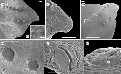
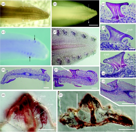
Regular scale patterning, restricted to the caudalmost tail and organized into two opposing rows on each side of the tail, is observed in few chondrichthyans. These evenly spaced scales, in dorsal and ventral rows, develop in an iterative sequence from the caudal tip, either side of the notochord. They are subsequently lost as a scattered pattern of placoid scales develops on the body and fins. An identical organized pattern is observed in tail scales of Scyliorhinus canicula (catshark), where the expression of sonic hedgehog signal is restricted to the epithelium of developing scales and remains localized to the scale pocket. Regulation of iterative scale position by sonic hedgehog is deeply conserved in vertebrate phylogeny. These scales also reveal an archaic histological structure of a dentine type found in the oldest known shark scales from the Ordovician and Silurian. This combination of regulated pattern and ancient dentine occurs only in the tail, representing the primary scalation. Scattered body scales in elasmobranchs such as S. canicula originate secondarily from differently regulated development, one with typical orthodentine around a central pulp cavity. These observations emphasize the modular nature of chondrichthyan scale development and illustrate previously undetected variation as an atavism in extant chondrichthyan dentine.
Figures
References
-
- Ballard W.W, Mellinger J, Lechenault H. A series of normal stages for development of Scyliorhinus canicula, the lesser spotted dogfish. J. Exp. Zool. 1993;267:318–336. doi:10.1002/jez.1402670309 - DOI
-
- Didier D.A. Phylogeny and classification of extant Holocephali. In: Carrier J.C, Musik J.A, Heithaus M.R, editors. Biology of sharks and their relatives. CRC Press; London, UK: 2004. pp. 115–135.
-
- Donoghue P.C.J. Evolution of development of the vertebrate dermal and oral skeletons: unravelling concepts, regulatory theories, and homologies. Paleobiology. 2002;28:474–507. doi:10.1666/0094-8373(2002)028<0474:EODOTV>2.0.CO;2 - DOI
-
- Eames B.F, Allen N, Young J, Kaplan A, Helms J.A, Schneider R.A. Skeletogenesis in the swell shark Cephaloscyllium ventriosum. J. Anat. 2007;210:542–554. doi:10.1111/j.1469-7580.2007.00723.x - DOI - PMC - PubMed
-
- Fraser G.J, Graham A, Smith M.M. Conserved deployment of genes during odontogenesis across osteichthyans. Proc. R. Soc. B. 2004;271:2311–2317. doi:10.1098/rspb.2004.2878 - DOI - PMC - PubMed
MeSH terms
Substances
LinkOut - more resources
Full Text Sources
Other Literature Sources
Molecular Biology Databases
Miscellaneous
