. 2007 Dec;171(6):1789-99.
doi: 10.2353/ajpath.2007.070398. Epub 2007 Nov 30.

The monocyte chemoattractant protein-1/cognate CC chemokine receptor 2 system affects cell motility in cultured human podocytes

Affiliations

The monocyte chemoattractant protein-1/cognate CC chemokine receptor 2 system affects cell motility in cultured human podocytes

Davina Burt et al. Am J Pathol. 2007 Dec.

Abstract

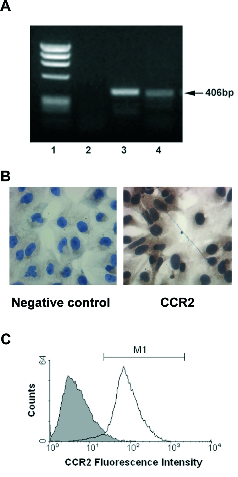
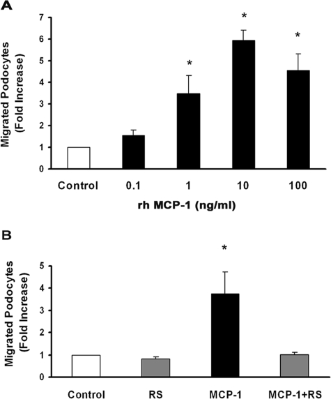
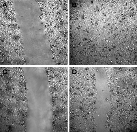
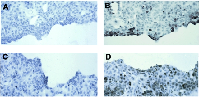
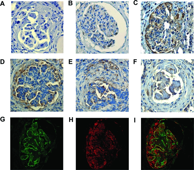
In crescentic glomerulonephritis (GN), monocyte chemoattractant protein-1 (MCP-1) is overexpressed within the glomeruli, and MCP-1 blockade has renoprotective effects. Adult podocytes are in a quiescent state, but acquisition of a migratory/proliferative phenotype has been described in crescentic GN and implicated in crescent formation. The cognate CC chemokine receptor 2 (CCR2), the MCP-1 receptor, is expressed by other cell types besides monocytes and has been implicated in both cell proliferation and migration. We investigated whether MCP-1 binding to CCR2 can induce a migratory/proliferative response in cultured podocytes. MCP-1 binding to CCR2 enhanced podocyte chemotaxis/haptotaxis in a concentration-dependent manner and had a modest effect on cell proliferation. Closure of a wounded podocyte monolayer was delayed by CCR2 blockade, and CCR2 was overexpressed at the wound edge, suggesting a role for CCR2 in driving podocyte migration. Immunohistochemical analysis of kidney biopsies from patients with crescentic GN demonstrated CCR2 expression in both podocytes and cellular crescents, confirming the clinical relevance of our in vitro findings. In conclusion, the MCP-1/CCR2 system is functionally active in podocytes and may be implicated in the migratory events triggered by podocyte injury in crescentic GN and other glomerular diseases.


Figures

Figure 1
Figure 1
Characterization of human podocytes. A: Representative scanning electron micrograph of podocytes showing typical cytoplasmatic projections. Original magnification, ×1200. B: Podocyte staining for nephrin by immunofluorescence. Original magnification, ×630.
Figure 2
Figure 2
CCR2 mRNA and protein is expressed by human podocytes. CCR2 mRNA expression was analyzed by RT-PCR and protein expression by cytofluorimetry and immunocytochemistry in cultured human podocytes as described in Materials and Methods. A: Representative 1.5% agarose gel stained with ethidium bromide. 1, molecular weight marker Φ174-HaeIII. 2, negative control. 3, positive control human monocytes. 4, human podocytes. B: Representative immunocytochemical staining of podocytes for CCR2 (right). The primary antibody was omitted in the negative control (left). Original magnification, ×400. C: Representative histogram showing surface CCR2 expression in podocytes (white histogram). Nonspecific staining (gray histogram) was determined using an isotype-specific control antibody.
Figure 3
Figure 3
MCP-1 binding to CCR2 induces podocyte chemotaxis. A chemotaxis assay was performed as described in Materials and Methods. Numbers of migrated cells per high-power field were counted, and results are expressed as fold increase over control. A: Podocytes were seeded in the upper chamber of a Boyden chamber, and increasing rh-MCP-1 concentrations (0.1, 1, 10, and 100 ng/ml) were placed in the lower chamber (n = 3). *P < 0.01 rh-MCP-1 at 1, 10, and 100 ng/ml versus control. B: Podocytes were preincubated with either the CCR2 antagonist RS102895 (6 μmol/L) or vehicle for 30 minutes, and then the chemotaxis assay was performed using rh-MCP-1 (10 ng/ml) as chemoattractant (n = 4). *P < 0.05 MCP-1 versus others.
Figure 4
Figure 4
MCP-1 induces podocyte haptotaxis via the CCR2 receptor. Podocytes were seeded in the upper chamber of a Boyden chamber and a chemotaxis assay performed using polycarbonate filters precoated with either 10 μg/ml rh-MCP-1 or bovine serum albumin (1 mg/ml) in the presence or the absence of RS102895 (6 μmol/L) or vehicle as described in Materials and Methods. Numbers of migrated cells per high-power field were counted, and results are expressed as fold increase over control (n = 3). *P < 0.01 rh-MCP-1 versus others.
Figure 5
Figure 5
CCR2 blockade delays the closure of a mechanically wounded podocyte monolayer. Podocyte confluent monolayers were wounded as described in Materials and Methods. Experiments were performed in the presence of RS102895 (6 μmol/L) (C and D) or vehicle (A and B). Wounded area was photographed at baseline (A and C) and after 24 hours (B and D). Representative images are shown of four independent experiments. Original magnification, ×40.
Figure 6
Figure 6
Effect of MCP-1 on podocyte cell number. Quiescent podocytes were exposed to rh-MCP-1 at concentrations of 1 ng/ml (□) and 10 ng/ml (▪) for 12, 24, 48, and 72 hours. Cell proliferation was assessed by cell counting and expressed as fold increase over control (dashed line) (n = 5). *P < 0.05 MCP-1 at 10 ng/ml versus control at 48 hours.
Figure 7
Figure 7
Injured podocytes overexpress the CCR2 receptor. CCR2 (B) and MCP-1 (D) expression was assessed in wounded podocyte monolayers by immunocytochemistry as described in the Materials and Methods. A and C: Negative controls: incubation with irrelevant isotype-specific control antibodies. Representative images are shown of three independent experiments. Original magnification, ×40.
Figure 8
Figure 8
Glomerular CCR2 staining in patients with crescentic glomerulonephritis. Glomerular staining for CCR2 by immunohistochemistry in a normal kidney (B) and kidney biopsies from patients with crescentic glomerulonephritis (C–F). Nonspecific staining was determined using a nonimmune isotypic control antibody (A). Double immunofluorescence for synaptopodin (G) and CCR2 (H) showed partial colocalization of the positive staining, as demonstrated by merging (I). Magnification, ×400.

References

    1. Pavenstädt H, Kriz W, Kretzler M. Cell biology of the glomerular podocyte. Physiol Rev. 2003;83:253–307. - PubMed
    1. Le Hir M, Keller C, Eschmann V, Hähnel B, Hosser H, Kriz W. Podocyte bridges between the tuft and Bowman’s capsule: an early event in experimental crescentic glomerulonephritis. J Am Soc Nephrol. 2001;12:2060–2071. - PubMed
    1. Moeller MJ, Soofi A, Hartmann I, Le Hir M, Wiggins R, Kriz W, Holzman LB. Podocytes populate cellular crescents in a murine model of inflammatory glomerulonephritis. J Am Soc Nephrol. 2004;15:61–67. - PubMed
    1. Reiser J, Oh J, Shirato I, Asanuma K, Hug A, Mundel TM, Honey K, Ishidoh K, Kominami E, Kreidberg JA, Tomino Y, Mundel P. Podocyte migration during nephrotic syndrome requires a coordinated interplay between cathepsin L and alpha3 integrin. J Biol Chem. 2004;279:34827–34832. - PubMed
    1. Kakizaki Y, Waga S, Sugimoto K, Tanaka H, Nukii K, Takeya M, Yoshimura T, Yokoyama M. Production of monocyte chemoattractant protein-1 by bovine glomerular endothelial cells. Kidney Int. 1995;48:1866–1874. - PubMed

Publication types