. 2007 Dec;171(6):1743-52.
doi: 10.2353/ajpath.2007.070184. Epub 2007 Nov 30.

Myeloperoxidase is critically involved in the induction of organ damage after renal ischemia reperfusion

Affiliations

Myeloperoxidase is critically involved in the induction of organ damage after renal ischemia reperfusion

Robert A Matthijsen et al. Am J Pathol. 2007 Dec.

Abstract

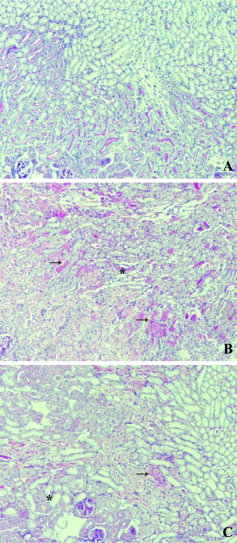
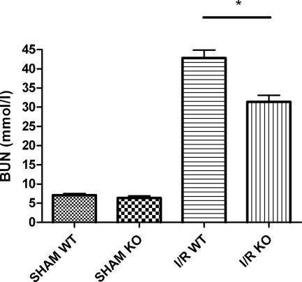
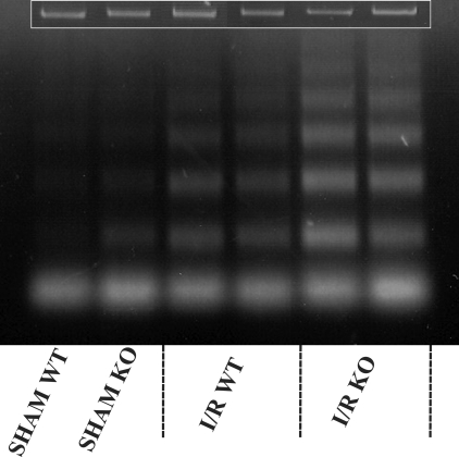
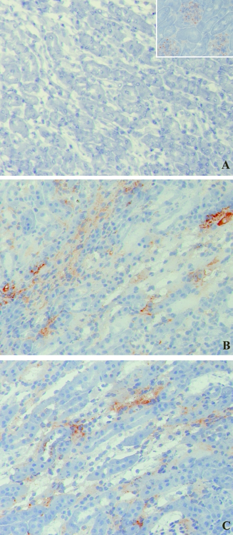
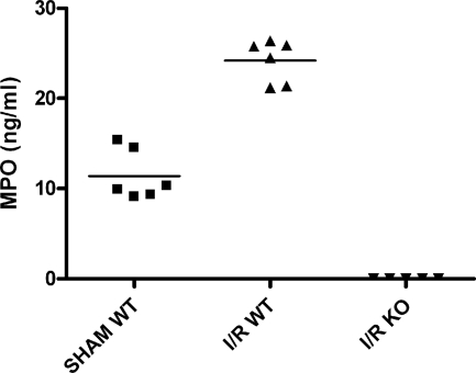
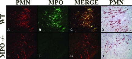
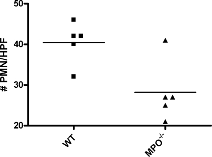
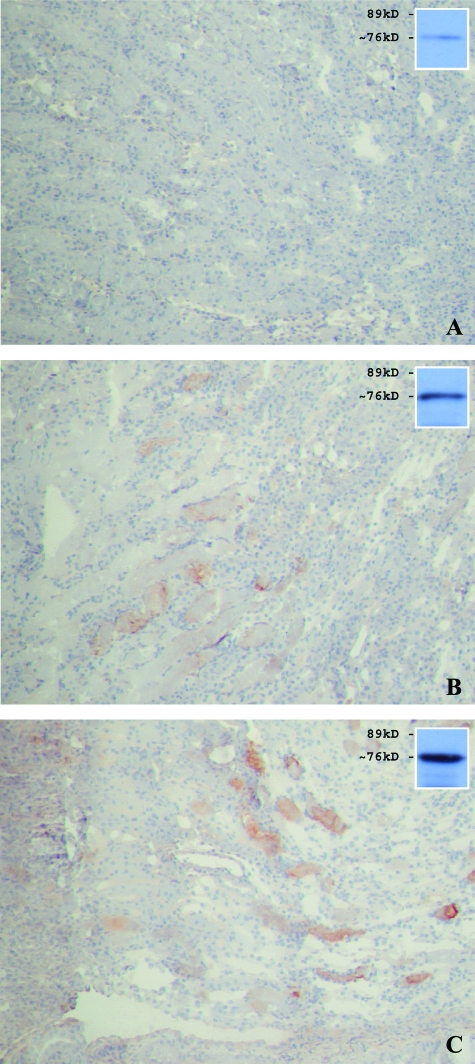
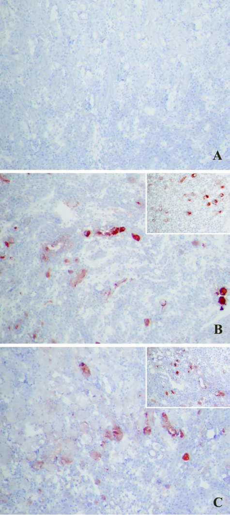
In this study the role of myeloperoxidase (MPO) in a murine (C57BL/6) model of ischemia and reperfusion (I/R)-induced renal failure was investigated. The renal function after I/R was analyzed in MPO-deficient (Mpo(-/-)) mice and compared with wild-type (WT) controls. A significant reduction in renal function loss (blood urea nitrogen) was observed after 24 hours of reperfusion of ischemically damaged kidneys in Mpo(-/-) mice compared with I/R WT controls (I/R Mpo(-/-) = 31.3 +/- 1.7 mmol/L versus I/R WT = 42.8 +/- 2.1 mmol/L, sham = 7.0 +/- 0.5 mmol/L; P = 0.003). The early reperfusion phase (2 hours of reperfusion) was characterized by a substantial increase in apoptosis and early complement activation, surprisingly similar in Mpo(-/-) and WT mice. Improved renal function in Mpo(-/-) mice after extended reperfusion was accompanied by a reduced neutrophil influx (P = 0.017) compared with WT controls. Activation and deposition of complement was not significantly reduced in Mpo(-/-) mice compared with WT controls after 24 hours of reperfusion, indicating no specific in vivo role for MPO in activating complement after renal I/R. Taken together, these results demonstrated an important contribution of MPO in the induction of organ damage after renal I/R by influencing critical factors such as neutrophil extravasation but not complement activation.


Figures

Figure 1
Figure 1
Similar renal morphology of ischemically damaged WT and KO kidneys after 24 hours of reperfusion. Microphotographs showing periodic acid-Schiff staining of kidney sections of sham-treated (A) as well as WT (B) and Mpo−/− (C) mice subjected to 40 minutes of ischemia followed by 24 hours of reperfusion. Shown are representative microphotographs of all groups. Original magnifications, ×100.
Figure 2
Figure 2
MPO deficiency significantly reduces renal function loss after renal I/R. Renal function after 24 hours of reperfusion as reflected by BUN concentration. Statistical significance of renal function in Mpo−/− mice as compared with WT animals was denoted at P = 0.003 (*).
Figure 3
Figure 3
MPO deficiency does not affect I/R-induced early apoptosis. Shown are two sham-treated animals as well as two representatives of each experimental group. Loading controls are given in the inset.
Figure 4
Figure 4
Two hours of reperfusion of ischemically damaged kidneys results in early MBL-C deposition, similar in Mpo−/− and WT mice. MBL-C binding in WT sham-treated (not shown) and KO sham-treated (A) animals was only observed in glomeruli. MBL-C deposition was evident in peritubular capillaries, the interstitium, and along the epithelial brush border of damaged tubules in both WT (B) and Mpo−/− (C) mice subjected to renal I/R. Shown are representative microphotographs of all groups. Original magnifications: ×200; ×600 (inset).
Figure 5
Figure 5
MPO plasma levels, representing total neutrophil activation, are increased 24 hours after reperfusion of ischemically damaged WT kidneys. No MPO was detected in Mpo−/− mice. Statistical significance of WT MPO levels as compared with sham-treated animals was denoted at P < 0.0001.
Figure 6
Figure 6
Immunofluorescent staining showing PMN sequestration (red, Alexa 568) and MPO presence (green, Alexa 488) in kidneys of WT (A–C) and Mpo−/− (E–G) mice subjected to I/R. Overlapping PMN and MPO data depict the fact that MPO is predominantly located around NIMP-R14-positive PMNs. D (WT) and H (Mpo−/−) show PMN infiltration in relation to structural changes. Shown are representative microphotographs of all groups. Original magnifications, ×200.
Figure 7
Figure 7
MPO deficiency reduces neutrophil infiltration after renal I/R. Statistical significance as compared with WT animals was denoted at P = 0.017.
Figure 8
Figure 8
MBL-C and C3 (insets) deposition in WT and Mpo−/− mice after 24 hours of reperfusion. MBL-C and C3 deposition was not different in WT (B) or Mpo−/− (C) mice. A: Virtually no MBL-C or C3 staining was detected in sham-treated WT (not shown) and Mpo−/− mice. Corresponding data were obtained for MBL-A deposition (data not shown). Shown are representative microphotographs of all groups. Original magnifications: ×200; ×100 (insets).
Figure 9
Figure 9
Common pathway activation as determined by immunohistochemical and Western blot analyses of C6 deposition in WT and Mpo−/− mice after 24 hours of reperfusion. C6 deposition in Mpo−/− mice (C) was similar as compared with WT (B) controls. A: No C6 staining was detected between WT sham (not shown) and Mpo−/− sham-treated mice. Shown are representative microphotographs of all groups. Western blot results for C6 (sham in A; WT in B; KO in C) under reducing conditions are shown in the insets. Original magnifications, ×100.
Figure 10
Figure 10
Similar common pathway activation between Mpo−/− (C) and WT (B) mice after 24 hours of reperfusion was confirmed by C9 immunostaining. WT sham (not shown) and Mpo−/− sham-treated mice (A) did not demonstrate any C9 staining. Original magnifications: ×100; ×200 (insets).

References

    1. Daemen MA, van de Ven MW, Heineman E, Buurman WA. Involvement of endogenous interleukin-10 and tumor necrosis factor-alpha in renal ischemia-reperfusion injury. Transplantation. 1999;67:792–800. - PubMed
    1. Daemen MA, van’t Veer C, Wolfs TG, Buurman WA. Ischemia/reperfusion-induced IFN-gamma up-regulation: involvement of IL-12 and IL-18. J Immunol. 1999;162:5506–5510. - PubMed
    1. Thakur ML, Gottschalk A, Zaret BL. Imaging experimental myocardial infarction with indium-111-labeled autologous leukocytes: effects of infarct age and residual regional myocardial blood flow. Circulation. 1979;60:297–305. - PubMed
    1. Zhou W, Farrar CA, Abe K, Pratt JR, Marsh JE, Wang Y, Stahl GL, Sacks SH. Predominant role for C5b-9 in renal ischemia/reperfusion injury. J Clin Invest. 2000;105:1363–1371. - PMC - PubMed
    1. Vakeva AP, Agah A, Rollins SA, Matis LA, Li L, Stahl GL. Myocardial infarction and apoptosis after myocardial ischemia and reperfusion: role of the terminal complement components and inhibition by anti-C5 therapy. Circulation. 1998;97:2259–2267. - PubMed

Publication types