. 2007 Dec;171(6):2021-32.
doi: 10.2353/ajpath.2007.061144. Epub 2007 Nov 30.

Nox1 expression determines cellular reactive oxygen and modulates c-fos-induced growth factor, interleukin-8, and Cav-1

Affiliations

Nox1 expression determines cellular reactive oxygen and modulates c-fos-induced growth factor, interleukin-8, and Cav-1

Rebecca S Arnold et al. Am J Pathol. 2007 Dec.

Abstract

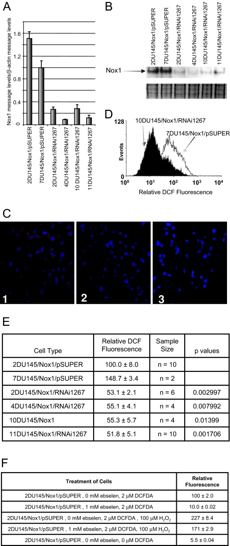
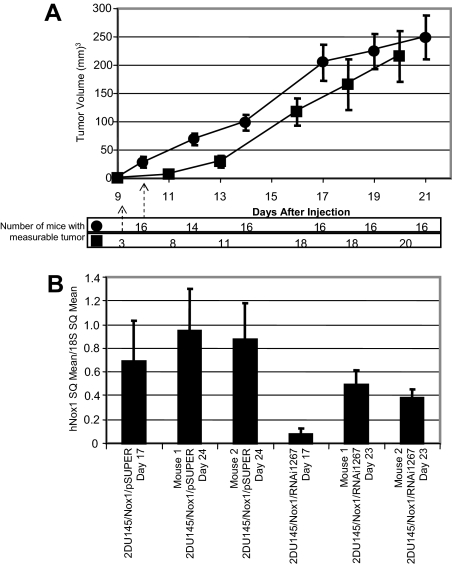
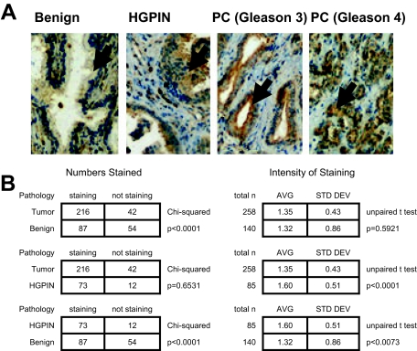
Increased cellular reactive oxygen species (ROS) can act as mitogenic signals in addition to damaging DNA and oxidizing lipids and proteins, implicating ROS in cancer development and progression. To analyze the effects of Nox1 expression and its relation to cellular ROS and signal transduction involved in cellular proliferation, Nox1RNAi constructs were transfected into DU145 prostate cancer cells overexpressing Nox1, causing decreased Nox1 message and protein levels in the Nox1RNAi cell lines. Increased ROS and tumor growth in the Nox1-overexpressing DU145 cells were reversed in the presence of the Nox1RNAi. Analysis and comparison of the message levels in the overexpression and RNAi cells demonstrated that Nox1 overexpression leads to changes in message levels of a variety of proteins including c-fos-induced growth factor, interleukin-8, and Cav-1. Finally, we found that Nox1 protein overexpression is an early event in the development of prostate cancer using a National Cancer Institute prostate cancer tissue microarray (CPCTR). Tumor (86%) was significantly more likely to have Nox1 staining than benign prostate tissue (62%) (P = 0.0001). These studies indicate that Nox1 overexpression may function as a reversible signal for cellular proliferation with relevance for a common human tumor.


Figures

Figure 1
Figure 1
Nox1RNAi1267 constructs decrease Nox1 message, protein, and ROS levels in DU145/Nox1 stably transfected cells. DU145 cells stably overexpressing human Nox1 were stably transfected with either pSUPER vector or with the Nox1RNAi1267 construct. Cells were harvested, and RNA or protein was prepared from individual clones as described in Materials and Methods. A: Real-time PCR analysis of clones was performed as described in Materials and Methods. B: Western blot analysis of several vector-transfected and Nox1RNAi1267-transfected cell lines. Two identical gels were run of protein. The top panel shows the Western blot and the decreased Nox1 protein levels in the Nox1RNAi1267-transfected cells. The bottom panel shows the Coomassie blue-stained gel demonstrating that equal amounts of protein were loaded per lane. The experiment was repeated 10 times with similar results. C: Immunofluorescence staining of 2 DU145/Nox1/pSUPER (1) and 11 DU145/Nox1/Nox1RNAI12676 cells (2). Cells were stained with a rabbit polyclonal antibody to Nox1. Staining of two DU145/Nox1/pSUPER cells with secondary antibody alone is also shown (3). D: Histogram analysis of DCFDA-treated DU145/Nox1/pSUPER and DU145/Nox1/RNAi1267 cell lines as described in Materials and Methods. DU145/Nox1/RNAi1267 cells demonstrated decreased relative DCF fluorescence compared with control cells. E: Reactive oxygen levels are decreased in NoxRNAi1267-transfected cells. Relative DCF fluorescence of various cells lines as described in Materials and Methods. Error represents the SEM of the sample size indicated in the third column. P values were calculated using the Wilcoxon two sample test. The values indicated are comparisons between the indicated Nox1 RNAi cell line and the 2DU145/Nox1/pSUPER cell line. The sample size for the 7DU145/Nox1/pSUPER was too small for comparison. F: DCF fluorescence is a result of increased hydrogen peroxide. Relative DCF fluorescence of 2DU145/Nox1/pSUPER cell line in the presence of various factors including DCF, ebselen, and hydrogen peroxide are shown. Error is the SEM of quadruplicate samples.
Figure 2
Figure 2
Delayed tumor growth in Nox1RNAi1267 xenografts. A: Mice were injected with 3.6 × 10⁶ cells each per mouse (•, 16 mice, 2DU145/Nox1/pSUPER; ▪, 21 mice, 2DU145/Nox1/RNAi1267). Mice were observed every other day. Tumor volume was measured as described in Materials and Methods. Error bars represent SEM of the tumor size for all mice in each set. Delay in tumor growth of 2DU145/Nox1/RNAi1267 mice compared with 2DU145/Nox1/pSUPER mice was statistically significant, P < 0.001 at 10 days. The number of mice in each subset that had measurable tumors at each time point measured is indicated below the graph. B: Total RNA was prepared from mouse tumors at day 17 and day 23, and Nox1 message levels were determined by quantitative RT-PCR as described above. Data are represented relative to 18S levels. Error bars represent the error calculated using the equation: propagation of error = [x/y ∗ square root((ςx/x)2 + (ςy/y)2)] where x = Nox1 SQ mean; y = 18S SQ mean, ς = SD.
Figure 3
Figure 3
Nox1 overexpression in HGPIN and cancer in human prostate tissue. A: Human prostate tissue was analyzed for Nox1 protein expression using immunohistochemical analysis as described in Materials and Methods. Arrows point to the prostate epithelial cells. Representative pictures of the normal prostate epithelial, HGPIN, and prostate cancer (Gleason 3 and Gleason 4) are shown. B: Overall NOX1 staining in benign prostate tissue, HGPIN, and prostate tumors. Analysis was performed as described in Materials and Methods. P values are a result of either χ2 or unpaired t-test analysis.

References

    1. Burdon RH. Control of cell proliferation by reactive oxygen species. Biochem Soc Trans. 1996;24:1028–1032. - PubMed
    1. Irani K, Xia Y, Zweier J, Sollott S, Der C, Rearon E, Sundaresan M, Finkel T, Goldschmidt-Clermont P. Mitogenic signaling mediated by oxidants in Ras-transformed fibroblasts. Science. 1997;275:1649–1652. - PubMed
    1. Nonaka Y, Iwagaki H, Kimura T, Fuchimoto S, Orita K. Effect of reactive oxygen intermediates on the in vitro invasive capacity of tumor cells and liver metastasis in mice. Int J Cancer. 1993;54:983–986. - PubMed
    1. Kundu N, Zhang S, Fulton AM. Sublethal oxidative stress inhibits tumor cell adhesion and enhances experimental metastasis of murine mammary carcinoma. Clin Exp Metastasis. 1995;13:16–22. - PubMed
    1. Suh YA, Arnold RS, Lassegue B, Shi J, Xu X, Sorescu D, Chung AB, Griendling KK, Lambeth JD. Cell transformation by the superoxide-generating oxidase Mox1. Nature. 1999;401:79–82. - PubMed

Publication types

MeSH terms