. 2007 Nov;19(11):3610-26.
doi: 10.1105/tpc.106.049775. Epub 2007 Nov 30.

A viroid RNA with a specific structural motif inhibits chloroplast development

Affiliations

A viroid RNA with a specific structural motif inhibits chloroplast development

Maria-Elena Rodio et al. Plant Cell. 2007 Nov.

Abstract

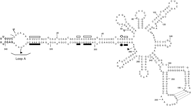
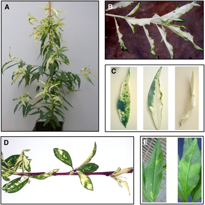
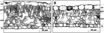
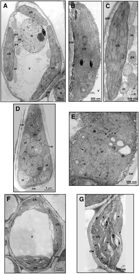
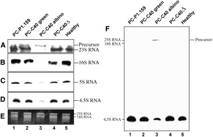
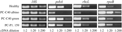
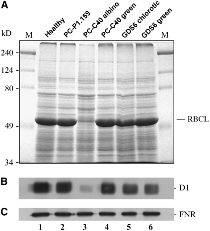
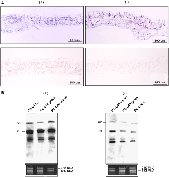
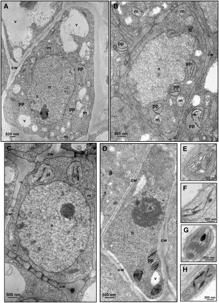
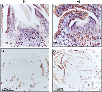
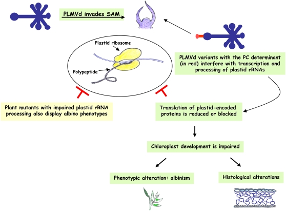
Peach latent mosaic viroid (PLMVd) is a chloroplast-replicating RNA that propagates in its natural host, peach (Prunus persica), as a complex mixture of variants, some of which are endowed with specific structural and pathogenic properties. This is the case of variant PC-C40, with an insertion of 12 to 13 nucleotides that folds into a hairpin capped by a U-rich loop, which is responsible for an albino-variegated phenotype known as peach calico (PC). We have applied a combination of ultrastructural, biochemical, and molecular approaches to dissect the pathogenic effects of PC-C40. Albino sectors of leaves infected with variant PC-C40 presented palisade cells that did not completely differentiate into a columnar layer and altered plastids with irregular shape and size and with rudimentary thylakoids, resembling proplastids. Furthermore, impaired processing and accumulation of plastid rRNAs and, consequently, of the plastid translation machinery was observed in the albino sectors of leaves infected with variant PC-C40 but not in the adjacent green areas or in leaves infected by mosaic-inducing or latent variants (including PC-C40Delta, in which the 12- to 13-nucleotide insertion was deleted). Protein gel blot and RT-PCR analyses showed that the altered plastids support the import of nucleus-encoded proteins, including a chloroplast RNA polymerase, the transcripts of which were detected. RNA gel blot and in situ hybridizations revealed that PLMVd replicates in the albino leaf sectors and that it can invade the shoot apical meristem and induce alterations in proplastids, bypassing the RNA surveillance system that restricts the entry of a nucleus-replicating viroid and most RNA viruses. Therefore, a non-protein-coding RNA with a specific structural motif can interfere with an early step of the chloroplast developmental program, leading ultimately to an albino-variegated phenotype resembling that of certain variegated mutants in which plastid rRNA maturation is also impaired. Our results highlight the potential of viroids for further dissection of RNA trafficking and pathogenesis in plants.


Figures

Figure 1.
Figure 1.
The PLMVd Reference Variant Inducing the Albino-Variegated Phenotype Known as PC. Primary and predicted secondary structures of the lowest free energy of PLMVd variant PC-C40 (Malfitano et al., 2003). The nucleotides of the characteristic insertion in loop A containing the PC determinant are highlighted in boldface. (+) and (−) self-cleaving domains are delimited by flags, residues conserved in most natural hammerhead structures are indicated by bars, and the self-cleavage sites are marked by arrows. Closed and open symbols refer to (+) and (−) polarities, respectively. Residues involved in a pseudoknot between positions 177 to 180 and 210 to 213, proposed on the basis of in vitro nuclease mapping and oligonucleotide binding shift assays (Bussière et al., 2000) and natural variability (Malfitano, 2000), are indicated by broken lines.
Figure 2.
Figure 2.
PC and Other PLMVd-Induced Phenotypes in GF-305 Peach Seedlings. (A) PC symptoms elicited by PLMVd variant PC-C40 (Malfitano et al., 2003). (B) Close view of an almost completely albino shoot from a PC-expressing plant inoculated with PLMVd variant PC-C40. (C) Distinct leaf patterns of the albino-variegated phenotype incited by variant PC-C40. (D) PC symptoms induced by variant PC-C40 in young expanding leaves of new flushes emerging after a dormant period. (E) Typical leaf mosaic induced by PLMVd variant GDS6 (Ambrós et al., 1999).
Figure 3.
Figure 3.
Light Microscopy Images of Mature GF-305 Peach Leaves. Transverse sections from a healthy leaf (A) and from the albino sector of a leaf infected by variant PC-C40 (B). In the latter, the palisade cells are not completely differentiated in a columnar layer (the height of which was approximately half that of green leaves from healthy and PLMVd-infected controls). ue, upper epidermis; pm, palisade mesophyll; sm, spongy mesophyll; le, lower epidermis.
Figure 4.
Figure 4.
Transmission Electron Microscopy Images of Cells and Plastids of Mature GF-305 Peach Leaves. (A) to (C) Mesophyll cell (A) and mature (B) and senescent (C) chloroplasts of a healthy control. (D) and (E) Mesophyll cell (D) and plastid resembling a proplastid and containing vesicles (E) observed in the albino sector of a leaf infected by variant PC-C40. Black arrows in (E) indicate rudimentary thylakoids. (F) and (G) Mesophyll cell (F) and altered chloroplast (G) from a green sector of a leaf infected by variant PC-C40. Arrowheads in (G) point to enlarged thylakoid interspaces. ch, chloroplast; cw, cell wall; m, mitochondrion; n, nucleus; pl, plastid; px, peroxisome; v, vacuole.
Figure 5.
Figure 5.
Accumulation Levels of Plastid rRNAs in GF-305 Peach Leaves. (A) to (D) RNA gel blot analyses of RNA preparations hybridized with cDNA probes specific for 23S, 16S, 5S, and 4.5S rRNAs after electrophoresis on denaturing agarose (1.2%) gels for detecting 23S and 16S rRNAs ([A] and [B]) or on denaturing polyacrylamide (5%) gels for detecting 5S and 4.5S rRNAs ([C] and [D]). Only the autoradiogram sections corresponding to each of the four rRNAs, and to a larger precursor observed exclusively in lane 3, are presented. (E) Ethidium bromide pattern of the upper section of the polyacrylamide gel showing equal intensity of the larger bands generated by the nucleus-encoded 25S and 18S rRNAs. The absence of the smaller bands in the RNA preparation from the albino leaf sector is consistent with the low accumulation of plastid rRNAs detected by RNA gel blot hybridization. (F) Overexposure of the membrane corresponding to (D) showing the presence of a larger RNA that most likely corresponds to the p23S-4.5S intermediate.
Figure 6.
Figure 6.
Transcript Accumulation in GF-305 Peach Leaves. RT-PCR estimation of the nucleus-encoded 18S rRNA, the transcripts from the plastid-encoded psbA and rbcL genes, and the transcript from the nucleus-encoded rpoB gene. The cDNAs, generated by random-primed reverse transcription of equalized RNA preparations, were serially diluted (1:2, 1:20, and 1:200) before amplification with gene-specific primer pairs. The amplified products were analyzed by agarose gel electrophoresis and ethidium bromide staining. Only gel sections containing the amplified cDNAs are presented. Results are representative of three replicates conducted on two biologically independent samples.
Figure 7.
Figure 7.
Accumulation Levels of Solubilized Proteins in GF-305 Peach Leaves. (A) Protein profiles after SDS-PAGE on a 12% gel and staining with Coomassie blue. The external lanes contain molecular markers (M), with their sizes in kilodaltons indicated at left. The position of the large subunit of the ribulose-1,5-bisphosphate carboxylase/oxygenase (RBCL), the accumulation of which is very much reduced in the albino sector of a leaf infected by variant PC-C40, is indicated at right. (B) and (C) Protein gel blot analyses with antibodies raised against the plastid-encoded D1 protein (B) and the nucleus-encoded and plastid-targeted FNR (C). Only the autoradiogram sections corresponding to each protein are shown.
Figure 8.
Figure 8.
Detection of PLMVd (+) and (−) Strands in GF-305 Peach Leaves Infected by Two Viroid Variants. (A) In situ hybridization of transverse sections from the albino sector of a leaf infected by variant PC-C40 (top) and from a healthy leaf (bottom). The sections were hybridized with riboprobes for detecting (+) (left) and (−) (right) PLMVd strands. Hybridization signals appear with a blue-violet color. (B) RNA gel blot hybridizations of different RNA preparations with riboprobes for detecting (+) (left) and (−) (right) PLMVd strands following fractionation by denaturing PAGE on a 5% gel. The positions of the monomeric circular (mc) and linear (ml) PLMVd RNAs are indicated at left. The bottom panels show the ethidium bromide pattern of the top section of the gels (for additional details, see the legend to Figure 5E). Results are representative of three independent experiments with similar outcome: the progeny of variant PC-C40 accumulated at a significantly higher level (50.5% ± 4.5%) in the albino sectors than in the green sectors of the same leaf. Loadings were equalized with respect to the nucleus-encoded rRNAs, which remain unaffected in symptomatic and asymptomatic tissues.
Figure 9.
Figure 9.
Transmission Electron Microscopy Images of Cells and Proplastids from the SAM of GF-305 Peach Seedlings. (A) SAM cell from an albino shoot infected by variant PC-C40. (B) SAM cell from a healthy control. (C) and (D) SAM cells from shoots infected by the latent variants PC-C40Δ (C) and PC-P1.159 (D). (E) to (H) Closer views of proplastids from the same tissue sections presented in (A) to (D), respectively. Proplastids from shoots infected by latent variants ([G] and [H]) do not show thylakoid alterations with respect to the healthy control (F), in contrast with thylakoids from the albino shoot infected by variant PC-C40, in which enlarged interspaces are clearly visible (E). cw, cell wall; m, mitochondrion; n, nucleus; pp, proplastid; v, vacuole.
Figure 10.
Figure 10.
Detection of PLMVd (+) and (−) Strands in the SAM of GF-305 Seedlings. In situ hybridizations with riboprobes for detecting (+) (left) and (−) (right) PLMVd strands in longitudinal SAM sections from albino shoots infected by variant PC-C40 ([A] and [B]) and from a healthy shoot ([C] and [D]). Hybridization signals appear with a blue-violet color different from the brown precipitates of unknown origin also visible in the healthy control.
Figure 11.
Figure 11.
Model for Pathogenesis Based on the Ability of PLMVd RNA to Invade the SAM and Alter the Proplastid Developmental Program.

Similar articles

Cited by

References

    1. Allison, L.A., Simon, L.D., and Maliga, P. (1996). Deletion of rpoB reveals a second distinct transcription system in plastids of higher plants. EMBO J. 15 2802–2809. - PMC - PubMed
    1. Ambrós, S., Hernández, C., Desvignes, J.C., and Flores, R. (1998). Genomic structure of three phenotypically different isolates of peach latent mosaic viroid: Implications of the existence of constraints limiting the heterogeneity of viroid quasi-species. J. Virol. 72 7397–7406. - PMC - PubMed
    1. Ambrós, S., Hernández, C., and Flores, R. (1999). Rapid generation of genetic heterogeneity in progenies from individual cDNA clones of peach latent mosaic viroid in its natural host. J. Gen. Virol. 80 2239–2252. - PubMed
    1. Barba, M., Cupidi, A., Loreti, S., Faggioli, F., and Martino, L. (1995). In vitro micrografting: A technique to eliminate peach latent mosaic viroid from peach. Acta Hortic. 386 531–535.
    1. Barkan, A. (1993). Nuclear mutants of maize with defects in chloroplast polysome assembly have altered chloroplast RNA metabolism. Plant Cell 5 389–402. - PMC - PubMed

Publication types

MeSH terms

LinkOut - more resources