. 2008 Feb;7(2):379-86.
doi: 10.1128/EC.00221-07. Epub 2007 Nov 30.

Combination of two regulatory elements in the Tetrahymena thermophila HSP70-1 gene controls heat shock activation

Affiliations

Combination of two regulatory elements in the Tetrahymena thermophila HSP70-1 gene controls heat shock activation

Sabrina Barchetta et al. Eukaryot Cell. 2008 Feb.

Abstract

The induction of heat shock genes (HSPs) is thought to be primarily regulated by heat shock transcription factors (HSFs), which bind target sequences on HSP promoters, called heat shock elements (HSEs). In this study, we investigated the 5' untranslated regions of the Tetrahymena thermophila HSP70-1 gene, and we found, in addition to the canonical and divergent HSEs, multiple sets of GATA elements that have not been reported previously in protozoa. By means of in vivo analysis of a green fluorescent protein reporter transgene driven by the HSP70-1 promoter, we demonstrate that HSEs do not represent the minimal regulatory elements for heat shock induction, since the HSP70-1 is tightly regulated by both HSE and GATA elements. Electrophoretic mobility shift assay also showed that HSFs are constitutively bound to the HSEs, whereas GATA elements are engaged only after heat shock. This is the first demonstration by in vivo analysis of functional HSE and GATA elements in protozoa. Furthermore, we provide evidence of a functional link between HSE and GATA elements in the activation of the heat shock response.


Figures

FIG. 1.
FIG. 1.
Partial nucleotide sequence of the 5′ UTR of the HSP70-1 gene. The sequences comprising the putative HSE I and HSE II are boxed, and the individual sequence motifs are in boldface. The cluster of GATA elements is shaded. The initiating ATG is shown in boldface italics and is followed by the codons of the first seven amino acids of the protein. In the plasmids used in this study, these seven codons were fused in frame with the initiating ATG of the GFP, by means of a BglII site. The synthetic primers used to amplify this sequence from the genomic DNA are underlined, and the restriction sites NotI and BglII, at the 5′ and 3′ ends, respectively, are in boldface.
FIG. 2.
FIG. 2.
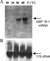
Northern blot analysis of total RNA samples extracted from T. thermophila cells after heat shock treatments performed at the indicated temperatures. (A) Samples of 20 μg of total RNA were transferred onto Hybond N membrane and assayed with labeled probe specific for the HSP70-1 mRNA. (B) Differences in sample loading were detected by reprobing the membrane with a labeled probe of the same length and complementary to the T. thermophila 17S rRNA.
FIG. 3.
FIG. 3.
Visualization of the fluorescence emission by T. thermophila cells transfected with the pWT (containing the wild-type 5′ flanking region of the T. thermophila HSP70-1). Samples of 20 ml of cell cultures were incubated at the indicated temperatures, and the GFP fluorescence emission was detected in vivo, throughout 1 h, by using an Olympus IX71 fluorescence microscope equipped with fluorescein isothiocyanate filters. Pictures were taken by the Olimpus U-CMAD3 digital camera at ×100 magnification. Two minutes prior to observation, the cells were treated with dibucaine hydrochloride at a final concentration of 0.3 mM to reduce the swimming speed.
FIG. 4.
FIG. 4.
Mutation analysis of the 5′ UTR of the T. thermophila HSP70-1 gene. (A) Schematic representation of the wild-type and mutated sequences used to produce the synthetic plasmids listed below: pWT, containing 900 bp of the wild-type T. thermophila HSP70-1 5′ UTR; pΔHSE I, derived from the pWT, lacking 16 nt containing the putative HSE I; pΔHSE II, derived from the pWT, lacking 28 nt containing the putative HSE II; pΔGATA I, derived from the pWT, lacking 45 nt containing the cluster of GATA motifs; pΔGATA II, derived from the pWT, maintaining the original sequence length and subjected to site-directed mutagenesis on the GATA sequences that were changed from GATAGATACATAGATAGATAGATAGATAGATAGATAAATAGATAGATG to GTTAAATACATAGATTAATTGTATGATCGAAACATAAATATTAAGATG. (B) Visualization of cell strains transfected with the corresponding plasmids of panel A. Cells lines, transfected with the listed plasmids, were grown to the density of 2 × 10⁵ cells/ml and heat shocked at different temperatures as described in Materials and Methods. To verify the fluorescence emission, samples of cells were collected throughout 1 h and observed under by fluorescence microscopy. The pictures shown were taken in vivo, after 15 min of heat shock at 38°C, by a fluorescence microscope Olympus IX71 equipped with a fluorescein isothiocyanate filter.
FIG. 5.
FIG. 5.
Detection of the GFP mRNA on transfected T. thermophila cells. Samples of 100 ng of cDNA were used in PCR analysis with primers designed on the GFP coding region and on the sequence of the 17S rRNA to amplify fragments of 750 and 300 bp, respectively. The reaction run in lane 1 contained no cDNA, while the reaction run in lane 2 contained cDNA obtained from not transfected cells. These reactions showed the specificity of the GFP primers. The other reactions contained cDNA obtained from cells transfected with the following plasmids: pWT, pΔHSE I, pΔHSE II, pΔGATA I, pΔGATA II (in lanes from 3 to 7, respectively). St., DNA standards.
FIG. 6.
FIG. 6.
EMSA of nuclear protein extracts from heat shocked and unshocked cells performed with targets containing HSE (A) or GATA elements (B). In both panels, binding reactions containing only the labeled wild-type target or the labeled wild-type target with the nuclear extracts from unshocked cells or from heat shocked cells are shown in lanes 1 to 3, respectively; 50- or 100-fold molar excesses of unlabeled wild-type target as cold competitor were added to binding reactions containing nuclear extracts from heat shocked cells (lanes 4 and 5, respectively). Lanes from 6 to 10 show binding reactions from experiments involving mutant targets as competitors. Specifically, in panel A binding reactions with nuclear extracts from shocked cells in the presence of labeled wild-type target with 50- or 100-fold molar excesses of the HSE mutated unlabeled target are shown in lanes 8 and 9, respectively; the binding reaction of HSE mutated labeled target with nuclear extracts from shocked cells is shown in lane 10; in lanes 6 and 7, binding reactions in conditions identical to those in lanes 2 and 3 were run as a reference. In panel B, binding reactions with nuclear extracts from shocked cells in the presence of GATA labeled wild-type target with 50- or 100-fold molar excesses of the GATA mutated unlabeled target are shown in lanes 8 and 9, respectively. The binding reaction of GATA mutated labeled probe with nuclear extracts from shocked cells is shown in lane 6; a binding reaction in conditions identical to those for lane 3 was run as a reference in lane 7. Asterisks indicate the DNA-protein complexes, and arrows indicate the free target.

Similar articles

Cited by

References

    1. Amaral, M. D., L. Galego, and C. Rodrigues-Pousada. 1988. Stress response of Tetrahymena pyriformis to arsenite and heat shock: differences and similarities. Eur. J. Biochem. 171463-470. - PubMed
    1. Amaral, M. D., L. Galero, and C. Rodrigues-Pousada. 1993. Heat-shock-induced protein synthesis is responsible for the switch-off of hsp 70 transcription in Tetrahymena. Biochim. Biophys. Acta 1174133-142. - PubMed
    1. Boorstein, W. R., T. Ziegelhoffer, and E. A. Craig. 1994. Molecular evolution of the HSP70 multigene family. J. Mol. Evol. 381-17. - PubMed
    1. Boye, J., J. Omichinsky, D. Clark, M. Pikaart, and G. Felsenfeld. 1998. Perturbation of nucleosome structure by the erythroid transcription factor GATA-1. J. Mol. Biol. 279529-544. - PubMed
    1. Bradford, M. M. 1976. A rapid and sensitive for the quantitation of microgram quantities of protein utilizing the principle of protein-dye binding. Anal. Biochem. 72248-254. - PubMed

Publication types

MeSH terms

Substances

LinkOut - more resources