

Elaiophore structure and oil secretion in flowers of Oncidium trulliferum Lindl. and Ornithophora radicans (Rchb.f.) Garay & Pabst (Oncidiinae: Orchidaceae)
- PMID: 18056056
- PMCID: PMC2701824
- DOI: 10.1093/aob/mcm297
Elaiophore structure and oil secretion in flowers of Oncidium trulliferum Lindl. and Ornithophora radicans (Rchb.f.) Garay & Pabst (Oncidiinae: Orchidaceae)
Abstract
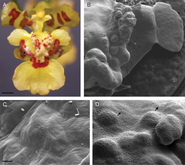
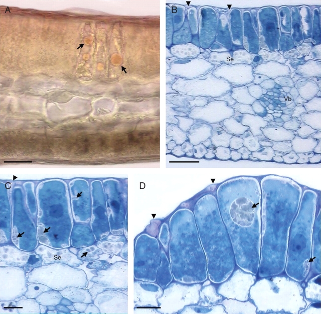
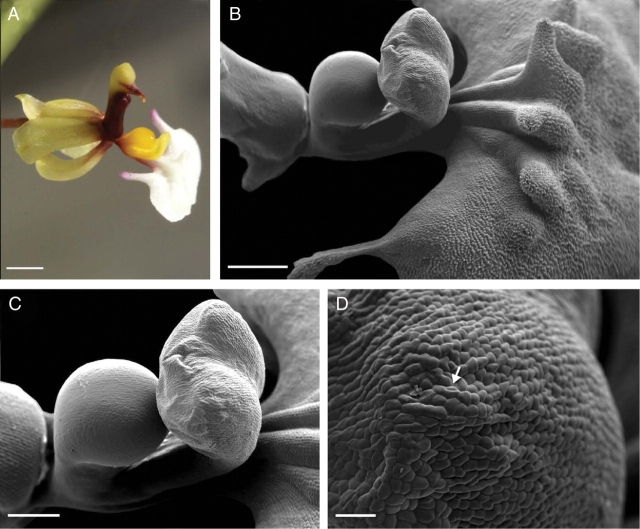
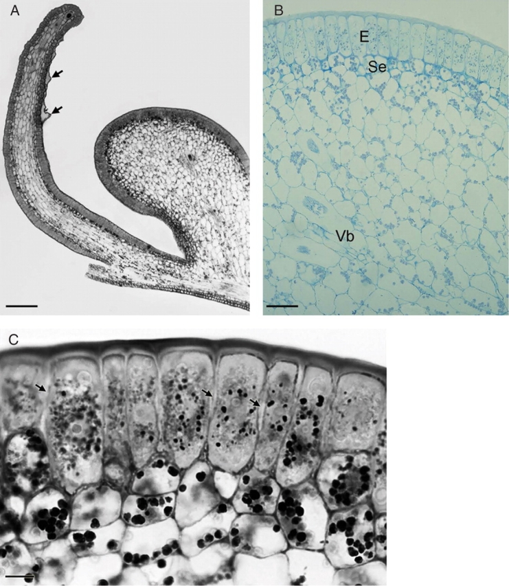
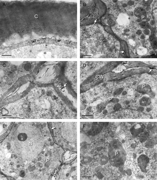
Background and aims: Many orchid flowers have glands called elaiophores and these reward pollinating insects with oil. In contrast to other reward-producing structures such as nectaries, the anatomy of the elaiophore and the process of oil secretion have not been extensively studied. In this paper, elaiophore structure is described for two members of Oncidiinae, Oncidium trulliferum Lindl. and Ornithophora radicans (Rchb.f.) Garay & Pabst.
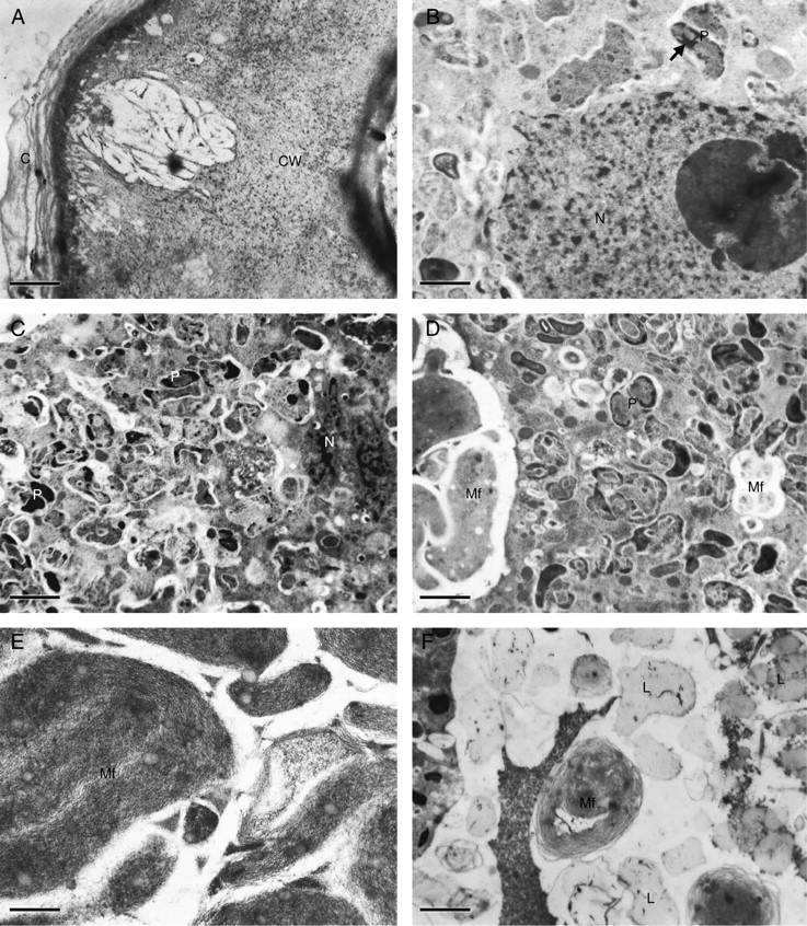
Methods: Elaiophores of both species were examined using light microscopy, scanning electron microscopy and transmission electron microscopy.
Key results and conclusions: In flowers of Oncidium trulliferum and Ornithophora radicans, oil is secreted by morphologically distinct elaiophores associated with the labellar callus. However, in O. trulliferum, elaiophores also occur on the lateral lobes of the labellum. In both these species, the epithelial elaiophores are composed of a single layer of palisade-like epidermal cells and a distinct subepithelial layer. Secretory elaiophore cells may contain numerous, starchless plastids, mitochondria and smooth endoplasmic reticulum profiles. In O. trulliferum, the cytoplasm contains myelin-like figures but these are absent from O. radicans. In the former species, cavities occur in the cell wall and these presumably facilitate the passage of oil onto the elaiophore surface. In O. radicans, the accumulation of oil between the outer tangential wall and the cuticle causes the latter to become distended. Since it is probable that the full discharge of oil from the elaiophores of O. radicans occurs only when the cuticle is ruptured by a visiting insect, this may contribute towards pollinator specificity. The structure of the elaiophore in these species resembles both that found in previously investigated species of Oncidiinae and that of certain members of the Malpighiaceae.
Figures
References
-
- Arumugasamy K, Udaiyan K, Manian S, Sugavanam V. Ultrastructure and oil secretion in Hiptage sericea Hook. Acta Societatis Botanicorum Poloniae. 1993;62:17–20.
-
- Attala NC, Machado SR. Anatomy and ultrastructure of Banisteriopsis variabilis Gates (Malpighiaceae) calyx glands. Acta Microscopica. 2003;12(Suppl. B):635–636.
-
- Bhatt JR. Development and structure of primary secretory ducts in the stem of Commiphora wightii (Burseraceae) Annals of Botany. 1987;60:405–416.
-
- Buchmann SL. The ecology of oil flowers and their bees. Annual Review of Ecology and Systematics. 1987;18:343–396.
-
- Cane JH, Eickwort GC, Wesley FR, Spielholz J. Foraging, grooming and mate-seeking behaviors of Macropis nuda (Hymenoptera, Melittidae) and use of Lysimachia ciliata (Primulaceae) oils in larval provisions and cell linings. American Midland Naturalist. 1983;110:257–264.
