. 2008 Jan;131(Pt 1):264-76.
doi: 10.1093/brain/awm267. Epub 2007 Dec 3.

OEG implantation and step training enhance hindlimb-stepping ability in adult spinal transected rats

Affiliations

OEG implantation and step training enhance hindlimb-stepping ability in adult spinal transected rats

Marc D Kubasak et al. Brain. 2008 Jan.

Abstract

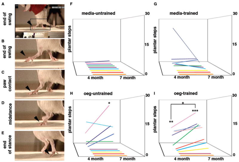
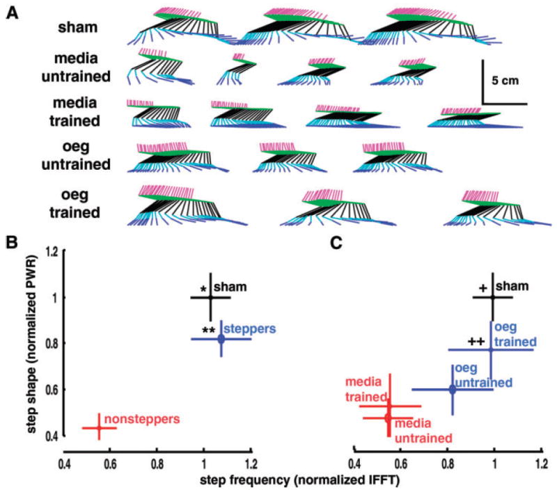
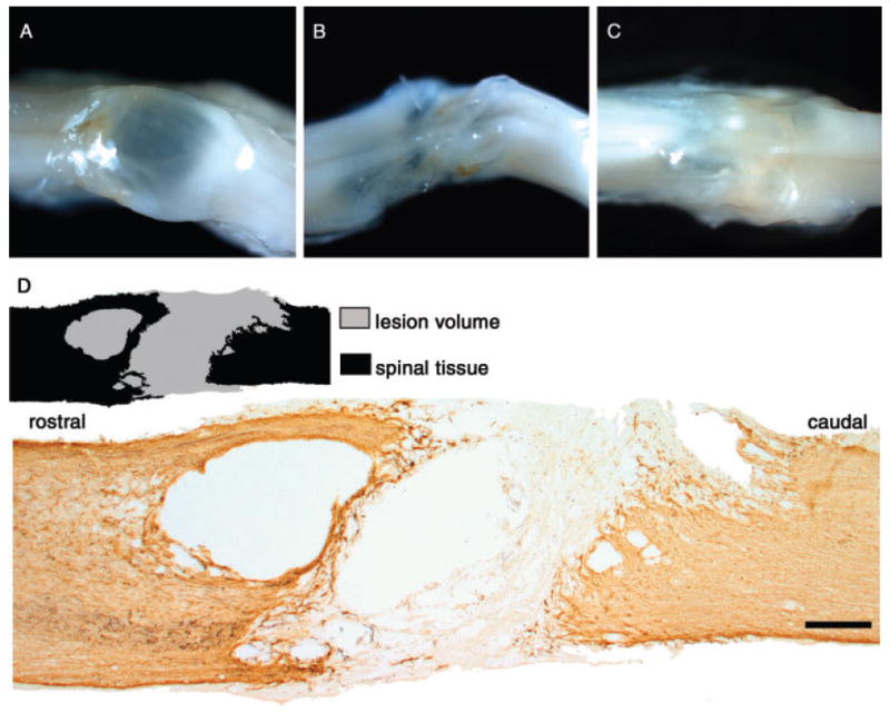
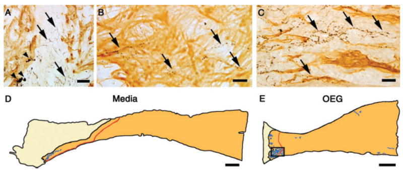
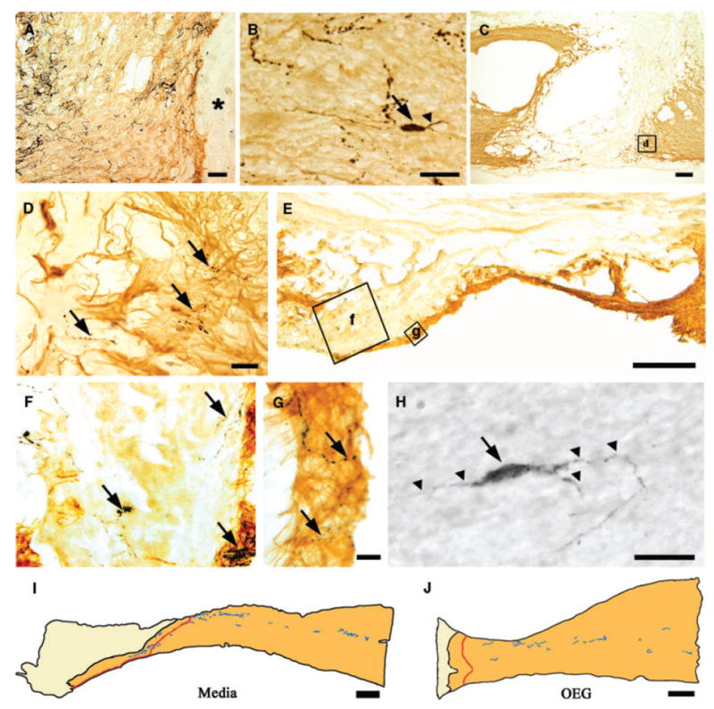
Numerous treatment strategies for spinal cord injury seek to maximize recovery of function and two strategies that show substantial promise are olfactory bulb-derived olfactory ensheathing glia (OEG) transplantation and treadmill step training. In this study we re-examined the issue of the effectiveness of OEG implantation but used objective, quantitative measures of motor performance to test if there is a complementary effect of long-term step training and olfactory bulb-derived OEG implantation. We studied complete mid-thoracic spinal cord transected adult female rats and compared four experimental groups: media-untrained, media-trained, OEG-untrained and OEG-trained. To assess the extent of hindlimb locomotor recovery at 4 and 7 months post-transection we used three quantitative measures of stepping ability: plantar stepping performance until failure, joint movement shape and movement frequency compared to sham controls. OEG transplantation alone significantly increased the number of plantar steps performed at 7 months post-transection, while training alone had no effect at either time point. Only OEG-injected rats plantar placed their hindpaws for more than two steps by the 7-month endpoint of the study. OEG transplantation combined with training resulted in the highest percentage of spinal rats per group that plantar stepped, and was the only group to significantly improve its stepping abilities between the 4- and 7-month evaluations. Additionally, OEG transplantation promoted tissue sparing at the transection site, regeneration of noradrenergic axons and serotonergic axons spanning the injury site. Interestingly, the caudal stump of media- and OEG-injected rats contained a similar density of serotonergic axons and occasional serotonin-labelled interneurons. These data demonstrate that olfactory bulb-derived OEG transplantation improves hindlimb stepping in paraplegic rats and further suggest that task-specific training may enhance this OEG effect.


Figures

Fig. 1
Fig. 1
OEG transplantation promotes plantar stepping in adult spinal rats and is augmented by step training. (A–E) An example of a plantar step from an OEG-untrained animal. (A, B) The plantar step shows extension of digits at the end of swing phase (arrowhead in B, enlarged from boxed area in A). (C-E) The plantar surface of the foot and toes (arrowheads) touch the treadmill at paw contact (C), and remain in contact with the treadmill through the midstance (D) and end of stance (E) phases. (F–I) Plantar step counts at 4 and 7 months post-injury for media-untrained (F, n = 9), media-trained (G, n = 9), OEG-untrained (H, n = 10) and OEG-trained (I, n = 10) rats. Significant differences in the mean number of plantar steps exist between the media-untrained and the OEG-untrained groups at 7 months (*P = 0.02) and between the media-untrained and OEG-trained groups at 4 (**P = 0.02) and 7 (***P ≤ 0.001) months. Plantar step counts are significantly different between OEG-trained animals evaluated at 4 and 7 months (+P=0.03). The number of rats with improvement in plantar stepping between the 4 and 7 month tests is significantly different between the OEG-trained and the media-untrained (P = 0.001), media-trained (P =0.001) and OEG-untrained (P = 0.03) groups.
Fig. 2
Fig. 2
The combination of OEG transplantation and step training improves stepping kinematics. (A) Stepping kinematics from representative trials that exhibit the IFFTand PWR values closest to the mean values for each of the five groups. Segments run from the iliac crest to the proximal femur (magenta), femur to the knee (green), knee to the ankle (black), ankle to the distal metatarsals (light blue) and distal metatarsals to the end of the toes (dark blue). Figures represent projection of kinematics onto the sagittal plane. (B,C) Step shape (represented by PWR values) is plotted against step frequency (IFFTvalues) and data are normalized to mean values for shams and represent the group means ( ± SEM). (B) Normalized IFFTand PWR scores for animals that took at least one plantar-placed step (steppers), animals that did not step (nonsteppers), and sham rats. IFFTand PWR scores of the nonstepping animals are significantly different from the sham (IFFT P < 0.01, PWR P < 0.01) and stepping (IFFT P < 0.01, PWR P < 0.01) animals. Stepping animals are not significantly different from the shams (IFFT P = 0.43, PWR P = 0.11). (C) IFFTand PWR scores of the media-untrained and media-trained animals are significantly different from sham (IFFT P = 0.01, PWR P < 0.01 for media-untrained; IFFT P = 0.01, PWR P = 0.02 for media-trained). OEG-untrained rats do not differ from shams in IFFT (P = 0.24) but show significantly lower PWR scores (P = 0.01). OEG-trained animals are not significantly different from shams (IFFT P = 0.48, PWR P = 0.11).
Fig. 3
Fig. 3
Tissue sparing at the transection site occurs with OEG transplantation. (A–C) Transected spinal cords demonstrate a range of cavitation. A spinal cord from a media-untrained (A) rat shows large transparent cavities and little evidence of regeneration, while much less cavitation is apparent in the injury site from a second media-untrained (B) rat. An OEG-trained animal demonstrates an opaque injury site devoid of pronounced cavitations (C). (D) A 25-μm-thick sagittal spinal cord section immunostained for GFAP (amber-brown) containing the transection site and a drawing illustrating the identification of the GFAP-positive tissue (black area in drawing) and the GFAP-negative transection site (gray) separating the rostral and caudal stumps. Scale bar: D, 400 μm.
Fig. 4
Fig. 4
Noradrenergic axons extend into and through the GFAP-negative scar. Sagittal sections are oriented with rostral to the left and dorsal to the top in this and the following figure. (A) DBH-positive axons (arrows) extend between the rostral GFAP-positive spinal cord and the GFAP-negative scar in a media-injected rat. Large bulbous endings (arrowheads) are present at the rostral border of the injury site. (B) In an OEG-injected animal, DBH-positive fibres (arrows) extend into the GFAP-positive caudal spinal cord. (C) DBH-positive fibres (arrows) course into the GFAP-positive caudal stump in an OEG-injected animal, with some fibres extending more than 250 μm from the caudal border of the GFAP-negative zone (C is enlarged from the boxed area in E) (D, E) Merged camera lucida drawings of seven 25-μm-thick sagittal sections from the caudal stump of a media-untrained (D) and an OEG-untrained (E) rat. The GFAP-negative scar tissue is light beige, the caudal GFAP-positive spinal cord is dark beige, and the red line demarcates a distance of 250 μm caudal to the GFAP-negative border. Many more DBH-positive fibres are detected caudal to the 250 μm boundary in OEG- compared to media-injected animals. Scale bars: A, 50 μm; B, C, 25 μm; D, E, 400 μm.
Fig. 5
Fig. 5
Serotonergic axons and cell bodies are found rostral (to the left) and caudal to the injury site. (A) 5-HT-positive (black) axons in the rostral GFAP-positive (amber-brown) spinal cord of a media-untrained animal terminate at the GFAP-negative scar (asterisk). (B) A 5-HT-positive interneuron (arrow) just rostral to the GFAP-negative zone of a media-injected rat. A branched process (arrowhead) extends from this bipolar neuron. (C, D) The injury site of an OEG-trained rat double-labelled for GFAP and 5-HT immunoreactivity. 5-HT-positive axons (D, arrows) course into the caudal GFAP-positive spinal cord (boxed area in C is enlarged in D). (E–G) 5-HT-positive fibres span the GFAP-negative scar of an OEG-untrained animal (boxed areas in E are enlarged in F and G). 5-HT-positive fibres (arrows) extend through the scar and enter the GFAP-positive caudal spinal cord. (H) Several processes (arrowheads) extend from this 5-HT-positive interneuron (arrow) found caudal to the transection site (~T10) in a media-injected rat. (I, J) Merged camera lucida drawings of seven 25-μm-thick sagittal sections of caudal spinal cord stumps from a media-untrained (I) and an OEG-untrained (J) animal demonstrate similar patterns of 5-HT-labelled axons. The GFAP-negative scar tissue is light beige, the caudal GFAP-positive spinal cord is dark beige, and the red line demarcates a distance of 250 μm from the GFAP-negative zone. Scale bars A, 50 μm; C, 200 μm; B, D, F, G, H, 25 μm; E, 100 μm; I, J, 400 μm.

Comment in

Similar articles

Cited by

References

    1. Barbeau H, Chau C, Rossignol S. Noradrenergic agonists and locomotor training affect locomotor recovery after cord transection in adult cats. Brain Res Bull. 1993;30:387–93. - PubMed
    1. Barbeau H, Rossignol S. Enhancement of locomotor recovery following spinal cord injury. Curr Opin Neurol. 1994;7:517–24. - PubMed
    1. Barbeau H, Rossignol S. Recovery of locomotion after chronic spinalization in the adult cat. Brain Res. 1987;412:84–95. - PubMed
    1. Barbeau H, Rossignol S. Initiation and modulation of the locomotor pattern in the adult chronic spinal cat by noradrenergic, serotonergic and dopaminergic drugs. Brain Res. 1991;546:250–60. - PubMed
    1. Basso DM, Beattie MS, Bresnahan JC. A sensitive and reliable locomotor rating scale for open field testing in rats. J Neurotrauma. 1995;12:1–21. - PubMed

Publication types