. 2007 Dec 15;179(12):8042-50.
doi: 10.4049/jimmunol.179.12.8042.

Passive immunization against the MHC class I molecule Mamu-AG disrupts rhesus placental development and endometrial responses

Affiliations

Passive immunization against the MHC class I molecule Mamu-AG disrupts rhesus placental development and endometrial responses

Gennadiy I Bondarenko et al. J Immunol. .

Abstract

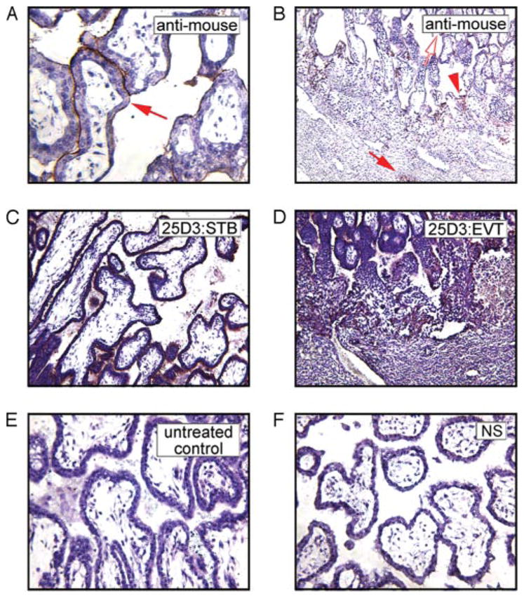
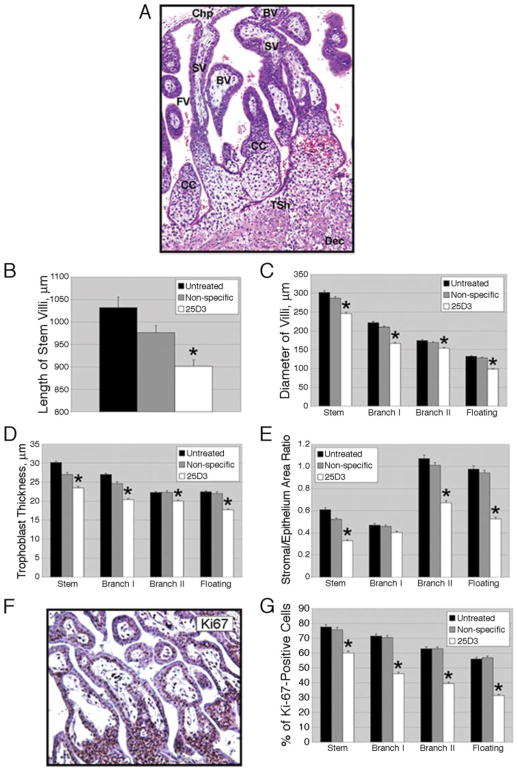
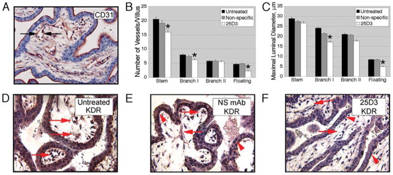
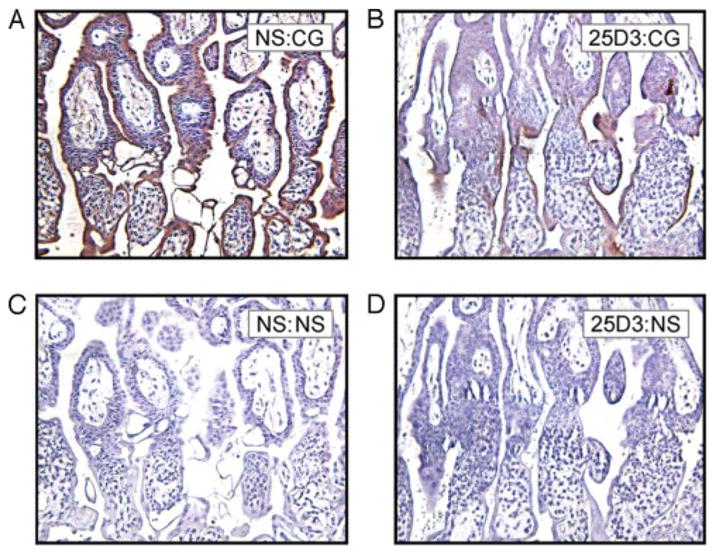
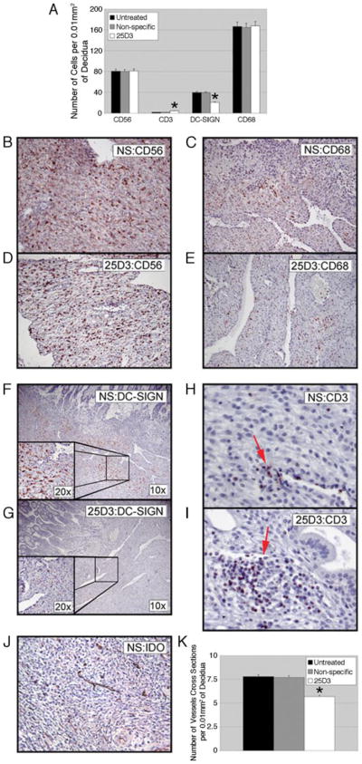
The unique MHC phenotype of the human and nonhuman primate placenta has suggested a potential role in maternal-fetal immune tolerance, pregnancy success, and maternal as well as fetal well-being. In the rhesus monkey (Macaca mulatta) a nonclassical MHC class I molecule, Mamu-AG, is a putative homologue of HLA-G and is hypothesized to play a role in maternal-fetal immune interactions during pregnancy. Rhesus monkeys were passively immunized during the second week after implantation with a mAb against Mamu-AG. Passive immunization altered the growth and vascularization of the fetal placenta, the placental modification of maternal endometrial vessels, the maternal leukocyte response to implantation, and the differentiation of epithelial and stromal cells in the endometrium. These data are the first to demonstrate in vivo the importance of MHC class I molecules expressed on primate trophoblasts in establishing an important environment for pregnancy success through coordinated interactions between endometrial and fetal tissues.


Conflict of interest statement

Disclosures

The authors have no financial conflict of interest.

Figures

FIGURE 1
FIGURE 1
Localization of anti-Mamu-AG (25D3) mAb in passively immunized monkeys. The mAb distribution in the rhesus monkey placenta at the implantation site in villous (A) and extravillous (B) trophoblasts visualized with anti-mouse IgG in the rhesus monkey placenta is similar to Mamu-AG expression detected in frozen sections with the 25D3 mAb in villous (C) and in extravillous (D) trophoblasts on day 24 in untreated control pregnancy. Untreated control (E) and NS-treated (F) groups demonstrated no mAb deposition as detected by immunostaining with a horse anti-mouse IgG.
FIGURE 2
FIGURE 2
Effects of passive immunization against Mamu-AG on placental growth and morphogenesis. A, Depiction of various villous types in the rhesus monkey placenta (untreated control, H&E staining). SV, Stem villus; BV, branching villus; CC, cytotrophoblast column; FV, floating villus; Chp, chorionic plate; Tsh, trophoblastic shell; Dec, decidua. B, Length of stem villi in untreated control, NS-treated, or 25D3-treated placentas. The mean + SEM of five independent samples from four placentas per group is shown; *, p < 0.01 compared with control groups in this and all other graphs in this figure. C, Diameter of classes of villi. D, Trophoblast thickness as determined from measurements on five villi in each class within each animal. E, Ratios of villous stromal area/trophoblast area. F, Representative IHC for Ki67. G, Proliferation index (percentage of Ki67-positive trophoblast nuclei) in each villous class.
FIGURE 3
FIGURE 3
Effects of passive immunization against Mamu-AG on placental vascularization. A, Representative IHC for CD31 to visualize villous stromal vessels. B, The number of vessels per villus. C, Maximal luminal vessel diameter (μm) in individual villous classes (see arrows in A).*, p < 0.01 in B and C. D–F, Representative IHC for KDR in untreated (D), NS-treated (E), or 25D3-treated (F) animals. Arrows indicate stromal endothelial cells and arrowheads indicate villous cytotrophoblasts.
FIGURE 4
FIGURE 4
IHC for monkey CG. Rhesus monkey placentas on day 24 of gestation, NS treated (A and C) or 25D3 treated (B and D), are shown. Immunostaining was performed on paraffin-embedded sections with anti-CG mAb (A and B) or a negative control primary Ab (C and D).
FIGURE 5
FIGURE 5
Effect of passive immunization on placental-decidual interactions. Placental lacunae in NS-treated (A) or 25D3-treated (B) monkeys. Paraffin-embedded sections with H&E staining are shown. Maternal RBC in A are indicated by arrows. Lacunae (L) at the placental-decidual border are indicated. C–H, Endovascular invasion of placental arterioles by EVCT. Nonspecific treated (C, E, and G) or 25D3-treated (D, F, and H) paraffin-embedded sections were immunostained with the indicated Abs as follows: anti-cytokeratin (CTK) (C and D; arrows indicate CTK-positive endovascular cytotrophoblasts); anti-von Willebrand factor (VWF) (E and F; arrows demonstrate lack of positive endothelial cells; see E, inset, for positively stained endometrial vessels); anti-smooth muscle actin (SMA) (G and H; arrows indicate discontinuous smooth muscle layer in NS-treated tissue (G) or intact smooth muscle layer in 25D3-treated tissue (H)).
FIGURE 6
FIGURE 6
Effects of passive immunization on decidual leukocytes and vascularization. Representative IHC for CD56 (B and D), CD68 (C and E), DC-SIGN (F and G), and CD3 (H and I) expression in the decidua are shown. Nonspecific treated (B, C, F, H, and J), and 25D3-treated (D, E, G, and I) tissues are shown. A, Quantitation of the indicated cell populations in five fields from each of four implantation sites per group. *, p < 0.01, indicating 25D3-treated groups that are significantly different from untreated or nonspecific treated animals. J, Representative IDO IHC for decidual vessels. K, Quantitation of the number of decidual vessels from five random fields of four animals per group.
FIGURE 7
FIGURE 7
Endometrial responses to passive immunization. A–C, Epithelial plaque reaction (A) and edema (B and C) at the margin of the rhesus monkey implantation site in NS specific-treated or 25D3-treated groups; H&E staining of paraffin sections is shown. Broad arrows in A delineate the zone of a persistent plaque reaction. D and E, Endometrial glands in the decidua of NS-treated (D) or 25D3-treated (E) animals. Paraffin-embedded sections were immunostained with anti-cytokeratin mAb (CTK).

Similar articles

Cited by

References

    1. McMaster MT, Librach CL, Zhou Y, Lim KH, Janatpour MJ, DeMars R, Kovats S, Damsky C, Fisher SJ. Human placental HLA-G expression is restricted to differentiated cytotrophoblasts. J Immunol. 1995;154:3771–3778. - PubMed
    1. Hunt JS. Stranger in a strange land. Immunol Rev. 2006;213:36–47. - PMC - PubMed
    1. Blaschitz A, Hutter H, Dohr G. HLA class I protein expression in the human placenta. Early Pregnancy. 2001;5:67–69. - PubMed
    1. Clements CS, Kjer-Nielsen L, Kostenko L, Hoare HL, Dunstone MA, Moses E, Freed K, Brooks AG, Rossjohn J, McCluskey J. Crystal structure of HLA-G: a nonclassical MHC class I molecule expressed at the fetal-maternal interface. Proc Natl Acad Sci USA. 2005;102:3360–3365. - PMC - PubMed
    1. Sanders SK, Giblin PA, Kavathas P. Cell-cell adhesion mediated by CD8 and human histocompatibility leukocyte antigen G, a nonclassical major histocompatibility complex class I molecule on cytotrophoblasts. J Exp Med. 1991;174:737–740. - PMC - PubMed

Publication types

Substances