

Selective inhibition of inducible NO synthase activity in vivo reverses inflammatory abnormalities in surfactant protein D-deficient mice
- PMID: 18056350
- PMCID: PMC4009628
- DOI: 10.4049/jimmunol.179.12.8090
Selective inhibition of inducible NO synthase activity in vivo reverses inflammatory abnormalities in surfactant protein D-deficient mice
Abstract
Surfactant protein D (SP-D)-deficient (SP-D-/-) mice exhibit early development of emphysema. Previously we have shown that SP-D deficiency results in increased production and activity of inducible NO synthase (iNOS). In this study, we examined whether treatment with the iNOS inhibitor 1400W could inhibit the inflammatory phenotype. Mice were treated with 1400W systemically for 7 wk from 3 wk of age. Treatment reduced total lung NO synthase activity to 14.7+/-6.1% of saline-treated 10-wk-old SP-D-/- littermates. Long-term administration of 1400W reduced lung inflammation and cellular infiltration; and significantly attenuated the increased levels of matrix metalloproteinases 2 and 9, chemokines (KC, TARC), and cytokines (IFN-gamma) seen in bronchoalveolar lavage (BAL) of SP-D-/- mice. Abrogation of these levels was associated with decreasing BAL chemotactic activity for RAW cells. Two weeks of treatment with 1400W reduced total lung NO synthase (NOS) activity to 12.7+/-6.3% of saline-treated SP-D-/- mice. Short-term iNOS inhibition resulted in attenuation of pulmonary inflammation within SP-D-/- mice as shown by decreases in total BAL cell count (63+/-6% of SP-D-/- control), macrophage size (>25 microm) within the BAL (62+/-10% of SP-D-/- control), and a percentage of BAL macrophages producing oxidants (76+/-9% of SP-D-/- control). These studies showed that s.c. delivery of 1400W can be achieved in vivo and can attenuate the inflammatory processes within SP-D deficiency. Our results represent the first report linking defects in the innate immune system in the lung with alterations in NO homeostasis.
Figures


 ), 8-wk-old (
 ), and 10-wk-old (█) time points are shown. #, p = 0.044 for significant difference from the corresponding WT level; and *, p = 0.022 from the corresponding saline level. Treatment of WT mice with saline or 1400W had no effect on lung NOS activity.
 ), 8-wk-old (
 ), and 10-wk-old (█) time points are shown. Data represent median values, with 5–20 samples in each group. #, p < 0.05 for significant difference from the corresponding WT level; and *, p < 0.05 from the corresponding saline level.
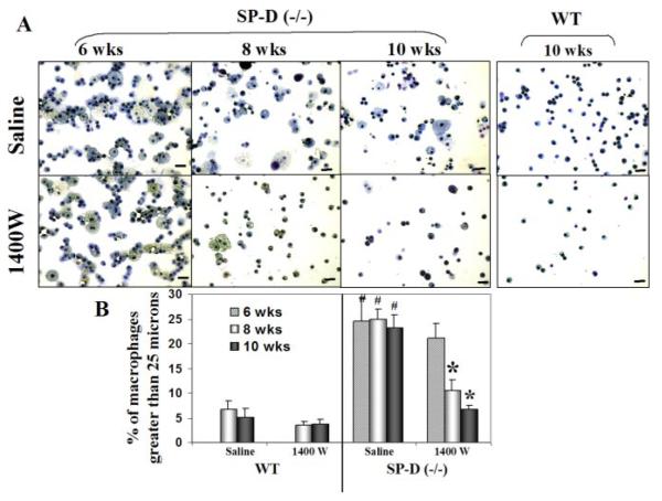
 ); 8-wk-old (
 ), and 10-wkold (█) time points are shown. #, p < 0.005 for significant difference from the corresponding WT level; and *, p < 0.0001 from the corresponding saline level.
 ), SP-D−/− saline-treated (
 ), and SP-D−/− 1400W-treated (█) mice are shown. #, p < 0.005 for significant difference from the corresponding WT level; and *, p < 0.0001 from the corresponding saline level. D, BAL from WT and SP-D−/− mice was assessed for their ability to induce RAW 264.7 macrophage migration using a modified Boyden chamber, following treatment with saline or 1400W. Data are expressed as the number of migrated cells per field. Results shown are mean ± SEM (n = 5–9 in each group). Saline-treated (
 ) and 1400W-treated (█) samples are shown. #, p < 0.005 for significant difference from the corresponding WT level; and *, p < 0.005 from the corresponding saline level.
 ) and 1400W-treated (█) samples are shown. #, p = 0.028 for significant difference from the corresponding WT level; and *, p = 0.032 from the corresponding saline level.
 ) and 1400W-treated (█) samples are shown. #, p < 0.0001 significant difference from the corresponding WT level; and *, p = 0.002 from the corresponding saline level. B, Diff-Quik staining of cytospins from SP-D−/− mice and littermate WT controls 2 wk after treatment with either saline or 1400W. C, Quantitation of macrophage size was done manually as described in Materials and Methods. Data are expressed as a percentage of macrophages >25 microns. Results shown are mean ± SEM (n = 3–15 animals in each group). #, p < 0.0001 significant difference from the corresponding WT level; and *, p < 0.008 from the corresponding saline level. Saline-treated (
 ) and 1400W-treated (█) samples are shown. D, BAL cells isolated from WT or SP-D−/− mice were incubated with DCF diacetate as described in Materials and Methods. Cells were harvested on ice and green fluorescence was measured by flow cytometry. Data are expressed as the percentage of macrophages producing oxidants. Results shown are mean ± SEM (n = 4 animals in each group). Saline-treated (
 ) and 1400W-treated (█) samples are shown. Two weeks of iNOS inhibition significantly reduced level of oxidants generated by macrophages in SP-D−/− mice. *, p = 0.031 significant difference from the corresponding saline level.
References
-
- Crouch EC. Structure, biologic properties, and expression of surfactant protein D (SP-D) Biochim. Biophys. Acta. 1998;1408:278–289. - PubMed
-
- LeVine AM, Whitsett JA, Gwozdz JA, Richardson TR, Fisher JH, Burhans MS, Korfhagen TR. Distinct effects of surfactant protein A or D deficiency during bacterial infection on the lung. J. Immunol. 2000;165:3934–3940. - PubMed
-
- Atochina EN, Beers MF, Hawgood S, Poulain F, Davis C, Fusaro T, Gow AJ. Surfactant protein-D, a mediator of innate lung immunity, alters the products of nitric oxide metabolism. Am. J. Respir. Cell Mol. Biol. 2004;30:271–279. - PubMed
Publication types
MeSH terms
Substances
Grants and funding
LinkOut - more resources
Full Text Sources
Medical
Molecular Biology Databases
