

Systemic and mucosal infection program protective memory CD8 T cells in the vaginal mucosa
- PMID: 18056354
- PMCID: PMC2648529
- DOI: 10.4049/jimmunol.179.12.8122
Systemic and mucosal infection program protective memory CD8 T cells in the vaginal mucosa
Abstract
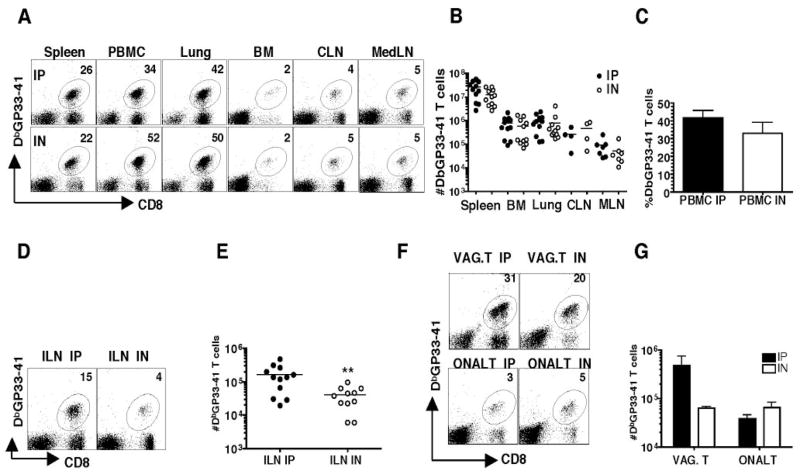
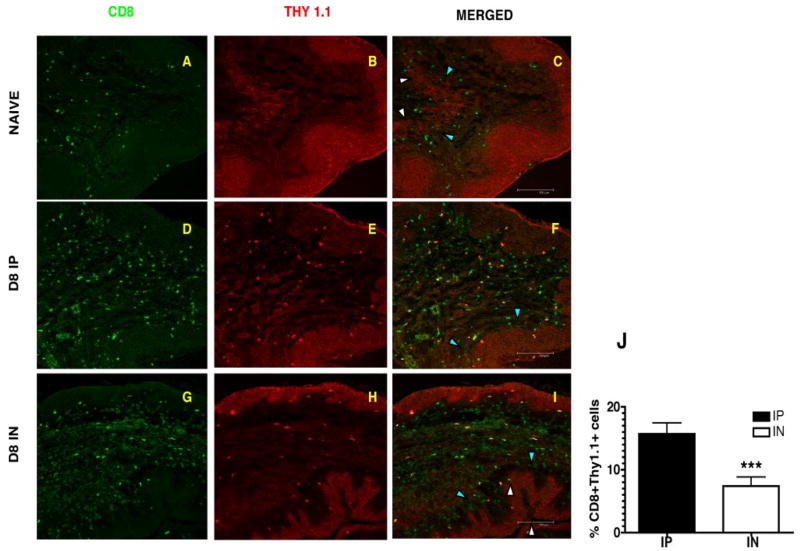
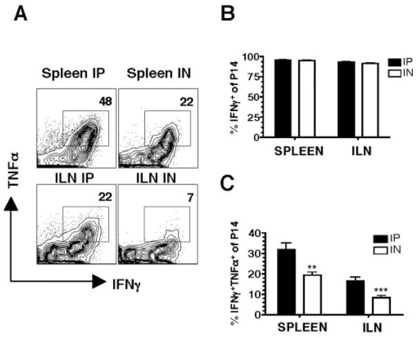
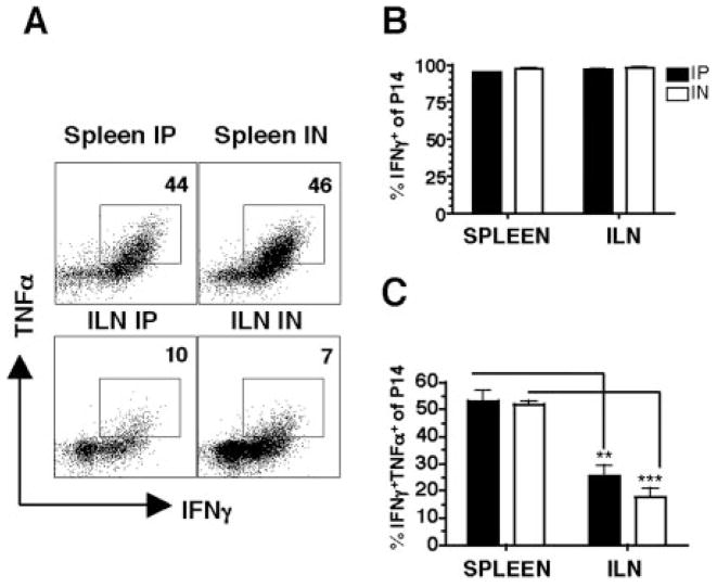
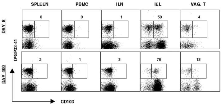
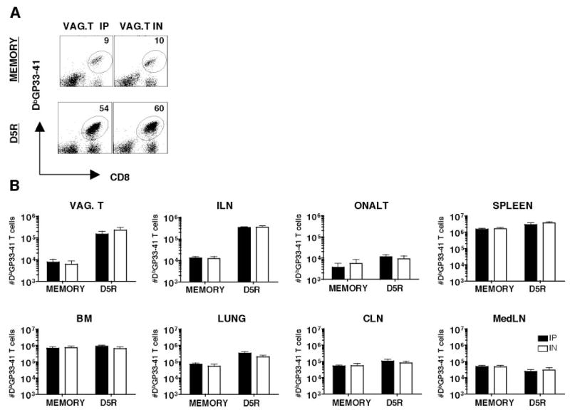
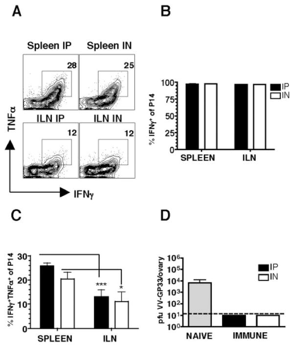
Whether mucosal immunization is required for optimal protective CD8 T cell memory at mucosal surfaces is controversial. In this study, using an adoptive transfer system, we compare the efficacy of two routes of acute lymphocytic choriomeningitis viral infection on the generation, maintenance, and localization of Ag-specific CD8 T cells in tissues, including the vaginal mucosa. Surprisingly, at day 8, i.p. infection results in higher numbers of Ag-specific CD8 T cells in the vaginal mucosa and iliac lymph node, as well as 2-3x more Ag-specific CD8 T cells that coexpress both IFN-gamma and TNF-alpha in comparison to the intranasal route of infection. Expression of the integrin/activation marker CD103 (alphaEbeta7) is low on vaginal mucosal Ag-specific CD8 T cells in comparison to gut mucosal intraepithelial lymphocytes. At memory, no differences are evident in the number, cytokine production, or protective function of Ag-specific CD8 T cells in the vaginal mucosa comparing the two routes of infection. However, differences persist in the cytokine profile of genital tract vs peripheral Ag-specific CD8 T cells. So although the initial route of infection, as well as tissue microenvironment, appear to influence both the magnitude and quality of the effector CD8 T cell response, both systemic and mucosal infection are equally effective in the differentiation of protective memory CD8 T cell responses against vaginal pathogenic challenge.
Figures
Similar articles
-
Circulating memory CD8+ T cells are limited in forming CD103+ tissue-resident memory T cells at mucosal sites after reinfection.Eur J Immunol. 2021 Jan;51(1):151-166. doi: 10.1002/eji.202048737. Epub 2020 Aug 31. Eur J Immunol. 2021. PMID: 32762051
-
Lymphocytic choriomeningitis virus persistence promotes effector-like memory differentiation and enhances mucosal T cell distribution.J Leukoc Biol. 2015 Feb;97(2):217-25. doi: 10.1189/jlb.1HI0314-154R. Epub 2014 Nov 13. J Leukoc Biol. 2015. PMID: 25395301 Free PMC article.
-
Retinoic acid as a vaccine adjuvant enhances CD8+ T cell response and mucosal protection from viral challenge.J Virol. 2011 Aug;85(16):8316-27. doi: 10.1128/JVI.00781-11. Epub 2011 Jun 8. J Virol. 2011. PMID: 21653670 Free PMC article.
-
Functional CD8+ CTLs in mucosal sites and HIV infection: moving forward toward a mucosal AIDS vaccine.Trends Immunol. 2008 Nov;29(11):574-85. doi: 10.1016/j.it.2008.07.010. Trends Immunol. 2008. PMID: 18838298 Review.
-
The Emerging Role of CD8+ Tissue Resident Memory T (TRM) Cells in Antitumor Immunity: A Unique Functional Contribution of the CD103 Integrin.Front Immunol. 2018 Aug 15;9:1904. doi: 10.3389/fimmu.2018.01904. eCollection 2018. Front Immunol. 2018. PMID: 30158938 Free PMC article. Review.
Cited by
-
Retinaldehyde dehydrogenase 2 as a molecular adjuvant for enhancement of mucosal immunity during DNA vaccination.Vaccine. 2016 Nov 4;34(46):5629-5635. doi: 10.1016/j.vaccine.2016.09.013. Epub 2016 Sep 23. Vaccine. 2016. PMID: 27670072 Free PMC article.
-
Intravaginal immunization with HPV vectors induces tissue-resident CD8+ T cell responses.J Clin Invest. 2012 Dec;122(12):4606-20. doi: 10.1172/JCI63287. Epub 2012 Nov 12. J Clin Invest. 2012. PMID: 23143305 Free PMC article.
-
Niches for the Long-Term Maintenance of Tissue-Resident Memory T Cells.Front Immunol. 2018 May 31;9:1214. doi: 10.3389/fimmu.2018.01214. eCollection 2018. Front Immunol. 2018. PMID: 29904388 Free PMC article. Review.
-
Long-lived tissue resident HIV-1 specific memory CD8+ T cells are generated by skin immunization with live virus vectored microneedle arrays.J Control Release. 2017 Dec 28;268:166-175. doi: 10.1016/j.jconrel.2017.10.026. Epub 2017 Oct 19. J Control Release. 2017. PMID: 29056444 Free PMC article.
-
Quantifying Memory CD8 T Cells Reveals Regionalization of Immunosurveillance.Cell. 2015 May 7;161(4):737-49. doi: 10.1016/j.cell.2015.03.031. Cell. 2015. PMID: 25957682 Free PMC article.
References
-
- Royce RA, Sena A, Cates W, Jr, Cohen MS. Sexual transmission of HIV. N Engl J Med. 1997;336:1072–1078. - PubMed
-
- Belyakov IM, Derby MA, Ahlers JD, Kelsall BL, Earl P, Moss B, Strober W, Berzofsky JA. Mucosal immunization with HIV-1 peptide vaccine induces mucosal and systemic cytotoxic T lymphocytes and protective immunity in mice against intrarectal recombinant HIV-vaccinia challenge. Proc Natl Acad Sci USA. 1998;95:1709–1714. - PMC - PubMed
-
- Belyakov IM, Hel Z, Kelsall B, Kuznetsov VA, Ahlers JD, Nacsa J, Watkins DI, Allen TM, Sette A, Altman J, et al. Mucosal AIDS vaccine reduces disease and viral load in gut reservoir and blood after mucosal infection of macaques. Nat Med. 2001;7:1320–1326. - PubMed
-
- Qimron U, Paul L, Bar-Haim E, Bloushtain N, Eisenbach L, Staats HF, Porgador A. Non-replicating mucosal and systemic vaccines: quantitative and qualitative differences in the Ag-specific CD8+ T cell population in different tissues. Vaccine. 2004;22:1390–1394. - PubMed
Publication types
MeSH terms
Substances
Grants and funding
LinkOut - more resources
Full Text Sources
Research Materials
