. 2007 Dec 15;179(12):8287-96.
doi: 10.4049/jimmunol.179.12.8287.

Prolonged (E)-4-hydroxy-3-methyl-but-2-enyl pyrophosphate-driven antimicrobial and cytotoxic responses of pulmonary and systemic Vgamma2Vdelta2 T cells in macaques

Affiliations

Prolonged (E)-4-hydroxy-3-methyl-but-2-enyl pyrophosphate-driven antimicrobial and cytotoxic responses of pulmonary and systemic Vgamma2Vdelta2 T cells in macaques

Zahida Ali et al. J Immunol. .

Abstract

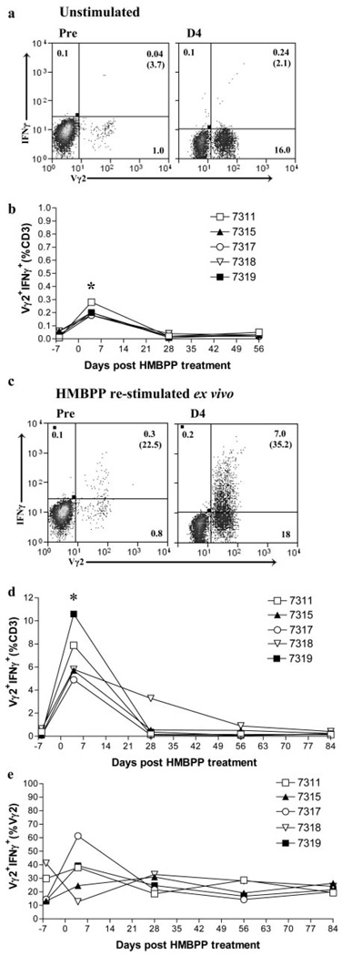
Although phosphoantigen-specific Vgamma2Vdelta2 T cells appear to play a role in antimicrobial and anticancer immunity, mucosal immune responses and effector functions of these gammadelta T cells during infection or phospholigand treatment remain poorly characterized. In this study, we demonstrate that the microbial phosphoantigen (E)-4-hydroxy-3-methyl-but-2-enyl pyrophosphate (HMBPP) plus IL-2 treatment of macaques induced a prolonged major expansion of circulating Vgamma2Vdelta2 T cells that expressed CD8 and produced cytotoxic perforin during their peak expansion. Interestingly, HMBPP-activated Vgamma2Vdelta2 T cells underwent an extraordinary pulmonary accumulation, which lasted for 3-4 mo, although circulating Vgamma2Vdelta2 T cells had returned to baseline levels weeks prior. The Vgamma2Vdelta2 T cells that accumulated in the lung following HMBPP/IL-2 cotreatment displayed an effector memory phenotype, as follows: CCR5+CCR7-CD45RA-CD27+ and were able to re-recognize phosphoantigen and produce copious amounts of IFN-gamma up to 15 wk after treatment. Furthermore, the capacity of massively expanded Vgamma2Vdelta2 T cells to produce cytokines in vivo coincided with an increase in numbers of CD4+ and CD8+ alphabeta T cells after HMBPP/IL-2 cotreatment as well as substantial perforin expression by CD3+Vgamma2- T cells. Thus, the prolonged HMBPP-driven antimicrobial and cytotoxic responses of pulmonary and systemic Vgamma2Vdelta2 T cells may confer immunotherapeutics against infectious diseases and cancers.


Conflict of interest statement

Disclosures

The authors have no financial conflict of interest.

Figures

FIGURE 1
FIGURE 1
HMBPP/IL-2 cotreatment induced a prolonged and massive expansion of circulating Vγ2Vδ2 T cells in vivo. Relative percentages of CD3+ T cells that express Vγ2Vδ2 after treatment with HMBPP or IL-2 alone are shown over time as the SEM for the number of indicated animals (a). Vγ2Vδ2 T cell expansion from pretreatment to day 4 in one representative cotreated animal is shown (b). Various γδ TCR-expressing lymphocytes in cotreated animals are shown over time as a relative percentage of total CD3+ T cells (c and e) and absolute numbers/µl blood (d and f). Vδ1 levels are shown as the SEM for the indicated animals.
FIGURE 2
FIGURE 2
HMBPP/IL-2 cotreatment induced an expansion of circulating CD8- and CD4-expressing T cells in vivo. Cotreated animals were treated with 50 mg/kg (open symbols) or 10 mg/kg (filled symbols) HMBPP plus IL-2. As controls, four animals received 50 mg/kg HMBPP only and two received IL-2 only. All animals were examined for CD8- and CD4-expressing lymphocytes. CD3-gated CD8+Vγ2+ cells are shown for one representative cotreated animal at day 7 (a) and over time for all examined animals (b). Absolute numbers of CD8+Vγ2+ (c), αβ CD8+Vγ2 (d), and αβ CD4+ (e) cells/µl blood are shown over time. Relative percentages of CD3+ T cells that are CD4+ or CD8+Vγ2 decreased in cotreated animals due to Vγ2Vδ2 T cell expansion (data not shown). Results for HMBPP- and IL-2 alone-treated animals are shown over time as the SEM for the number of indicated animals (b–e). Asterisks indicate statistically different T cell levels between cotreated and control groups at those time points (p< 0.05) (b–e).
FIGURE 3
FIGURE 3
HMBPP/IL-2 cotreatment induced a prolonged accumulation of Vγ2Vδ2 T cells and a transient increase in αβ T cells in pulmonary mucosa. Freshly collected cells from BAL fluid of animals treated with 50 mg/kg (open symbols) or 10 mg/kg (filled symbols) HMBPP plus IL-2, or HMBPP (50 mg/kg) or IL-2 alone, were stained over time for Vγ2Vδ2 (b and c)-, αβ CD8+Vγ2 (d)-, and αβ CD4+ (e)-expressing T cells. Vγ2Vδ2 T cell accumulation in pulmonary mucosa from pretreatment to day 11 in one representative cotreated animal (a) and over time for all examined animals (b) is shown from CD3-gated cells. Absolute numbers of Vγ2Vδ2 (c), αβ CD8+Vγ2 (d), and αβ CD4+ (e) cells are shown over time. Relative percentages of CD3+ T cells that are CD4+ or CD8+Vγ2 decreased in cotreated animals due to Vγ2Vδ2 T cell expansion (data not shown). T cell levels for HMBPP- and IL-2 alone-treated animals are shown as the SEM. Asterisks indicate statistically different T cell levels in cotreated animals from pretreatment (day 0) (p < 0.05) (b).
FIGURE 4
FIGURE 4
HMBPP/IL-2 cotreatment induced a short-term increase in Vγ2Vδ2 T cells in gingival and rectal mucosae. Freshly collected cells from gingival mucosa (a and b) and rectal mucosal biopsies (c and d) from animals cotreated with 50 mg/kg (open symbols) or 10 mg/kg (filled symbols) HMBPP plus IL-2, or with HMBPP alone (50 mg/kg) were stained over time for Vγ2Vδ2-expressing CD3+ T cells. Vγ2Vδ2 T cell levels in gingival (a and b) and rectal (c and d) mucosae from pretreatment to day 4 in one representative cotreated animal (a and c) and over time for all examined animals (b and d) are shown from CD3-gated cells. Gingival and rectal Vγ2Vδ2 T cell levels for HMBPP alone-treated animals are shown as the SEM (b and d).
FIGURE 5
FIGURE 5
Migration and memory marker expression by pulmonary and circulating Vγ2Vδ2 T cells. BAL fluid and whole blood from the following cotreated animals: 7311, 7315, 7317, 7318, and 7319 were examined. CD3- and Vγ2-gated BAL cells or PBL were examined for α4 integrin, β7 integrin, CCR5, and CCR7 expression before (a) and at 16 wk post-HMBPP treatment (b) and are shown as the SEM. Vγ2-gated BAL cells (c and d) or PBL (e) from five cotreated animals were examined for CD27 and CD45RA expression over time, and the relative percentages (c and e) or absolute numbers (d) of Vγ2 cells are shown as the SEM. All four subpopulations in the blood were statistically different from pretreatment (day 0) to day 4 (p < 0.05) (e). The expression of the following markers was statistically different between lung and blood Vγ2 T cells, as follows: CD45RA+CD27+ at days 0 and 56, CD45RACD27+ at days 0 and 4, and CD45RACD27 at day 4 (c and e).
FIGURE 6
FIGURE 6
Cytotoxic responses of circulating Vγ2Vβ2 T cells. Freshly collected PBL from cotreated animals were stimulated for 6 h with HMBPP (c–e) or medium alone (a and b) plus anti-CD28 and anti-CD49d mAbs before staining. Perforin expression by CD3-gated Vγ2+ and Vγ2 T cells is shown as the SEM for five examined animals after incubation with medium alone (a) or HMBPP restimulation (c). The percentages of Vγ2 T cells that express perforin are indicated within parentheses in the dot plots. Percentages of CD3+ cells that are Vγ2+perforin+ after incubation with medium alone are shown for each individual animal examined over time (b). Percentages of CD3+ (d) or Vγ2+ (e) cells that are Vγ2+perforin+ after HMBPP restimulation for each animal are also shown. Asterisks indicate statistically different T cell levels from pretreat-ment (day 0) (p < 0.05) (b, d, and e). Vγ2+Perforin+ cell levels were statistically different between medium-alone and HMBPP-restimulated groups at day 4 (p< 0.05) (b and d).
FIGURE 7
FIGURE 7
Antimicrobial responses of circulating Vγ2Vδ2 T cells. Freshly collected PBL from cotreated animals were stimulated for 6 h with HMBPP (c–e) or medium alone (a and b) plus anti-CD28 and anti-CD49d mAbs before staining. IFN-γ expression by CD3-gated Vγ2+ and Vγ2 T cells is shown as the SEM for five examined animals after incubation with medium alone (a) or HMBPP restimulation (c). The percentages of Vγ2 T cells that express IFN-7 are indicated within parentheses in the dot plots. The percentages of CD3+ cells that are Vγ2+IFN-γ+ after incubation with medium alone are shown for each individual animal examined over time(b). The percentages of CD3+ (d) or Vγ2+ (e) cells that are Vγ2+IFN-γ+ after HMBPP restimulation in each animal are also shown. Asterisks indicate statistically different T cell levels from pretreatment (day 0) (p< 0.05) (b and d). Vγ2+IFN-γ+ cell levels were statistically different between medium-alone and HMBPP-restimulated groups at day 4 (p< 0.05) (b and d).
FIGURE 8
FIGURE 8
Massive antimicrobial responses of long-lived pulmonary Vγ2Vδ2 T cells. Freshly collected BAL cells from cotreated animals at 12 and 15 wk post-in vivo HMBPP treatment were incubated for 6 h with HMBPP plus anti-CD28 and anti-CD49d mAbs before staining. IFN-γ expression by CD3-gated Vγ2+ and Vγ2 T cells is shown for each examined animal on week 12 (a) or as the SEM for four examined animals on week 15 (b). Perforin expression by CD3-gated Vγ2+ and Vγ2 T cells is shown as the SEM for the number of indicated animals on weeks 12 and 15 (c). The percentages of Vγ2 T cells that express IFN-γ or perforin are indicated within parentheses in the dot plots.

References

    1. Shen Y, Zhou D, Qiu L, Lai X, Simon M, Shen L, Kou Z, Wang Q, Jiang L, Estep J, et al. Adaptive immune response of Vγ2Vδ2+ T cells during mycobacterial infections. Science. 2002;295:2255–2258. - PMC - PubMed
    1. Wang L, Kamath A, Das H, Li L, Bukowski JF. Antibacterial effect of human Vγ2Vδ2 T cells in vivo. J. Clin. Invest. 2001;108:1349–1357. - PMC - PubMed
    1. Poccia F, Battistini L, Cipriani B, Mancino G, Martini F, Gougeon ML, Colizzi V. Phosphoantigen-reactive Vγ9Vδ2 T lymphocytes suppress in vitro human immunodeficiency virus type 1 replication by cell-released antiviral factors including CC chemokines. J. Infect. Dis. 1999;180:858–861. - PubMed
    1. Dieli F, Troye-Blomberg M, Ivanyi J, Fournie JJ, Bonneville M, Peyrat MA, Sireci G, Salerno A. Vγ9/Vδ2 T lymphocytes reduce the viability of intracellular Mycobacterium tuberculosis. Eur. J. Immunol. 2000;30:1512–1519. - PubMed
    1. Ottones F, Dornand J, Naroeni A, Liautard JP, Favero J. Vγ9Vδ2 T cells impair intracellular multiplication of Brucella suis in autologous monocytes through soluble factor release and contact-dependent cytotoxic effect. J. Immunol. 2000;165:7133–7139. - PubMed

Publication types

MeSH terms