. 2007 Dec 3;179(5):911-22.
doi: 10.1083/jcb.200709083.

Megf10 regulates the progression of the satellite cell myogenic program

Affiliations

Megf10 regulates the progression of the satellite cell myogenic program

Chet E Holterman et al. J Cell Biol. .

Abstract

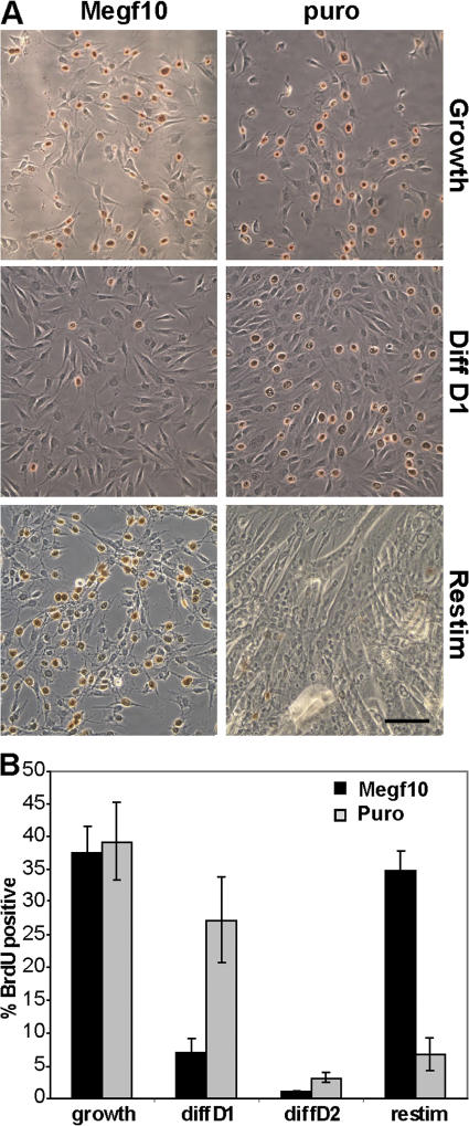
We identify here the multiple epidermal growth factor repeat transmembrane protein Megf10 as a quiescent satellite cell marker that is also expressed in skeletal myoblasts but not in differentiated myofibers. Retroviral expression of Megf10 in myoblasts results in enhanced proliferation and inhibited differentiation. Infected myoblasts that fail to differentiate undergo cell cycle arrest and can reenter the cell cycle upon serum restimulation. Moreover, experimental modulations of Megf10 alter the expression levels of Pax7 and the myogenic regulatory factors. In contrast, Megf10 silencing in activated satellite cells on individual fibers or in cultured myoblasts results in a dramatic reduction in the cell number, caused by myogenin activation and precocious differentiation as well as a depletion of the self-renewing Pax7+/MyoD- population. Additionally, Megf10 silencing in MyoD-/- myoblasts results in down-regulation of Notch signaling components. We conclude that Megf10 represents a novel transmembrane protein that impinges on Notch signaling to regulate the satellite cell population balance between proliferation and differentiation.


Figures

Figure 1.
Figure 1.
Megf10 is expressed in proliferating myoblasts and resting adult skeletal muscle. (A) Quantitative PCR was performed on samples from wild-type and MyoD/ primary myoblasts cultured under growth (Gr) or differentiation (D1–D5) conditions. Samples were normalized to GAPDH and then to wild-type D5, which was set to a baseline expression of 1. Megf10 expression is down-regulated in wild-type myoblasts after serum withdrawal, whereas MyoD/ myoblasts maintain Megf10 expression after serum withdrawal (n = 3). Error bars represent SEM. (B) Quantitative PCR demonstrating the relative expression of Megf10 in mouse tissues. All samples were normalized to GAPDH, and the lowest expressing tissue (liver) was set to a baseline expression of 1. High levels of Megf10 expression are detected in the brain and in regenerating (3 d after ctx injection) skeletal muscle (n = 3). Error bars represent SEM. Br, brain; H, heart; K, kidney; Li, liver; Lu, lung; Sk, skeletal muscle; SkRg, regenerating skeletal muscle; Sp, spleen. (C) Quantitative PCR showing the relative up-regulation of Pax7 and Megf10 gene expression levels during activation of satellite cells, RNA samples were extracted from freshly isolated FACS-sorted α7integrin+, CD31/Sca1/CD45 satellite cells (Quiescent) and in vitro–cultured myoblasts (Activated; n = 3). Error bars represent SEM. (D) In situ hybridization was performed with Megf10 antisense riboprobe on tibialis anterior muscle from 2-mo-old mice. Transcripts are located in fiber-associated cells in a position relative to satellite cells (arrowheads).
Figure 2.
Figure 2.
Megf10 is expressed in quiescent and activated satellite cells. (A) Immunohistochemistry on frozen sections of tibialis anterior muscles from 2-mo-old mice revealed that Megf10 was coexpressed with syndecan 4 in resting skeletal muscle. Arrowheads point to double-positive cells. Bar, 10 μm. (B) Individual myofibers isolated from the EDL muscle of adult mice were costained for Megf10, Pax7, and syndecan 4 or M-cadherin. Megf10 expression was limited to cells that also expressed Pax7 and syndecan 4 or M-cadherin (arrowheads), demonstrating that Megf10 expression is limited to quiescent satellite cells in resting skeletal muscle. Bar, 10 μm. (C) Individual fibers from the EDL muscle of Myf5-Cre*ROSA-YFP reporter mice were isolated, freshly fixed, and stained for YFP, Pax7, and Megf10 expression. Megf10 expression was detected in Pax7+/YFP+ quiescent satellite cells but never in Pax7+/YFP quiescent satellite cells. Arrow points to a Pax7+/YFP/Megf10 satellite cell. (D) Myofibers were cultured for 3 d and stained for Pax7, MyoD, and myogenin in the same color to label all satellite cells. Counterstaining with the Megf10 antibody demonstrated that all (99.5%; n > 1,000) activated satellite cells express Megf10. Bar, 50 μm. (E) The presence of a Pax7+/YFP+/Megf10+ progenitor cell (arrowhead) and a Pax7+/YFP/Megf10 stem cell (arrow) on the same fiber after isolation. After 3 d in culture, both Pax7+/YFP (arrow) and Pax7+/YFP+ (arrowhead) proliferating cells express Megf10. Bars, 50 μm.
Figure 3.
Figure 3.
Overexpression of Megf10 enhances proliferation of myogenic cells. (A) C2C12 myoblasts were infected with Megf10. Western blot with α-HA reveals Megf10 expression in the plasma membrane and cytoplasmic fractions. Fraction purity was examined by blotting for integrin α4 (plasma membrane), Grb2 (cytoplasm), Lamin A (nuclear membrane), and TBP (nuclear). Whole cell extract was used as a positive control. (B) Immunocytochemistry on infected cells demonstrated cytoplasmic and potential plasma membrane localization of Megf10 in infected cells but not controls. Bar, 10 μm. (C) C2C12 cells infected with Megf10 or empty vector puro controls were counted at either 24, 48, or 72 h after plating (n = 9). Error bars represent SEM.
Figure 4.
Figure 4.
Overexpression of Megf10 inhibits differentiation of myogenic cells. (A) C2C12 cells infected with Megf10 or empty vector puro controls were stained with α-MyHC. Cells overexpressing Megf10 displayed a dramatic decrease in differentiation as compared with controls. (B) Western blots reveal a dramatic decrease in MyHC expression at 5 d after serum withdrawal in cells overexpressing Megf10. (C) The number of nuclei per MyHC-positive body was examined and plotted to demonstrate that although <5% of MyHC-positive control cells remained mononuclear, 50% of MyHC-positive Megf10 cells failed to fuse to form multinucleated myotubes (n = 3). Error bars represent SEM.
Figure 5.
Figure 5.
Megf10 stimulates quiescence after serum withdrawal. (A) C2C12 myoblasts infected with Megf10 or empty vector puro control were maintained in growth media or differentiated for 1 to 5 d. Cells were stained for BrdU incorporation. Approximately 30% of cells incorporate BrdU under growth conditions. Within 24 h of serum withdrawal, only 8% of Megf10-expressing cells incorporate BrdU, indicating withdrawal from the cell cycle, whereas 25% of control cells incorporate BrdU. A large percentage of cells expressing Megf10 that were subjected to low serum were able to reenter the cell cycle, whereas the majority of control cells had terminally differentiated (n = 3). Bar, 100 mm. (B) Histogram demonstrating the number of cells that incorporated BrdU during a 30-min pulse after 120 h of differentiation and 24 h of restimulation (n = 3). Error bars represent SEM.
Figure 6.
Figure 6.
Overexpression of Megf10 alters the levels of key myogenic proteins. (A) Western blot of protein extracts from Megf10-overexpressing cells and empty vector puro controls. Myf5 levels are elevated in Megf10-overexpressing cells, whereas MyoD and Pax7 are down-regulated under growth conditions. Cells overexpressing Megf10 fail to up-regulate myogenin during differentiation. (B) Quantitative PCR analysis reveals that although Myf5 and Pax7 RNA levels are altered in Megf10-expressing cells, alterations in MyoD are occurring mainly at the protein level (*, P < 0.02). Transcript levels are normalized to GAPDH and control levels are set to 1 (n = 3). Error bars represent SEM.
Figure 7.
Figure 7.
Megf10 regulates satellite cell activation and proliferation. (A) Individual fibers isolated from EDL muscles were transfected with Megf10 siRNA, cultured for 3 d, and stained for Pax7 and Megf10. siRNA knockdown induces a loss of Megf10 protein expression by the majority of the activated satellite cells (n = 3). Bar, 50 μm. (B) Histograms depicting individual counts of the total numbers of activated satellite cells (labeled with Pax7 and MyoD/myogenin) on fibers transfected with Megf10 siRNA. The mean number of myogenic cells per fiber is reduced by a mean of 30% as compared with scrambled siRNA controls (n = 3). (C) Knockdown of Megf10 dramatically reduces the number of self-renewing progenitors (Pax7+/MyoD) from 19 to 7% and respectively increases the number of Pax7+/MyoD+ cells (n = 3; **, P = 0.008). Error bars represent SEM. (D) Although the number of activated progenitor cells is reduced, the overall ratio of stem cell (YFP) to progenitor cell (YFP+) remains unchanged (n = 3). Error bars represent SEM.
Figure 8.
Figure 8.
Loss of Megf10 results in precocious differentiation. (A) EDL fibers were transfected with Megf10 siRNA, cultured for 60 h, and stained for Pax7 and myogenin. Megf10 knockdown induces three times more cells to undergo precocious differentiation (n = 3; **, P = 0.004). Error bars represent SEM. Bar, 50 μm. (B) Quantitative PCR analysis demonstrates up-regulation of myogenin and MyHC and down-regulation of Pax7, MyoD, and Myf5 in wild-type primary myoblasts in which Megf10 has been knocked down (n = 6; *, P < 0.05; **, P < 0.01). Error bars represent SEM. (C) Quantitative PCR demonstrates down-regulation of components of the Notch signaling pathway after shRNA knockdown of Megf10 in MyoD/ primary myoblasts (n = 3; *, P < 0.05; **, P < 0.01). Error bars represent SEM.

References

    1. Anderson, J.E. 2000. A role for nitric oxide in muscle repair: nitric oxide-mediated activation of muscle satellite cells. Mol. Biol. Cell. 11:1859–1874. - PMC - PubMed
    1. Beauchamp, J.R., L. Heslop, D.S. Yu, S. Tajbakhsh, R.G. Kelly, A. Wernig, M.E. Buckingham, T.A. Partridge, and P.S. Zammit. 2000. Expression of CD34 and Myf5 defines the majority of quiescent adult skeletal muscle satellite cells. J. Cell Biol. 151:1221–1234. - PMC - PubMed
    1. Bischoff, R. 1994. The satellite cell and muscle regeneration. In Myogenesis. A.G. Engel and C. Franszini-Armstrong, editors. McGraw-Hill, New York. 97–118.
    1. Braissant, O., and W. Wahli. 1998. Differential expression of peroxisome proliferator-activated receptor-alpha, -beta, and -gamma during rat embryonic development. Endocrinology. 139:2748–2754. - PubMed
    1. Charge, S.B., and M.A. Rudnicki. 2004. Cellular and molecular regulation of muscle regeneration. Physiol. Rev. 84:209–238. - PubMed

Publication types