. 2007 Dec 1;21(23):3061-6.
doi: 10.1101/gad.1600707.

Atg7-dependent autophagy promotes neuronal health, stress tolerance, and longevity but is dispensable for metamorphosis in Drosophila

Affiliations

Atg7-dependent autophagy promotes neuronal health, stress tolerance, and longevity but is dispensable for metamorphosis in Drosophila

Gábor Juhász et al. Genes Dev. .

Abstract

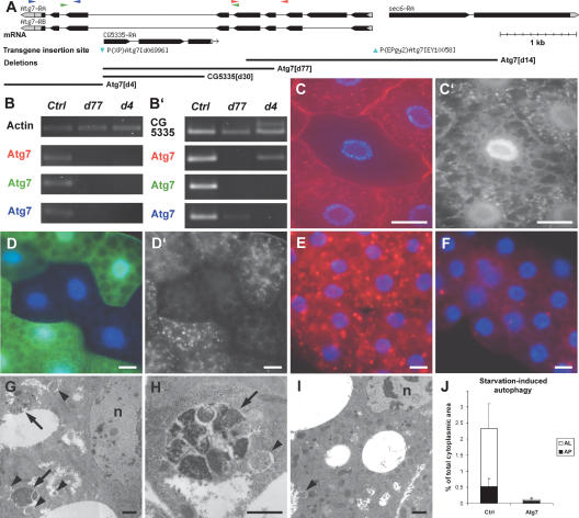
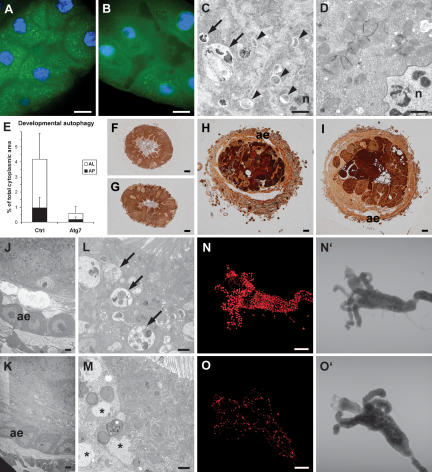
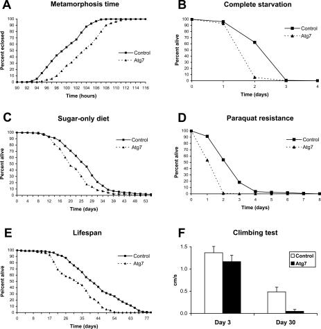
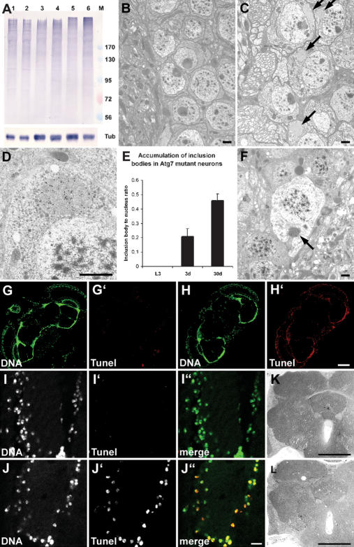
Autophagy, a cellular process of cytoplasmic degradation and recycling, is induced in Drosophila larval tissues during metamorphosis, potentially contributing to their destruction or reorganization. Unexpectedly, we find that flies lacking the core autophagy regulator Atg7 are viable, despite severe defects in autophagy. Although metamorphic cell death is perturbed in Atg7 mutants, the larval-adult midgut transition proceeds normally, with extended pupal development compensating for reduced autophagy. Atg7-/- adults are short-lived, hypersensitive to nutrient and oxidative stress, and accumulate ubiquitin-positive aggregates in degenerating neurons. Thus, normal levels of autophagy are crucial for stress survival and continuous cellular renewal, but not metamorphosis.


Figures

Figure 1.
Figure 1.
Atg7 is required for starvation-induced autophagy in the fat body. (A) The genomic region of Atg7. The two P-element insertions used to generate deletions (blue arrowheads), and four deletions are shown. (B,B′) Atg7 mRNA expression was analyzed by RT–PCR in control and mutant third instar larvae. (B) No product is seen after 25 amplification cycles in Atg7 mutants, indicating strongly reduced Atg7 expression. (B′) Thirty cycles reveal the expression of C-terminal Atg7 sequences in Atg7d77 and N-terminal sequences in Atg7d4 larvae. As expected, deleted parts are not expressed in the mutants. See A for Atg7 primer locations. (CD′) Starvation-induced autophagy is severely impaired in Atg7d4 mutant fat body clones, as shown by GFP-Atg8a labeling (C′) and Lysotracker Red staining (D′). Mutant cells are marked by lack of myrRFP (red, C) and GFP (green, D) expression, respectively. (EJ) Atg7d77 larval fat bodies show a strongly reduced autophagy in response to starvation. Very little Lysotracker staining is seen in Atg7d77 mutant fat bodies (F), while controls accumulate numerous Lysotracker-positive dots (E). Transmission EM reveals many autophagosomes (arrowheads) and autolysosomes (arrows) in control (G,H), but not in Atg7d77 fat body cells following a 3-h starvation (I). J shows a morphometric evaluation of EM images. (AP) Autophagosome; (AL) autolysosome. Error bars represent standard deviation. Bars: CF, 10 μm; GI, 1 μm. (B,E,G,H,J) Control: CG5335d30/Atg7d14. (B) Atg7d4: Atg7d4/Atg7d14. (B,F,I,J) Atg7d77: Atg7d77/Atg7d14. (C) FRT42D Atg7d4/CgGAL4 UAS-GFP-Atg8a FRT42D UAS-myrRFP. (D) FRT42D Atg7d4/UAS-2XeGFP FRT42D fb-GAL4.
Figure 2.
Figure 2.
Atg7 is required for developmental autophagy and apoptotic DNA fragmentation in the midgut. (AE) Developmental autophagy defects in Atg7d77 mutant larval midguts. (A) Developmental autophagy results in punctate GFP-Atg8a localization in control midguts. (B) Atg7d77 mutant midguts show a more uniform GFP-Atg8a signal with very few dots. Ultrastructural analysis reveals numerous autophagosomes and autolysosomes in control (C), but not in mutant midguts (D). E shows a morphometric evaluation of EM images; labels as in Figure 1. (FM) Adult epithelium is formed normally in Atg7d77 mutants during metamorphosis. (F,G) The larval midgut is composed mostly of large, polyploid cells, with intercalated small, diploid imaginal cells in control and mutant animals at the time of puparium formation (white prepupal stage). (HK) The adult epithelium is formed from proliferating imaginal cells by 5 h RPF, accompanied by shortening and thickening of the entire midgut (note increased diameter in cross-section between FH or GI), and shedding of the larval cell layer into the lumen (dark cell masses in H,I). (I) Although appearing less condensed at this time, larval cells of Atg7 mutants are also shed into the lumen. (J,K) Formation of the adult midgut epithelium (ae) proceeds normally in both control and mutant animals. Aberrant structures reminiscent of protein aggregates were seen in the apical area of mutant midgut cells at 5 h RPF (M), in the region where control cells accumulated numerous large autolysosomes (L). TUNEL staining of white prepupal midguts reveals defects in DNA fragmentation in dying Atg7d77 larval cells (O), when compared with the robust nuclear staining seen in controls (N). Bars: A,B,FI, 10 μm; C,D,JM, 1 μm; N,O, 200 μm. (A,C,E,F,H,J,L,N) Control: CG5335d30/Atg7d14. (B,D,E,G,I,K,M,O) Atg7d77: Atg7d77/Atg7d14.
Figure 3.
Figure 3.
Atg7 mutants show delayed metamorphosis, shorter life span, and stress sensitivity. Atg7d77 mutants eclose later than age-matched controls (A), and emerging adults die faster than control flies on complete starvation (B) or if fed a sugar-only diet (C). (D) Including 30 mM paraquat in the medium to induce oxidative stress also kills mutants faster than controls. Atg7d77 mutants show a reduced life span (E), and 30-d-old mutant flies score poorly in a climbing test relative to controls (−90.4% change, P < 10−15), while they perform almost as well as control flies at day 3 (−14.6% change, P = 0.00004) (F). (All panels) Control: CG5335d30/Atg7d14; Atg7d77: Atg7d77/Atg7d14.
Figure 4.
Figure 4.
Ubiquitinated proteins accumulate in Atg7 mutant brains, associated with progressive neurodegeneration. (A) A Western blot shows accumulation of ubiquitinated high-molecular-weight proteins in aging Atg7d77 fly heads. No difference is seen between control (lane 1) and mutant (lane 2) late L3 larval samples. Accumulation of ubiquitinated proteins is already visible in 3-d-old Atg7d77 adult head extracts (lane 4, cf. control in lane 3), and becomes very obvious by day 30 (lane 6, cf. control in lane 5). Numbers next to the marker (M) refer to molecular weights in kilodaltons. β-Tubulin is shown as a loading control. (BF) Electron micrographs reveal the presence of inclusion bodies in 30-d-old Atg7d77 (arrows in C), but not in control neurons (shown in B). (D) Immunoelectron microscopy confirms that these inclusion bodies contain ubiquitinated proteins. E shows progressive accumulation of aberrant protein aggregates in Atg7d77 mutants: No inclusion bodies are seen in late third instar larval brains, while they appear in neurons of 3-d-old males, and further accumulate by age 30 d. (F) Dead neurons are readily identified in 30-d-old mutant brains, many still containing ubiquitin-positive inclusion bodies in the degenerating cytoplasm (arrow). (GJ) TUNEL staining of horizontal brain sections shows widespread DNA fragmentation in Atg7d77 brains. (H,J) Most cells show DNA fragmentation in 30-d-old Atg7d77 brains, (G,I) while no staining is seen in controls. Moderate vacuolization is seen in 30-d-old Atg7d77 mutant brains (L), while no sign of degeneration is observed in controls (K). Bars: BF, 1 μm; I,J, 10 μm; G,H,K,L, 150 μm. (B,G,I,K) Control: CG5335d30/Atg7d14. (C,D,F,H,J,L) Atg7d77: Atg7d77/Atg7d14.

References

    1. Butterworth F.M., Forrest E.C., Forrest E.C. Ultrastructure of the preparative phase of cell death in the larval fat body of Drosophila melanogaster. Tissue Cell. 1984;16:237–250. - PubMed
    1. Cuervo A.M., Bergamini E., Brunk U.T., Droge W., Ffrench M., Terman A., Bergamini E., Brunk U.T., Droge W., Ffrench M., Terman A., Brunk U.T., Droge W., Ffrench M., Terman A., Droge W., Ffrench M., Terman A., Ffrench M., Terman A., Terman A. Autophagy and aging: The importance of maintaining ‘clean’ cells. Autophagy. 2005;1:131–140. - PubMed
    1. Hara T., Nakamura K., Matsui M., Yamamoto A., Nakahara Y., Suzuki-Migishima R., Yokoyama M., Mishima K., Saito I., Okano H., Nakamura K., Matsui M., Yamamoto A., Nakahara Y., Suzuki-Migishima R., Yokoyama M., Mishima K., Saito I., Okano H., Matsui M., Yamamoto A., Nakahara Y., Suzuki-Migishima R., Yokoyama M., Mishima K., Saito I., Okano H., Yamamoto A., Nakahara Y., Suzuki-Migishima R., Yokoyama M., Mishima K., Saito I., Okano H., Nakahara Y., Suzuki-Migishima R., Yokoyama M., Mishima K., Saito I., Okano H., Suzuki-Migishima R., Yokoyama M., Mishima K., Saito I., Okano H., Yokoyama M., Mishima K., Saito I., Okano H., Mishima K., Saito I., Okano H., Saito I., Okano H., Okano H., et al. Suppression of basal autophagy in neural cells causes neurodegenerative disease in mice. Nature. 2006;441:885–889. - PubMed
    1. Klionsky D.J., Cregg J.M., Dunn W.A., Emr S.D., Sakai Y., Sandoval I.V., Sibirny A., Subramani S., Thumm M., Veenhuis M., Cregg J.M., Dunn W.A., Emr S.D., Sakai Y., Sandoval I.V., Sibirny A., Subramani S., Thumm M., Veenhuis M., Dunn W.A., Emr S.D., Sakai Y., Sandoval I.V., Sibirny A., Subramani S., Thumm M., Veenhuis M., Emr S.D., Sakai Y., Sandoval I.V., Sibirny A., Subramani S., Thumm M., Veenhuis M., Sakai Y., Sandoval I.V., Sibirny A., Subramani S., Thumm M., Veenhuis M., Sandoval I.V., Sibirny A., Subramani S., Thumm M., Veenhuis M., Sibirny A., Subramani S., Thumm M., Veenhuis M., Subramani S., Thumm M., Veenhuis M., Thumm M., Veenhuis M., Veenhuis M., et al. A unified nomenclature for yeast autophagy-related genes. Dev. Cell. 2003;5:539–545. - PubMed
    1. Komatsu M., Waguri S., Ueno T., Iwata J., Murata S., Tanida I., Ezaki J., Mizushima N., Ohsumi Y., Uchiyama Y., Waguri S., Ueno T., Iwata J., Murata S., Tanida I., Ezaki J., Mizushima N., Ohsumi Y., Uchiyama Y., Ueno T., Iwata J., Murata S., Tanida I., Ezaki J., Mizushima N., Ohsumi Y., Uchiyama Y., Iwata J., Murata S., Tanida I., Ezaki J., Mizushima N., Ohsumi Y., Uchiyama Y., Murata S., Tanida I., Ezaki J., Mizushima N., Ohsumi Y., Uchiyama Y., Tanida I., Ezaki J., Mizushima N., Ohsumi Y., Uchiyama Y., Ezaki J., Mizushima N., Ohsumi Y., Uchiyama Y., Mizushima N., Ohsumi Y., Uchiyama Y., Ohsumi Y., Uchiyama Y., Uchiyama Y., et al. Impairment of starvation-induced and constitutive autophagy in Atg7-deficient mice. J. Cell Biol. 2005;169:425–434. - PMC - PubMed

Publication types

MeSH terms

Substances

LinkOut - more resources