

Amino-biphosphonate-mediated MMP-9 inhibition breaks the tumor-bone marrow axis responsible for myeloid-derived suppressor cell expansion and macrophage infiltration in tumor stroma
- PMID: 18056472
- PMCID: PMC2646404
- DOI: 10.1158/0008-5472.CAN-07-1882
Amino-biphosphonate-mediated MMP-9 inhibition breaks the tumor-bone marrow axis responsible for myeloid-derived suppressor cell expansion and macrophage infiltration in tumor stroma
Abstract
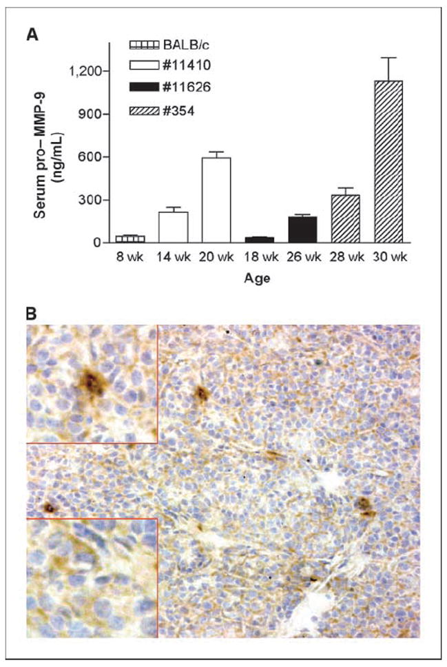
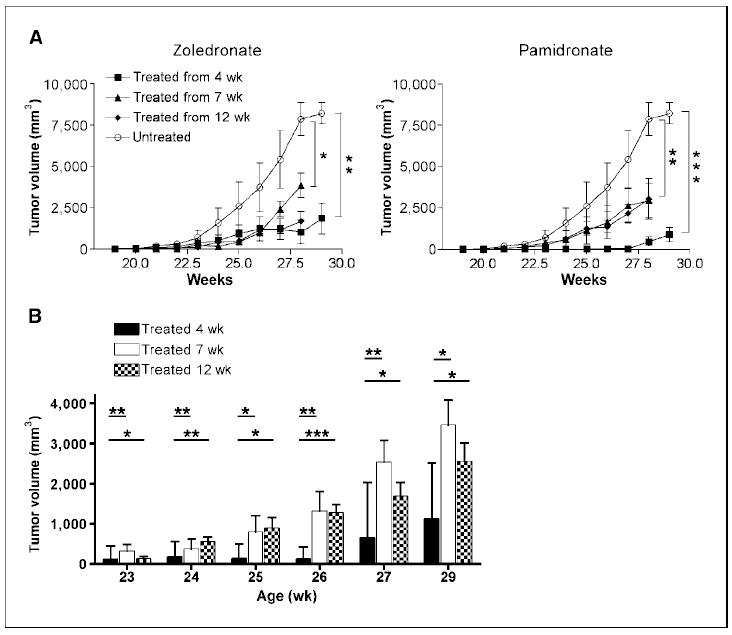
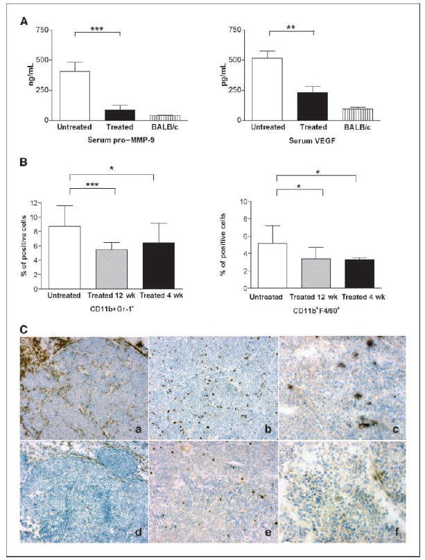
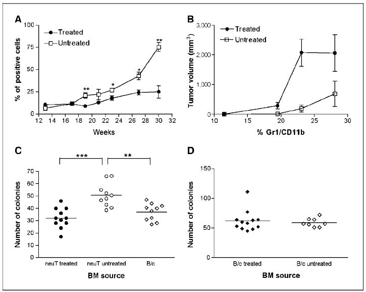
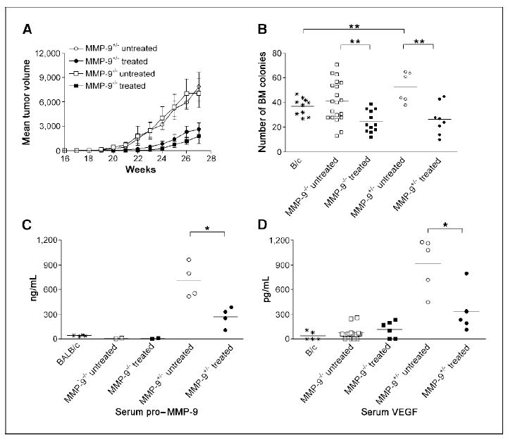
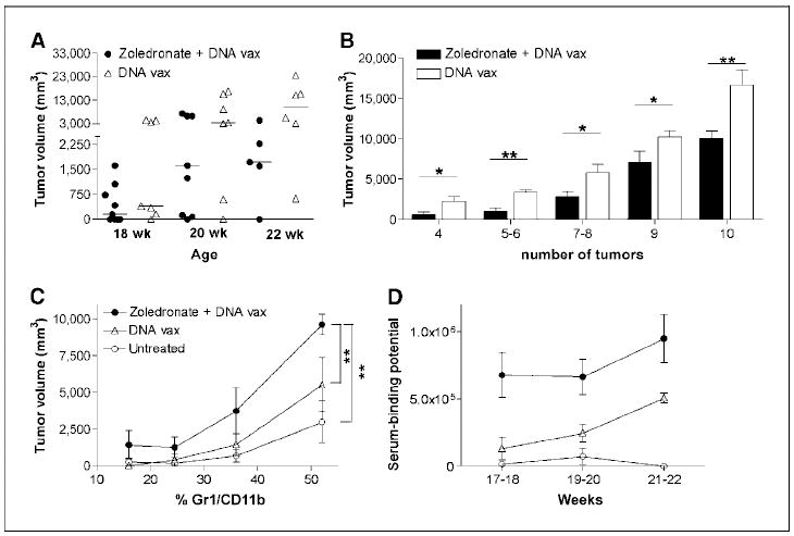
BALB-neuT mice expressing an activated rat c-erbB-2/neu transgene under the mouse mammary tumor virus long terminal repeat show enhanced hematopoiesis with hyperproduction of myeloid-derived suppressor cells (MDSC) because of vascular endothelial growth factor (VEGF) secreted by the tumor. Here, we show that both tumor and stromal cells express matrix metalloproteinase-9 (MMP-9), thereby increasing the levels of pro-MMP-9 in the sera of tumor-bearing mice. Treatment with amino-biphosphonates impaired tumor growth, significantly decreased MMP-9 expression and the number of macrophages in tumor stroma, and reduced MDSC expansion both in bone marrow and peripheral blood by dropping serum pro-MMP-9 and VEGF. We dissected the role of tumor-derived MMP-9 from that secreted by stromal leukocytes by transplanting bone marrow from MMP-9 knockout mice into BALB-neuT mice. Although bone marrow progenitor-derived MMP-9 had a major role in driving MDSC expansion, amino-biphosphonate treatment of bone marrow chimeras further reduced both myelopoiesis and the supportive tumor stroma, thus enhancing tumor necrosis. Moreover, by reducing MDSC, amino-biphosphonates overcome the tumor-induced immune suppression and improved the generation and maintenance of antitumor immune response induced by immunization against the p185/HER-2. Our data reveal that suppression of MMP-9 activity breaks the vicious loop linking tumor growth and myeloid cell expansion, thus reducing immunosuppression. Amino-biphosphonates disclose a specific MMP-9 inhibitory activity that may broaden their application above their current usage.
Figures
Similar articles
-
Matrix metalloproteinase-9 is required for tumor vasculogenesis but not for angiogenesis: role of bone marrow-derived myelomonocytic cells.Cancer Cell. 2008 Mar;13(3):193-205. doi: 10.1016/j.ccr.2007.11.032. Cancer Cell. 2008. PMID: 18328424 Free PMC article.
-
Myeloid cell expansion elicited by the progression of spontaneous mammary carcinomas in c-erbB-2 transgenic BALB/c mice suppresses immune reactivity.Blood. 2003 Sep 15;102(6):2138-45. doi: 10.1182/blood-2003-01-0190. Epub 2003 May 15. Blood. 2003. PMID: 12750171
-
The contribution of bone marrow-derived cells to the tumor vasculature in neuroblastoma is matrix metalloproteinase-9 dependent.Cancer Res. 2005 Apr 15;65(8):3200-8. doi: 10.1158/0008-5472.CAN-04-3770. Cancer Res. 2005. PMID: 15833851
-
Targeting immune suppressing myeloid-derived suppressor cells in oncology.Crit Rev Oncol Hematol. 2011 Jan;77(1):12-9. doi: 10.1016/j.critrevonc.2010.02.004. Epub 2010 Mar 20. Crit Rev Oncol Hematol. 2011. PMID: 20304669 Free PMC article. Review.
-
Influence of bone marrow-derived hematopoietic cells on the tumor response to radiotherapy: experimental models and clinical perspectives.Cell Cycle. 2009 Apr 1;8(7):970-6. doi: 10.4161/cc.8.7.8075. Epub 2009 Apr 4. Cell Cycle. 2009. PMID: 19270527 Free PMC article. Review.
Cited by
-
Myeloid-Derived Suppressor Cells and Therapeutic Strategies in Cancer.Mediators Inflamm. 2015;2015:159269. doi: 10.1155/2015/159269. Epub 2015 May 19. Mediators Inflamm. 2015. PMID: 26078490 Free PMC article. Review.
-
Regulatory cells and the effect of cancer immunotherapy.Mol Cancer. 2023 Feb 4;22(1):26. doi: 10.1186/s12943-023-01714-0. Mol Cancer. 2023. PMID: 36739406 Free PMC article. Review.
-
Zoledronic acid impairs myeloid differentiation to tumour-associated macrophages in mesothelioma.Br J Cancer. 2010 Aug 24;103(5):629-41. doi: 10.1038/sj.bjc.6605814. Epub 2010 Jul 27. Br J Cancer. 2010. PMID: 20664588 Free PMC article.
-
Anti-angiogenesis immunotherapy.Hum Vaccin. 2011 Sep;7(9):976-81. doi: 10.4161/hv.7.9.16407. Epub 2011 Sep 1. Hum Vaccin. 2011. PMID: 21860259 Free PMC article.
-
Immunological dysregulation in multiple myeloma microenvironment.Biomed Res Int. 2014;2014:198539. doi: 10.1155/2014/198539. Epub 2014 Jun 11. Biomed Res Int. 2014. PMID: 25013764 Free PMC article. Review.
References
-
- Gabrilovich DI, Chen HL, Girgis KR, et al. Production of vascular endothelial growth factor by human tumors inhibits the functional maturation of dendritic cells. Nat Med. 1996;2:1096–103. - PubMed
-
- Melani C, Chiodoni C, Forni G, Colombo MP. Myeloid cell expansion elicited by the progression of spontaneous mammary carcinomas in c-erbB-2 transgenic BALB/ c mice suppresses immune reactivity. Blood. 2003;102:2138–45. - PubMed
-
- Yang L, DeBusk LM, Fukuda K, et al. Expansion of myeloid immune suppressor Gr+CD11b+ cells in tumor-bearing host directly promotes tumor angiogenesis. Cancer Cell. 2004;6:409–21. - PubMed
Publication types
MeSH terms
Substances
Grants and funding
LinkOut - more resources
Full Text Sources
Other Literature Sources
Medical
Molecular Biology Databases
Research Materials
Miscellaneous
