. 2007 Dec 18;104(51):20302-7.
doi: 10.1073/pnas.0710091104. Epub 2007 Dec 3.

Mammary epithelial-specific disruption of the focal adhesion kinase blocks mammary tumor progression

Affiliations

Mammary epithelial-specific disruption of the focal adhesion kinase blocks mammary tumor progression

Hicham Lahlou et al. Proc Natl Acad Sci U S A. .

Abstract

Elevated expression and activation of the focal adhesion kinase (FAK) occurs in a large proportion of human breast cancers. Although several studies have implicated FAK as an important signaling molecule in cell culture systems, evidence supporting a role for FAK in mammary tumor progression is lacking. To directly assess the role of FAK in this process, we have used the Cre/loxP recombination system to disrupt FAK function in the mammary epithelium of a transgenic model of breast cancer. Using this approach, we demonstrate that FAK expression is required for the transition of premalignant hyperplasias to carcinomas and their subsequent metastases. This dramatic block in tumor progression was further correlated with impaired mammary epithelial proliferation. These observations provide direct evidence that FAK plays a critical role in mammary tumor progression.


Conflict of interest statement

The authors declare no conflict of interest.

Figures

Fig. 1.
Fig. 1.
Ablation of FAK expression increases the latency of mammary tumor formation and results in fewer hyperplastic lesions in MMTV-PyVmT mice. (A) Kinetics of mammary tumor onset in FAKwt/wt/-, FAKflox/wt/-, and FAKflox/flox/MMTV-Cre/-PyVmT mice. The age indicated is that at which a mammary tumor is first palpable in each transgenic strain. T50 denotes the age at which a tumor is palpated in 50% of the mice. *, P < 0.001 vs. FAKwt/wt mice, Student's t test. (B) Representative mammary gland whole mounts from 10-week-old virgin FAKwt/wt/- (Left), FAKflox/wt/- (Center), and FAKflox/flox/- (Right) MMTV-Cre/-PyVmT mice. (Scale bar: 5 mm.) Average mass (±SEM) of inguinal mammary glands from six, eight, and seven mice, respectively, for each strain is included. *, P < 0.01 vs. FAKwt/wt mice, Student's t test. (C) Quantification of the area occupied by hyperplastic lesions expressed as a percentage (±SEM) of the total mammary gland surface. Quantitative image analysis of the mammary gland whole mounts was performed by using the Aperio ImageScope software. *, P < 0.001 vs. FAKwt/wt mice, Student's t test.
Fig. 2.
Fig. 2.
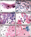
FAK is not required for oncogenic transformation of the mammary epithelium in the MMTV-PyVmT mouse model. (A) Mammary gland whole mount from a 10-week-old FAKflox/flox/MMTV-Cre/-PyVmT/GTRosa26 mouse (representative of n = 4 animals) X-Gal-stained in situ for Cre-mediated β-galactosidase activity. Cre-expressing cells (blue) are present in untransformed ductal structures (arrows). (Scale bar: 5 mm.) (B) A higher magnification of mammary ducts showing hyperplastic nodules both expressing a functional Cre (arrowhead) or not (arrow). (Scale bar: 500 μm.) Representative sections of X-Gal-stained mammary glands from 10-week-old FAKwt/wt/MMTV-Cre/-PyVmT/GTRosa26 (n = 3) (C) and FAKflox/flox/MMTV-Cre/-PyVmT/GTRosa26 (n = 4) (D) mice. Solid nests of tumor cells (asterisks) and hyperplastic regions of the epithelium (arrowheads) are indicated. (Scale bar: 5 μm.) (E and F) Representative X-Gal-stained cryosections of tumors from 12-week-old FAKflox/flox/MMTV-Cre/-PyVmT/GTRosa26 mice (n = 5). Unstained tumoral structures are indicated by asterisks. Stained nontransformed ducts (arrows, E) and a group of stained preneoplastic structures (arrowhead, F) are also indicated. (Scale bars: 10 μm.)
Fig. 3.
Fig. 3.
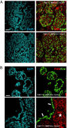
FAK deletion is restricted to MMTV-PyVmT hyperplasias. (A) Immunohistofluorescence analysis of Cre and FAK expression on paraffin sections of mammary glands from 10-week-old FAKwt/wt/- (A Upper), FAKflox/wt/- (A Lower), and FAKflox/flox/MMTV-Cre/-PyVmT (B) mice. Data are representative of at least five animals of each genotype. FAK is coexpressed with Cre throughout the MMTV-PyVmT-derived solid tumor tissue (A Upper). Note the reduced level of FAK expression in the Cre-positive/FAKflox/wt tumoral cells. In FAKflox/flox glands, Cre-positive/FAK-negative cells are detectable only in hyperplastic (B Upper) or nontransformed ductal structures (B Lower, arrows). FAK expressing cells are present in solid nests of tumor cells (B Lower, asterisk). (Scale bars: 20 μm.)
Fig. 4.
Fig. 4.
FAK-deleted cells do not contribute to lung metastasis in the MMTV-PyVmT mouse model. Representative sections of X-Gal-stained lung lobes from FAKwt/wt/- (Left) (n = 8) and FAKflox/flox/- (Right) (n = 6) MMTV-Cre/-PyVmT/GTRosa26 mice bearing late stage tumors. β-Gal-negative metastases are indicated by arrowheads. Lower shows X-Gal-stained metastases at higher magnification. [Scale bars: 50 μm (Upper), 500 μm (Lower).]
Fig. 5.
Fig. 5.
FAK-negative mammary epithelial neoplastic cells show a reduced proliferative capacity but do not exhibit any increase in apoptosis. Paraffin sections of mammary gland from 6-week-old FAKwt/wt/-, FAKflox/wt/-, and FAKflox/flox/MMTV-Cre/-PyVmT mice were submitted to immunofluorescence analysis of Cre and Ki67 (A) or activated caspase-3 (B) expression. [Scale bars: 50 μm (A), 20 μm (B).] Graphical representation of the immunostaining shown in A (C) and B (D). Percentages (±SEM) were calculated after counting multiple fields of at least five animals of each genotype. *, P < 0.01 vs. FAKwt/wt mice, Student's t test.

References

    1. Guo W, Giancotti FG. Nat Rev Mol Cell Biol. 2004;5:816–826. - PubMed
    1. Hannigan G, Troussard AA, Dedhar S. Nat Rev Cancer. 2005;5:51–63. - PubMed
    1. Playford MP, Schaller MD. Oncogene. 2004;23:7928–7946. - PubMed
    1. Mitra SK, Schlaepfer DD. Curr Opin Cell Biol. 2006;18:516–523. - PubMed
    1. White DE, Kurpios NA, Zuo D, Hassell JA, Blaess S, Mueller U, Muller WJ. Cancer Cell. 2004;6:159–170. - PubMed

Publication types

Substances

LinkOut - more resources