

Transcranial magnetic stimulation in child neurology: current and future directions
- PMID: 18056688
- PMCID: PMC2539109
- DOI: 10.1177/0883073807307972
Transcranial magnetic stimulation in child neurology: current and future directions
Abstract
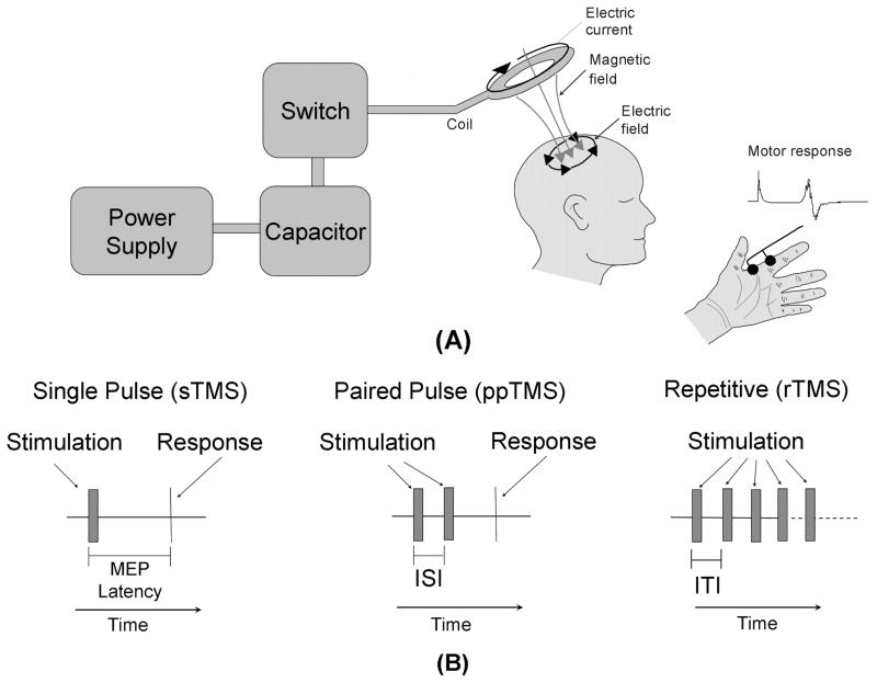
Transcranial magnetic stimulation (TMS) is a method for focal brain stimulation based on the principle of electromagnetic induction, where small intracranial electric currents are generated by a powerful, rapidly changing extracranial magnetic field. Over the past 2 decades TMS has shown promise in the diagnosis, monitoring, and treatment of neurological and psychiatric disease in adults, but has been used on a more limited basis in children. We reviewed the literature to identify potential diagnostic and therapeutic applications of TMS in child neurology and also its safety in pediatrics. Although TMS has not been associated with any serious side effects in children and appears to be well tolerated, general safety guidelines should be established. The potential for applications of TMS in child neurology and psychiatry is significant. Given its excellent safety profile and possible therapeutic effect, this technique should develop as an important tool in pediatric neurology over the next decade.
Figures
References
-
- Kobayashi M, Pascual-Leone A. Transcranial magnetic stimulation in neurology. Lancet Neurol. 2003;2:145–156. - PubMed
-
- Pascual-Leone A, Walsh V. Transcranial magnetic simulation. In: Toga A, Mazziotta J, editors. Brain Mapping: The Methods. San Diego, CA: Academic Press; 2002. pp. 255–290.
-
- Stefan K, Kunesch E, Cohen LG, et al. Induction of plasticity in the human motor cortex by paired associative stimulation. Brain. 2000;123:572–584. - PubMed
-
- McKay DR, Ridding MC, Thompson PD, et al. Induction of persistent changes in the organization of the human motor cortex. Exp Brain Res. 2002;143:342–349. - PubMed
-
- Uy J, Ridding MC. Increased cortical excitability induced by transcranial DC and peripheral nerve stimulation. J Neurosci Methods. 2003;127:193–197. - PubMed
Publication types
MeSH terms
Grants and funding
LinkOut - more resources
Full Text Sources
Other Literature Sources
Medical
Miscellaneous
