. 2008 Jan;135(2):217-25.
doi: 10.1242/dev.011031. Epub 2007 Dec 5.

The serine protease Corin is a novel modifier of the Agouti pathway

Affiliations

The serine protease Corin is a novel modifier of the Agouti pathway

David Enshell-Seijffers et al. Development. 2008 Jan.

Abstract

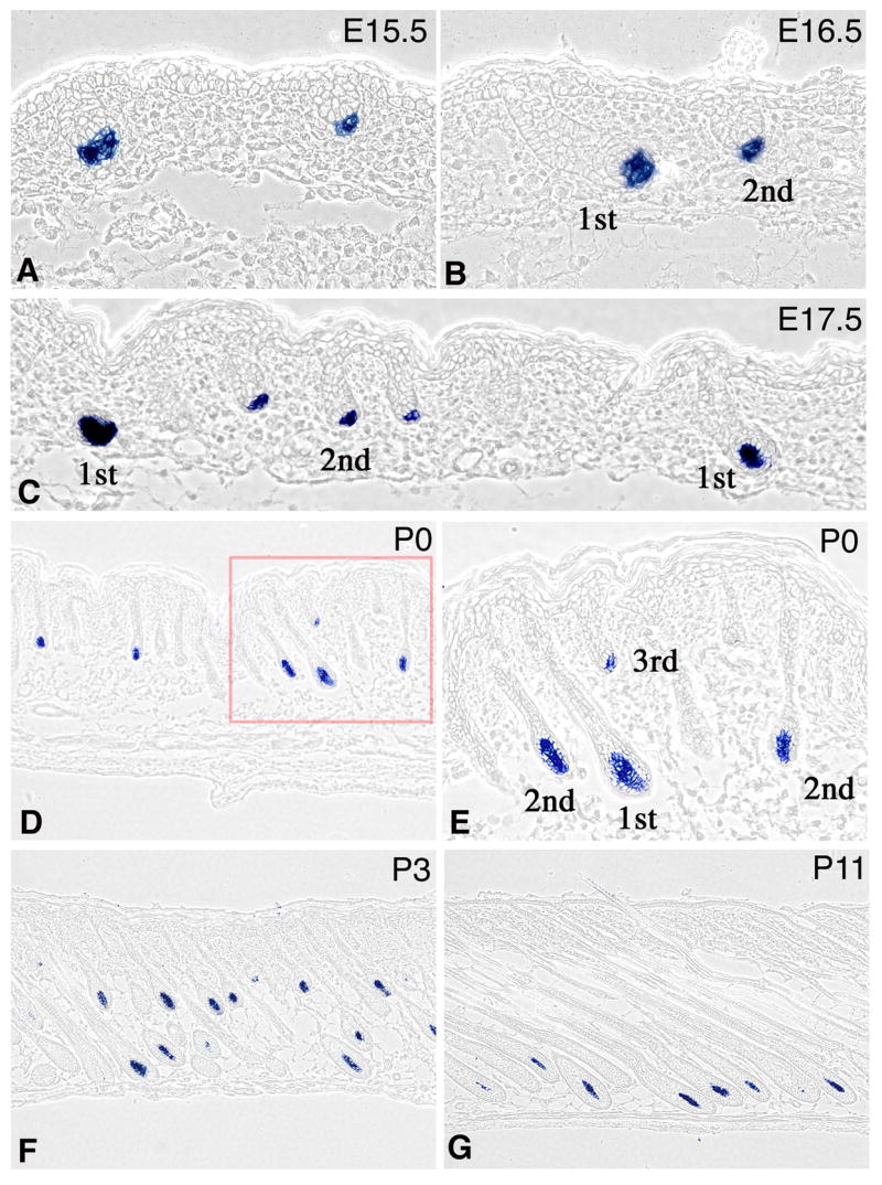
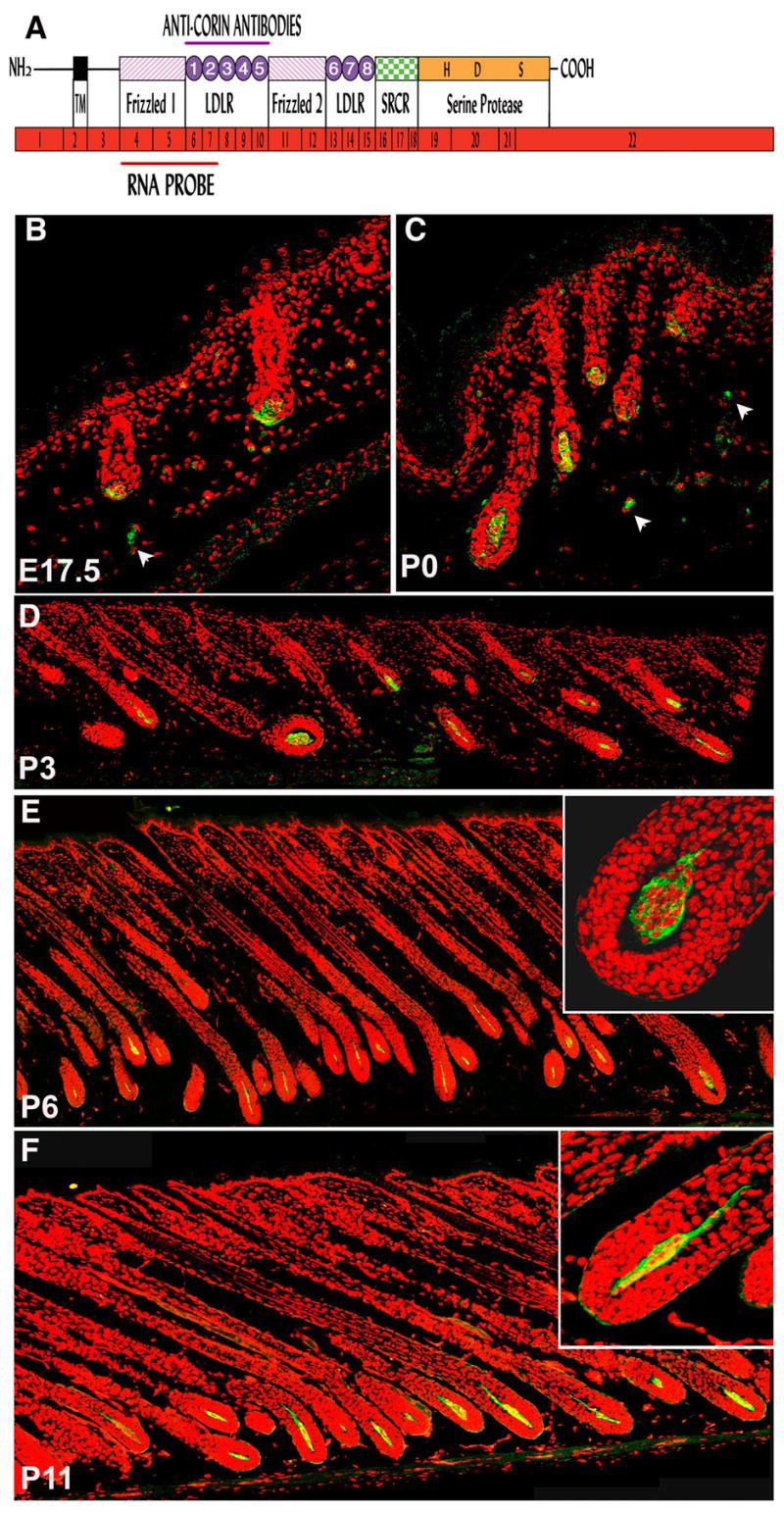
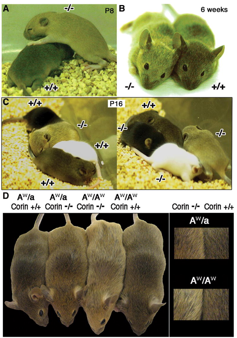
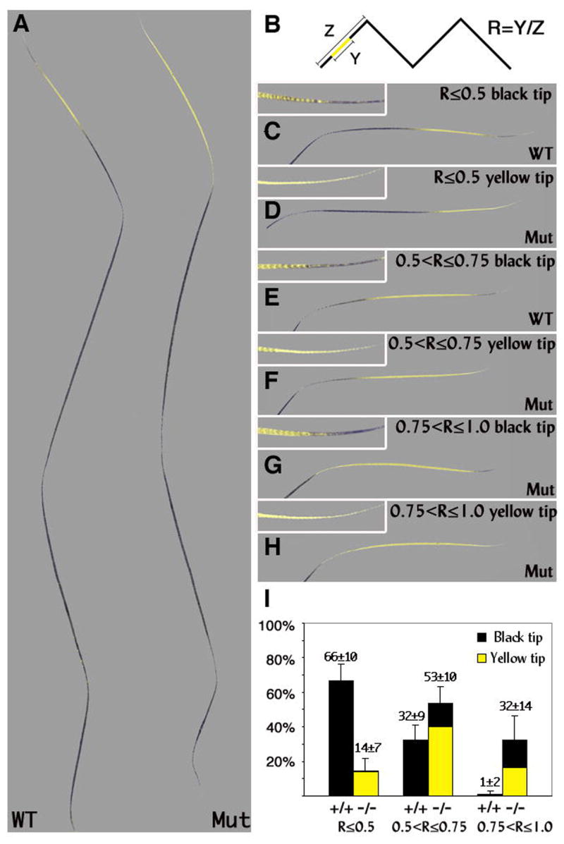
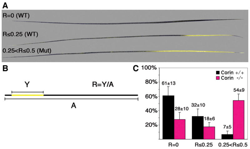
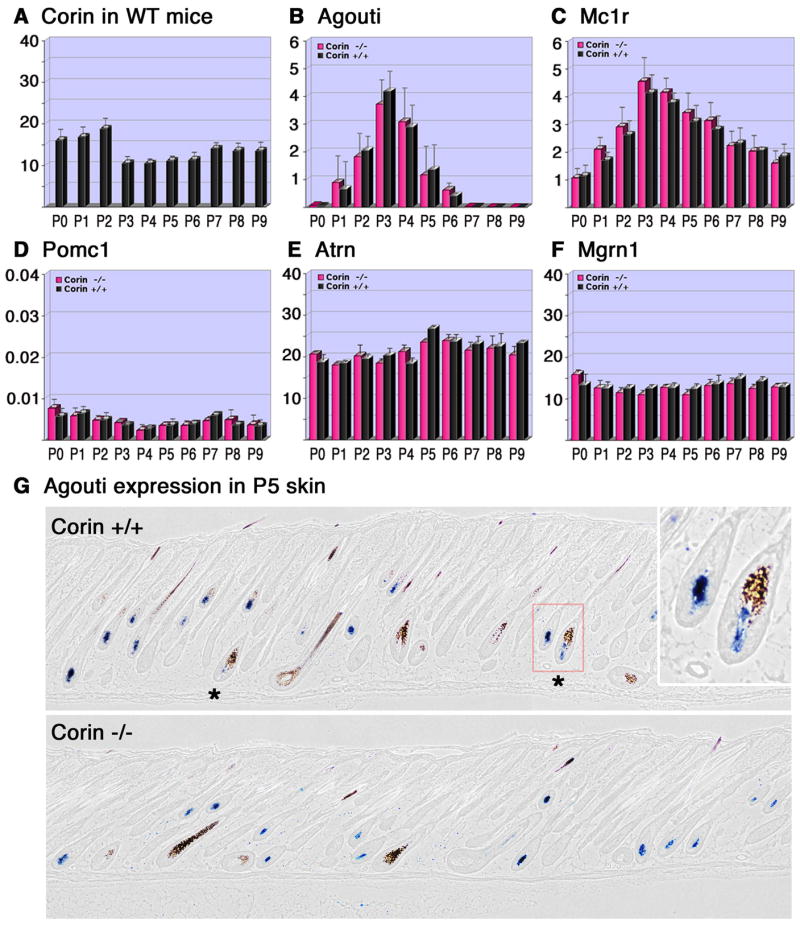
The hair follicle is a model system for studying epithelial-mesenchymal interactions during organogenesis. Although analysis of the epithelial contribution to these interactions has progressed rapidly, the lack of tools to manipulate gene expression in the mesenchymal component, the dermal papilla, has hampered progress towards understanding the contribution of these cells. In this work, Corin was identified in a screen to detect genes specifically expressed in the dermal papilla. It is expressed in the dermal papilla of all pelage hair follicle types from the earliest stages of their formation, but is not expressed elsewhere in the skin. Mutation of the Corin gene reveals that it is not required for morphogenesis of the hair follicle. However, analysis of the ;dirty blonde' phenotype of these mice reveals that the transmembrane protease encoded by Corin plays a critical role in specifying coat color and acts downstream of agouti gene expression as a suppressor of the agouti pathway.


Figures

Figure 1
Figure 1. Corin expression is confined to the DP
In-situ hybridization to detect Corin transcripts (blue) in FVB mice. Corin is first detected when the dermal condensate segregates from surrounding dermis during the first (A, E15.5) and second (B, E16.5) waves of follicle formation. Follicles derived from the first, second and third waves of follicle formation are indicated. Throughout the growth phase of the hair cycle, Corin transcripts are expressed in the DP and not detected elsewhere in the skin. Samples from E17.5 (C) P0 (D,E), P3 (F) and P11 (G) are shown. The field enclosed by the red square in D is shown in higher magnification in panel E to reveal that Corin is expressed throughout the DP and not in the surrounding hair matrix.
Figure 2
Figure 2. Expression of Corin protein coincides with Corin transcript accumulation
(A) Schematic representation of corin mRNA and protein. The mRNA of Corin (shown in red) comprises 22 exons encoding a type-II transmembrane serine protease. The single-pass transmembrane domain (TM) of Corin resides in close proximity to the N-terminus, and the large extracellular portion includes two frizzled-like cysteine-rich motifs (designated Frizzled 1 and 2), eight LDL receptor repeats (LDLR 1-8), a macrophage scavenger receptor-like domain (SRCR) and a catalytic domain of trypsin-like serine protease at the C-terminus. The active site residues of the catalytic triad (H, D and S) are shown. The regions corresponding to the RNA probe and the fragment of Corin protein used to immunize rabbits are indicated. (B–F) Immonuhistochemical detection of Corin (green) in DP at E17.5 (B), P0 (C), P3 (D), P6 (E) and P11 (F). Red stain highlights nuclei. Occasional staining was observed outside the DP (white arrowheads) but this was also detected in controls in which only the secondary antibody was used (data not shown and Fig. S2). In E and F, higher magnification of a bulb region is shown (upper right) to reveal that the majority of the protein is localized at the periphery of DP cells.
Figure 3
Figure 3. The generation of Corin knock-out mice
(A) Gene targeting approach used for corin ablation. A YFP-Neo cassette replaced 64bp downstream of the ATG in exon 1(red box). Primers (arrowheads, see table S1) and expected PCR fragments are indicated. Primer WT51 binds in the deleted-64bp segment and the primers SC3 and SC5 bind outside the targeting construct. The NcoI fragments used for Southern analysis are indicated. The FRT flanked pgkNeoR cassette was oriented in parallel to Corin transcription. (B,C,D) Genomic analysis of wild-type, heterozygous and homozygous Corin mice. (B) Southern analysis of NcoI-digested genomic DNA. The 5'-probe reveals 18.6kb and 10kb bands from wild-type and mutant alleles respectively. (C) PCR with primers SCNEO5 and SC3 generates a 6075bp band from the targeted allele (upper panel) while WT51 and SC3 generate a 5266bp fragment from wild type. (D) PCR with WT51, WT31, KILY5 and SEQFP1 detects both wild-type (312bp) and mutant (478bp) alleles for routine genotyping. (E,F) Immunostaining of frozen P3 skin-sections from wild-type and mutant mice with anti-Corin antibodies. Corin-green; Nuclei-red.
Figure 4
Figure 4. Coat-color phenotype of Corin mutants
Mice lacking Corin (−/−) exhibit lighter coat-color than corresponding wild type (+/+) on an Agouti background. (A,B) Mice during the first (A) and second (B) hair cycles are shown. (C) Two litters at P16 are shown each containing one a/a (black), one tyrC/tyrC (white), and two A/A with Corin genotypes indicated. (D) Pairs of mice homozygous and heterozygous for a functional agouti allele and wild-type or mutant for Corin are shown with higher magnification views at right. On both Agouti genotypes lack of Corin leads to a lighter coat color and in the absence of Corin, AW/AW are lighter than AW/a.
Figure 5
Figure 5. Yellow band extension in Corin mutant zigzag hairs
(A) The longest yellow band found in zigzag hairs lacking Corin (Mut) and the shortest subapical yellow band found in wild-type zigzag hairs (WT) are shown. (B) The approach used to quantify the differences between wild-type and mutant zigzag hairs. The ratio (R) between the length of the yellow band (Y) and the length of the apical segment (Z) was scored and assigned to three categories: R≤0.5 (C and D), 0.5
Figure 6
Figure 6. Yellow band extension in Corin mutant awl hairs
(A) Representative examples of three categories of awl hairs used in this analysis. In contrast to zigzag hairs, awl hairs may lack a subapical yellow band. (B) The ratio (R) between the length of the yellow band (Y) and the length of the whole hair (A) was calculated and assigned to the categories: R=0, R≤0.25, 0.25
Figure 7
Figure 7. Analysis of the RNA levels of Corin, agouti, Mc1r, Pomc1, Atrn and Mgrn1 throughout the anagen phase of the hair cycle
(A–F) The relative RNA levels determined by Real-time PCR (y-axis) of Corin (A), agouti (B), Mc1r (C), Pomc1 (D), Atrn (E) and Mgrn1 (F) are shown from birth to P9 (x-axis) for Corin +/+ (black) and Corin −/− (pink). The RNA levels of all genes tested were normalized to the same units, but different scales employed in A,E,F vs. B,C vs. D. Thus the level of Atrn mRNA is about 5 fold higher than the RNA levels of agouti at its peak (B and E), while Pomc1 levels are similar to those of agouti at its baseline levels from P7 on (B and D). (G) In-situ hybridization of agouti in P5 skin. Stars indicate hair follicle bulbs with agouti expression in the DP and eumelanin deposition. Note that in the absence of Corin, eumelanin deposition has not been detected in the bulb region surrounding agouti-expressing DPs. In the right upper corner, higher magnification of the region enclosed by the red square is shown.

References

    1. Barsh G. From Agouti to Pomc--100 years of fat blonde mice. Nat Med. 1999;5:984–5. - PubMed
    1. Barsh G, Gunn T, He L, Schlossman S, Duke-Cohan J. Biochemical and genetic studies of pigment-type switching. Pigment Cell Res. 2000;13(Suppl 8):48–53. - PubMed
    1. Byrne C, Tainsky M, Fuchs E. Programming gene expression in developing epidermis. Development. 1994;120:2369–83. - PubMed
    1. Chai BX, Neubig RR, Millhauser GL, Thompson DA, Jackson PJ, Barsh GS, Dickinson CJ, Li JY, Lai YM, Gantz I. Inverse agonist activity of agouti and agouti-related protein. Peptides. 2003;24:603–9. - PubMed
    1. Challis BG, Coll AP, Yeo GS, Pinnock SB, Dickson SL, Thresher RR, Dixon J, Zahn D, Rochford JJ, White A, et al. Mice lacking pro-opiomelanocortin are sensitive to high-fat feeding but respond normally to the acute anorectic effects of peptide-YY(3-36) Proc Natl Acad Sci U S A. 2004;101:4695–700. - PMC - PubMed

Publication types