. 2007 Dec 5;27(49):13481-90.
doi: 10.1523/JNEUROSCI.4158-07.2007.

EphB receptors regulate stem/progenitor cell proliferation, migration, and polarity during hippocampal neurogenesis

Affiliations

EphB receptors regulate stem/progenitor cell proliferation, migration, and polarity during hippocampal neurogenesis

Michael J Chumley et al. J Neurosci. .

Abstract

The adult brain maintains two regions of neurogenesis from which new neurons are born, migrate to their appropriate location, and become incorporated into the circuitry of the CNS. One of these, the subgranular zone of the hippocampal dentate gyrus, is of primary interest because of the role of this region in learning and memory. We show that mice lacking EphB1, and more profoundly EphB1 and EphB2, have significantly fewer neural progenitors in the hippocampus. Furthermore, other aspects of neurogenesis, such as polarity, cell positioning, and proliferation are disrupted in animals lacking the EphB1 receptor or its cognate ephrin-B3 ligand. Our data strongly suggest that EphB1 and ephrin-B3 cooperatively regulate the proliferation and migration of neural progenitors in the hippocampus and should be added to a short list of candidate target molecules for modulating the production and integration of new neurons as a treatment for neurodegenerative diseases or brain injury.


Figures

Figure 1.
Figure 1.
EphB1 is expressed in the adult hippocampal neural stem/progenitor cell population. a, Low- and high-magnification bright-field coronal images of the DG from a 4-week-old EphB1lacZ/+ mouse reacted with X-gal shows expression of the knocked-in β-gal reporter (blue stain) in the SGZ. b, Confocal IF labeling of an 8- to 10-week-old adult DG from nestin-eGFP transgenic reporter mice using GFP (green), DCX (red), and NeuN (blue) antibodies identifies type I (green), type IIa (yellow), and type IIb (red) stem/progenitor cells in the SGZ and mature neurons (blue) in the GL. c, Confocal IF of an 8- to 10-week-old EphB1lacZ/+ adult using β-gal (green) and DCX (red) antibodies demonstrates coexpression of EphB1 in DCX+ cells within the SGZ (arrows). d, Confocal IF using DCX (red) and PSA-NCAM (green) antibodies demonstrates coexpression of both markers on adult stem/progenitors (yellow). e, Flow cytometry (FACS) of cells isolated from nestin-eGFP transgenic mice after microdissection of the DG from 2-week-old animals and sorted for eGFP fluorescence and PSA-NCAM immunoreactivity. Arrows represent developmental progression of the neural progenitor cells, and boxes represent sorted populations. All sorted populations resulted in ≥95% purity after postsort analysis. f, RT-PCR from total RNA collected from sorted populations in e detects EphB1 transcripts in all three neural progenitor groups but not in the nonprogenitor population (GFP/PSA-NCAM).
Figure 2.
Figure 2.
Reduced numbers and ectopic localization of neural stem/progenitor cells in mice lacking EphB1. a, Representative low- and high-magnification confocal IF images of the DG from 8- to 10-week-old adult wild-type and EphB1−/− null mutants carrying the nestin-eGFP transgene immunolabeled as indicated. Type I (green), type IIa (yellow), and type IIb (red) neural stem/progenitor cells are located in the SGZ in wild-type animals and extend cell processes radially through the mature neurons in the GL (blue) and into the ML. EphB1−/− mutants have fewer type I (green) and type IIa (yellow) cells and show mislocalized stem/progenitors within the GL (arrows). b, The numbers of nestin/eGFP+ cells in adult mice were counted in a set of four coronal sections along the rostral–caudal range of each hippocampus/DG. Compared with wild-type mice, the mean number of nestin/eGFP+ progenitors is significantly reduced in the EphB1−/− mutants throughout the rostral–caudal range of the DG (p < 0.0001; 2-way ANOVA). Bonferroni corrected comparisons indicate significant differences at each anatomical position (*p < 0.001). Error bars represent ±SEM. c, Confocal images acquired at 7, 14, and 21 d of age indicate that EphB1−/− mutants already exhibit fewer nestin/eGFP+ (green) cells. Flow cytometry (FACS) of cells isolated from nestin-eGFP transgenic mice after microdissection of the DG at 14 and 21 d of age was used to determine the percentage of eGFP+ cells and is presented as green numerals in each respective image. The EphB1−/− mutants exhibited 50% fewer nestin/eGFP+ cells at each time point.
Figure 3.
Figure 3.
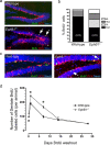
Ectopic localization of proliferating cells in the DG of mice lacking EphB1. a, A single intraperitoneal injection of BrdU was used to label proliferating cells in 8- to 10-week-old mice. After a 2 h washout, hippocampi were collected and processed for confocal IF to detect cells that incorporated BrdU (green) and expressed DCX (red). In wild-type animals, most proliferating cells are DCX+ and are localized in a tight band of cells that comprise the SGZ, whereas in EphB1−/− mutants, numerous proliferating cells are detected outside of the SGZ (arrows). The ectopic proliferating cells in EphB1−/− mutants are often DCX+ or found in close association with DCX+ cells, suggesting active sites of neurogenesis. All DNA is labeled with TOTO-3 (blue). b, Quantitative cell counts were compared using Student's t test and indicate that EphB1−/− animals have a significantly lower percentage of total proliferating (BrdU+) cells in the SGZ (p = 0.0002) and have a significantly higher percentage in the GL (p = 0.0078), H (p = 0.0376), and ML (p = 0.0048). c, Confocal IF for Ki-67 (green) also detects proliferating cells outside of the SGZ and in close association with mislocalized DCX+ cells (red) in the EphB1−/− mutants. d, BrdU “birth dating” suggests more proliferating cells in the DG of EphB1−/− mutants. Three injections of BrdU separated by 2 h were used to label a substantial portion of cells in S-phase. Comparisons of total BrdU+ cells were performed after 2 and 24 h washouts as well as 7, 14, and 28 d after the last BrdU injection. The total number of BrdU+ cells is presented for six coronal sections from each animal. A Student's t test indicates that EphB1−/− animals have significantly more cells in S-phase at 2 and 24 h, as well as 7 d after BrdU administration (*p < 0.05). Error bars represent ±SEM for n = 3–7 animals per genotype at each time point.
Figure 4.
Figure 4.
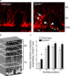
Premature branching and random polarization of the DCX+ dendritic outgrowth in adult mice lacking EphB1. a, Confocal IF images of DCX+ cells (red) reveal in the EphB1−/− mutants premature branching of the dendritic outgrowth within the GL (large arrowheads), random nonradial orientation of the outgrowth (small arrowheads), and numerous cellular processes that encroach into the hilus (asterisk). Ectopic positioned cells are indicated (arrows). b, Counts of DCX+ apical dendrites in the GL and three different molecular layers (ML 1–3) involved the generation of ROIs through the z-plane of 30 μm sections and manual counting of DCX+ outgrowths within the ROI. Shown is a representative stacked confocal image of DCX+ cells/processes from a wild-type brain. c, Consistent with premature branching, the number of dendritic projections per DCX+ cell in EphB1−/− mutants is significantly increased from one (no branches) to two (one branch) in the GL of the dentate compared with wild-type specimens (p = 0.0005, Student's t test). The graph represents the mean ± SEM number of projections per DCX+ cell at each of the ROIs indicated in b. A total of 270 wild-type and 248 EphB1−/− mutant processes were counted.
Figure 5.
Figure 5.
Ephrin-B3 exhibits ligand-like activity to control neural stem/progenitor positioning in the SGZ and prevent cellular processes from invading the hilus. a, X-gal staining of an adult ephrin-B3lacZ/lacZ hippocampus demonstrates intense expression of the ephrin-B3-β-gal fusion protein (blue) in the H and IML, with relatively weaker labeling of the OML. b, Confocal IF for β-gal (blue) and DCX (red) in the adult dentate of an ephrin-B3lacZ/lacZ homozygote shows intense expression of the ephrin-B3-β-gal fusion protein in the H and IML, with little or no expression in the SGZ and GL. Note that DCX+ cells (red) primarily reside in the SGZ and lie immediately adjacent to high levels of ephrin-B3 in the H. High-magnification confocal z-series (30 μm) of boxed section indicates normal positioning of DCX+ cells in the SGZ as well as normal morphology of the dendritic outgrowth in the ephrin-B3lacZ/lacZ homozygote. c, Confocal IF for the nestin-eGFP transgene (green), DCX (red), and NeuN (blue) in the adult dentate of an ephrin-B3−/− protein-null homozygote shows mislocalized neural stem/progenitor cells in the GL (arrows) and H (d, asterisk). d, High-magnification confocal images of the SGZ in wild-type and ephrin-B3−/− adult animals demonstrate an increase in DCX+ basal cellular projections that penetrate the H (red) in the null mutant.
Figure 6.
Figure 6.
Combined loss of EphB1 and EphB2 results in even more dramatic neurogenic defects in the dentate gyrus. a, RT-PCR from total RNA collected from 2-week-old microdissected dentate cells containing the nestin-eGFP transgene and sorted for eGFP fluorescence demonstrates that EphB2 transcripts are found specifically in the GFP+ stem/progenitor population. In contrast, ephrin-B3 is primarily expressed in the GFP population, which is mainly comprised of mature granule cell neurons and astrocytes in the hilus. b, Nissl stains of hippocampi from indicated adult mice demonstrate that the EphB1−/−; EphB2−/− compound null and EphB1−/−; EphB2lacZ/lacZ compound mutant deficient for EphB2 forward signaling have a smaller DG than the EphB1−/− single mutant. c, Estimated DG volume for wild-type, EphB1−/−, EphB1−/−; EphB2−/− compound null and EphB1−/−; EphB2lacZ/lacZ compound mutant animals (n = 3 for each). ANOVA indicates that DG volume is significantly affected by loss of one or more EphB receptors (p < 0.0001), and post hoc comparisons demonstrate that the DG volumes of EphB1−/−; EphB2−/− and EphB1−/−; EphB2lacZ/lacZ animals are significantly smaller than wild-type (*p < 0.001, Bonferroni corrected). The defect is more severe in the EphB1−/−; EphB2lacZ/lacZ than in the EphB1−/−; EphB2−/− animal (¥p < 0.05, Bonferroni corrected). d, Confocal IF images using GFP (green), DCX (red), and NeuN (blue) antibodies show that the compound null has a more dramatic decrease in the numbers of stem/progenitor cells as well as mature granule cell neurons than the EphB1−/− single mutant.

References

    1. Abrous DN, Koehl M, Le Moal M. Adult neurogenesis: from precursors to network and physiology. Physiol Rev. 2005;85:523–569. - PubMed
    1. Ahn S, Joyner AL. In vivo analysis of quiescent adult neural stem cells responding to Sonic hedgehog. Nature. 2005;437:894–897. - PubMed
    1. Bilder D. Epithelial polarity and proliferation control: links from the Drosophila neoplastic tumor suppressors. Genes Dev. 2004;18:1909–1925. - PubMed
    1. Brambilla R, Schnapp A, Casagranada F, Labrador JP, Bergemann AD, Flanagan JG, Pasquale EB, Klein R. Membrane-bound LERK2 ligand can signal through three different Eph-related receptor tyrosine kinases. EMBO J. 1995;14:3116–3126. - PMC - PubMed
    1. Cappello S, Attardo A, Wu X, Iwasato T, Itohara S, Wilsch-Brauninger M, Eilken HM, Rieger MA, Schroeder TT, Huttner WB, Brakebusch C, Gotz M. The Rho-GTPase cdc42 regulates neural progenitor fate at the apical surface. Nat Neurosci. 2006;9:1099–1107. - PubMed

Publication types

MeSH terms