. 2008 Feb;82(4):1759-76.
doi: 10.1128/JVI.00873-07. Epub 2007 Dec 5.

Kaposi's sarcoma-associated herpesvirus induces sustained levels of vascular endothelial growth factors A and C early during in vitro infection of human microvascular dermal endothelial cells: biological implications

Affiliations

Kaposi's sarcoma-associated herpesvirus induces sustained levels of vascular endothelial growth factors A and C early during in vitro infection of human microvascular dermal endothelial cells: biological implications

Ramu Sivakumar et al. J Virol. 2008 Feb.

Abstract

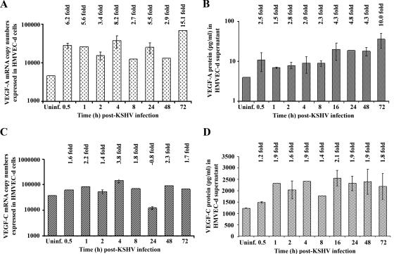
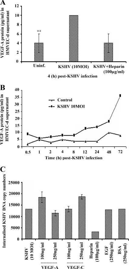
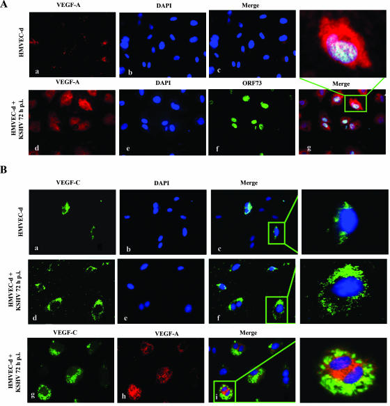
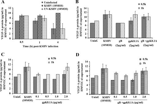
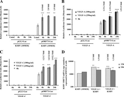
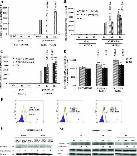
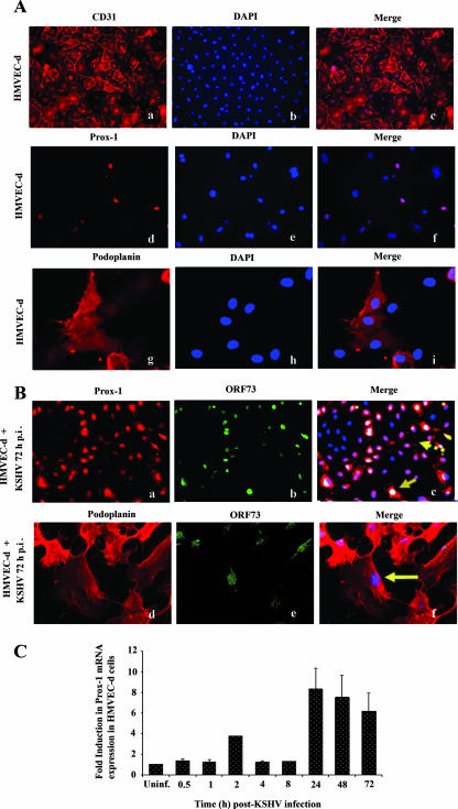
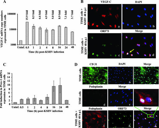
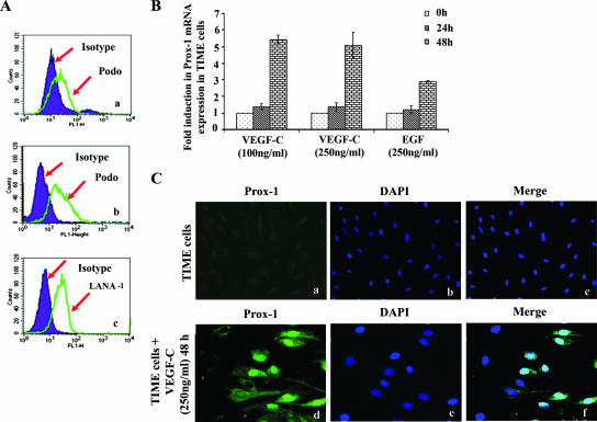
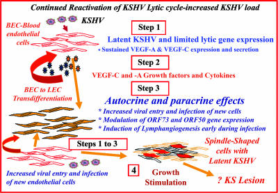
Kaposi's sarcoma (KS), a vascular tumor associated with human immunodeficiency virus type 1 infection, is characterized by spindle-shaped endothelial cells, inflammatory cells, cytokines, growth and angiogenic factors, and angiogenesis. KS spindle cells are believed to be of the lymphatic endothelial cell (LEC) type. Kaposi's sarcoma-associated herpesvirus (KSHV, or human herpesvirus 8) is etiologically linked to KS, and in vitro KSHV infection of primary human dermal microvascular endothelial cells (HMVEC-d) is characterized by the induction of preexisting host signal cascades, sustained expression of latency-associated genes, transient expression of a limited number of lytic genes, sustained induction of NF-kappaB and several cytokines, and growth and angiogenic factors. KSHV induced robust vascular endothelial growth factor A (VEGF-A) and VEGF-C gene expression as early as 30 min postinfection (p.i.) in serum-starved HMVEC-d, which was sustained throughout the observation period of 72 h p.i. Significant amounts of VEGF-A and -C were also detected in the culture supernatant of infected cells. VEGF-A and -C were also induced by UV-inactivated KSHV and envelope glycoprotein gpK8.1A, thus suggesting a role for virus entry stages in the early induction of VEGF and requirement of KSHV viral gene expression for sustained induction. Exogenous addition of VEGF-A and -C increased KSHV DNA entry into target cells and moderately increased latent ORF73 and lytic ORF50 promoter activation and gene expression. KSHV infection also induced the expression of lymphatic markers Prox-1 and podoplanin as early as 8 h p.i., and a paracrine effect was seen in the neighboring uninfected cells. Similar observations were also made in the pure blood endothelial cell (BEC)-TIME cells, thus suggesting that commitment to the LEC phenotype is induced early during KSHV infection of blood endothelial cells. Treatment with VEGF-C alone also induced Prox-1 expression in the BEC-TIME cells. Collectively, these studies show that the in vitro microenvironments of KSHV-infected endothelial cells are enriched, with VEGF-A and -C molecules playing key roles in KSHV biology, such as increased infection and gene expression, as well as in angiogenesis and lymphangiogenesis, thus recapitulating the microenvironment of early KS lesions.


Figures

FIG. 1.
FIG. 1.
Detection of VEGF-A and VEGF-C mRNA and protein in KSHV-infected HMVEC-d cells. HMVEC-d cells grown to 80 to 90% confluence were serum starved for 8 h and infected with KSHV at an MOI of 10. (A and C) Infected and uninfected cells were washed and lysed, and total RNA was prepared. DNase I-treated RNA (250 ng) was subjected to real-time RT-PCR with VEGF-A and VEGF-C gene-specific primers. Known concentrations of DNase I-treated, in vitro-transcribed VEGF-A and VEGF-C transcripts were used in a real-time RT-PCR to construct a standard graph from which the relative copy numbers of transcripts were calculated and normalized, with GAPDH used as the internal control. Each reaction was done in duplicate, and each bar represents the average ± standard deviation from three independent experiments. The VEGF-A and VEGF-C levels normalized to GAPDH in the uninfected cells were considered as 1 for comparison. (B and D) The levels of VEGF-A and VEGF-C proteins released in the cell-free culture supernatants were measured by enzyme-linked immunosorbent assay. The data were normalized to a 1-mg/ml total protein concentration in the supernatant. Each reaction was done in duplicate, and each point represents the average ± standard deviation from three independent experiments. VEGF-A and VEGF-C released from uninfected cells were considered as 1 for comparison, and the induction levels in infected cells are indicated.
FIG. 2.
FIG. 2.
Blocking KSHV binding by heparin inhibits VEGF-A expression. (A) KSHV was incubated at 37°C for 1 h with DMEM containing 100 μg/ml of soluble heparin. This mixture was then added to a serum-starved (8 h) HMVEC-d cell monolayer and incubated for 4 h at 37°C. The levels of VEGF-A released in the cell-free culture supernatants were measured by enzyme-linked immunosorbent assay. **, statistically significant (P < 0.02). (B) HMVEC-d cells grown to 80 to 90% confluence were serum starved for 8 h and either uninfected (control) or infected with KSHV at an MOI of 10. The levels of VEGF-A proteins released in the cell-free culture supernatants collected at the indicated time points were measured by ELISA. The data were normalized to a 1-mg/ml total protein concentration in the supernatant. Each reaction was done in duplicate, and each point represents the average ± standard deviation from three independent experiments. (C) VEGF-A and VEGF-C enhance KSHV entry. HMVEC-d cells were serum starved for 8 h and either untreated or treated with VEGF-A or VEGF-C (100 and 250 ng/ml) or with human EGF or BSA (250 ng/ml) for 1 h at 37°C in serum-free EBM2 medium and infected (10 DNA copies per cell) for 2 h. Cells were washed, treated with trypsin-EDTA (0.25% trypsin and 5 mM EDTA) for 5 min, washed, and collected, and total DNA was prepared. The KSHV ORF73 gene in 100 ng of DNA was amplified by real-time DNA PCR, and the copy numbers were calculated from the standard graph generated by the real-time PCR using known concentrations of a cloned ORF73 gene. Each reaction was done in duplicate, and each point represents the average ± standard deviation of three experiments.
FIG. 3.
FIG. 3.
Immunofluorescent detection of KSHV infection-induced VEGF-A and -C protein expression in HMVEC-d cells. (A and B) Uninfected HMVEC-d cells (panels a, b, and c) or cells infected with KSHV (MOI, 10) for 72 h (panels d, e, and f) were permeabilized and stained with anti-VEGF-A mouse monoclonal or VEGF-C goat polyclonal antibody, respectively, and detected by Alexa-594-coupled anti-mouse antibody or Alexa-488-coupled anti-goat antibody. Infected HMVEC-d cells were permeabilized and also stained with anti-ORF73 rabbit polyclonal antibody (A, panel f). Nuclei were counterstained with DAPI (b and e). The inset in panel g indicates nuclear staining for ORF73 in a cell with cytoplasmic staining for VEGF-A. KSHV-infected HMVEC-d cells express both VEGF-C (B, panel g) and VEGF-A (B, panel h). The inset demonstrates coexpression of VEGF-C and VEGF-A in the cytoplasm (i) of infected HMVEC-d cells. Magnification, ×40.
FIG. 4.
FIG. 4.
Induction of VEGF-A by UV-inactivated KSHV and viral glycoproteins gB and gpK8.1A. (A) HMVEC-d cells (80 to 90% confluence) serum starved for 8 h were uninfected or infected with either live KSHV or UV-irradiated KSHV for the indicated times at an MOI of 10 per cell. (B) Serum-starved HMVEC-d cells were uninfected, infected with KSHV (MOI, 10), or induced with KSHV glycoproteins gB and gpK8.1A alone or together for the indicated times. (C) Serum-starved HMVEC-d cells were uninfected, infected with KSHV (MOI, 10), or induced with KSHV glycoprotein gpK8.1A at the indicated concentrations for 0.5 and 1 h. (D) Serum-starved HMVEC-d cells were uninfected, infected with KSHV (MOI, 10), or induced with KSHV glycoproteins gB and gpK8.1A together at the indicated concentrations for 0.5 and 1 h. The levels of VEGF-A released in the cell-free culture supernatants were measured by ELISA. Each reaction was done in duplicate, and each point represents the average ± standard deviation from three independent experiments.
FIG. 5.
FIG. 5.
Exogenous addition of VEGF-A or VEGF-C activates KSHV ORF73 promoter and gene expression. 293 cells were transfected with control pGL3-Luc or ORF73 promoter-luciferase construct and after 24 h cells were either uninfected or infected with KSHV at an MOI of 10 for 4 h, 8 h, and 24 h (A) or treated with exogenous VEGF-A (B) and -C (C) (100 ng/ml and 250 ng/ml). At the indicated time points, cells were harvested, lysed, and assayed for firefly luciferase activity. The data represent the mean relative luciferase units after normalizing with the cotransfected Renilla luciferase activity. (D) HMVEC-d cells (80 to 90% confluence) were serum starved for 8 h, either untreated or treated with VEGF-A or VEGF-C (100 ng/ml) for 1 h at 37°C, and infected with KSHV at an MOI of 10. Expression of ORF73 mRNA was monitored at the indicated time points. DNase I-treated RNA (250 ng) was subjected to real-time RT-PCR with an ORF73 gene-specific primer. Known concentrations of DNase I-treated, in vitro-transcribed ORF73 transcripts were used to construct a standard graph from which the relative copy numbers of transcripts were calculated and normalized, with GAPDH used as the internal control. Each reaction was done in duplicate, and each bar represents the average ± standard deviation from three independent experiments. **, ***, ****, statistically significant at P < 0.02, P < 0.01, and P < 0.001, respectively.
FIG. 6.
FIG. 6.
Exogenous addition of VEGF-A or VEGF-C activates KSHV ORF50 promoter and gene expression. 293 cells were transfected with control p-Luc or ORF50 promoter-Luciferase constructs and after 24 h, cells were either uninfected or infected with KSHV at an MOI of 10 for 4 h and 8 h (A) or treated with exogenous VEGF-A (B) and C (C) (100 ng/ml and 250 ng/ml). At the indicated time points cells were harvested, lysed, and assayed for firefly luciferase activity. The data represent the mean relative luciferase units after normalizing with the cotransfected Renilla luciferase activity. (D) HMVEC-d cells (80 to 90% confluence) were serum starved for 8 h, either untreated or treated with VEGF-A or VEGF-C (100 ng/ml) for 1 h at 37°C, and infected with KSHV at an MOI of 10. Expression of ORF50 mRNA was monitored at the indicated time points. DNase I-treated RNA (250 ng) was subjected to real-time RT-PCR with an ORF50 gene-specific primer. Known concentrations of DNase I-treated, in vitro-transcribed ORF50 transcripts were used to construct a standard graph from which the relative copy numbers of transcripts were calculated and normalized, with GAPDH used as the internal control. Each reaction was done in duplicate, and each bar represents the average ± standard deviation from three independent experiments. **, ***, ****, -statistically significant at P < 0.02, P < 0.01, and P < 0.001, respectively. (E) Exogenous addition of VEGF-A or VEGF-C induces KSHV LANA-1 expression at the protein level. HMVEC-d cells (80 to 90% confluence) were serum starved for 8 h, either untreated or treated with VEGF-A or VEGF-C (250 ng/ml) for 1 h at 37°C, and infected with KSHV at an MOI of 10. Cells were then harvested for flow cytometry after 48 h postinfection and detached from the plate with 0.25% trypsin-EDTA, and viability was assessed by trypan blue exclusion. Nuclear staining to identify KSHV-infected cells was performed using rat monoclonal antibody (ABI) to the KSHV LANA-1 protein. Flow cytometry was performed with a FACSCalibur flow cytometer and analyzed with CellQuest Pro software. Expression of LANA-1 at 48 h p.i. is shown as a green line for KSHV alone (a), a red line for VEGF-A-treated and KSHV-infected cells (b), a black line for VEGF-C-treated and KSHV-infected cells (c) compared to the isotype control, which is shown in a shaded purple histogram. (F) HMVEC-d cells (80 to 90% confluence) were serum starved for 8 h, either untreated or treated with VEGF-A or VEGF-C (250 ng/ml) for 1 h at 37°C, and infected with KSHV at an MOI of 10. Expression of LANA-1 protein was monitored at the indicated time points by Western blotting. Equal amounts of protein samples were resolved by SDS-7.5% PAGE, subjected to Western blotting, and reacted with rabbit polyclonal antibody against KSHV LANA-1. To confirm equal protein loading, blots were also reacted with antibodies against human β-actin. Each blot is representative of at least three independent experiments. The LANA-1 level in the untreated but KSHV-infected cells was considered as 1 for comparison. (G) Exogenous addition of VEGF-A or VEGF-C activates KSHV ORF73-luciferase expression. 293 cells were transfected with control pGL3-Luc or the ORF73 promoter-luciferase construct along with pcDNA-GFP as a transfection efficiency control. After 24 h, cells were either untreated (NT) or treated with exogenous VEGF-A or VEGF-C (250 ng/ml). At the indicated time points, cells were harvested and lysed, and total cell lysates were immunoblotted with antiluciferase primary antibodies. Transfection efficiency was detected by using anti-GFP mouse monoclonal antibody. To confirm equal protein loading, blots were also reacted with antibodies against human β-actin. Each blot is representative of at least three independent experiments. The luciferase level in the untreated cells was considered as 1 for comparison.
FIG. 7.
FIG. 7.
KSHV infection of HMVEC-d cells induces expression of lymphatic lineage-specific markers Prox-1 and podoplanin. (A) Uninfected HMVEC-d cells (a to i) or (B) cells infected with KSHV (MOI, 10) for 72 h (a to f) were permeabilized and stained with anti-CD31, Prox-1, or podoplanin antibodies and detected with Alexa 594-coupled anti-mouse antibodies. Nuclei were counterstained with DAPI. (B) Infected HMVEC-d cells were permeabilized and also stained with anti-ORF73 polyclonal antibody and detected by Alexa 488-coupled anti-rabbit antibodies. Solid arrows indicate nuclei demonstrating positive staining for ORF73 in cells showing nuclear staining for Prox-1 (B, panel c). The solid arrow (B, panel f) shows nuclei demonstrating positive staining for ORF73 in cells showing staining for podoplanin. The broken arrow (B, panel c) indicates ORF73-negative cells positive for Prox-1 staining. Magnification, ×20 (A, panels a to f; B, panels a to c) or ×40 (A, panels g to i; B, panels d to f). (C) HMVEC-d cells grown to 80 to 90% confluence were serum starved for 8 h and infected with KSHV at an MOI of 10. Infected and uninfected cells were washed and lysed, and total RNA was prepared. After cDNA synthesis, real-time RT-PCR was performed for Prox-1 gene expression using the Sybr green detection protocol. The data were normalized with the expression of GAPDH. The data shown represent the average ± standard deviation from three independent experiments. The Prox-1 level normalized to GAPDH in the uninfected cells was considered 1 for comparison.
FIG. 8.
FIG. 8.
KSHV infection of TIME cells induced the expression VEGF-C and lymphatic markers. (A) TIME cells grown to 80 to 90% confluence were serum starved for 8 h and infected with KSHV at an MOI of 10, and the kinetics of VEGF-C mRNA induction was quantitated as per procedures described in the Fig. 1 legend. The VEGF-C level normalized to GAPDH in the uninfected cells was considered 1 for comparison. (B) Cells infected with KSHV (MOI, 10) for 48 h (a, b, c, and d) were permeabilized and stained with goat anti-VEGF-C antibodies and detected by Alexa 594-coupled anti-goat antibodies. Infected cells were also permeabilized and stained with anti-ORF73 polyclonal antibody and detected by Alexa 488-coupled anti-rabbit antibodies (c). Nuclei were counterstained with DAPI (b). The solid arrow indicates cytoplasmic staining for VEGF-C in cells infected with KSHV (d). Magnification, ×40. (C) Prox-1 mRNA from uninfected and KSHV-infected TIME cells was quantified by real-time RT-PCR as per the procedures described in the Fig. 7C legend. The Prox-1 level normalized to GAPDH in the uninfected cells was considered 1 for comparison. (D) Uninfected TIME cells (a to e) or cells infected with KSHV (MOI, 10) for 48 h (f to h) were permeabilized, stained with anti-CD31, and detected by Alexa 488- or Alexa 594-coupled anti-mouse antibodies. Magnification, ×20 (a to c) or ×40 (d and e). Infected cells were permeabilized and also stained with anti-ORF73 polyclonal antibody and detected by Alexa 488-coupled anti-rabbit antibodies (g). Nuclei were counterstained with DAPI (b). The inset in panel h indicates membrane staining for podoplanin in infected TIME cells. Magnification, ×40.
FIG. 9.
FIG. 9.
Exogenous VEGF-C induces Prox-1 mRNA in TIME cells. (A) TIME cells were infected with KSHV (MOI, 10) and harvested for flow cytometry at 24 h (a) and 48 h postinfection (b). Cells were detached from the plate with 0.25% trypsin-EDTA, and viability was assessed by trypan blue exclusion. For podoplanin, cell surface staining was carried out for 1 h at 4°C. Nuclear staining to identify KSHV-infected cells was performed using rat monoclonal antibody to the KSHV LANA-1 protein (c). Flow cytometry was performed with a FACSCalibur flow cytometer and analyzed with CellQuest Pro software. Expression of podoplanin and LANA-1 at 24 h and 48 h p.i. is shown by the green line and compared to the isotype control, which is shown in the shaded purple histogram. (B) TIME cells were incubated with serum-free EBM2 containing different concentrations (100 and 250 ng/ml) of recombinant VEGF-C for 24 h and 48 h at 37°C. As a control, cells were treated for corresponding times with 250 ng/ml human EGF. Prox-1 mRNA was then quantified by real-time RT-PCR as per the procedures described in the Fig. 7C legend. The Prox-1 level normalized to GAPDH in the untreated cells was considered 1 for comparison. (C) Untreated (a to c) or VEGF-C-treated (250 ng/ml for 48 h) TIME cells (d to f) were permeabilized and stained with Prox-1 antibodies and detected with Alexa 488-coupled anti-mouse antibodies. Nuclei were counterstained with DAPI. Magnification, ×20 (a to c).
FIG. 10.
FIG. 10.
Schematic model depicting the potential implications of VEGF-A and VEGF-C induction during in vitro KSHV infection of endothelial cells. KSHV has been shown to be reactivated under immunosuppression conditions, resulting in increased circulating virus. Similar to in vitro infection of HMVEC-d cells, primary infection of endothelial cells in vivo could also result in the induction of preexisting host signal cascades, sustained expression of latency-associated genes, transient expression of a limited number of lytic genes, sustained induction of NF-κB and several cytokines and growth and angiogenic factors, including VEGF-A and -C (step 1). These factors when released to the extracellular environment of infected cells could act in an autocrine or paracrine fashion on the infected cells as well as neighboring cells (step 2). In the microenvironment of KS lesions, the presence of VEGF-A and -C may potentially be facilitating (i) an increase in infection of uninfected neighboring cells by KSHV released from the B cells and monocytes in the inflammatory cell pool, (ii) modulation of viral gene expression, and (iii) induction of lymphangiogenesis early during infection (step 3). Continual repetitions of these steps in an immunosuppressed individual could result in an increase in new infection of endothelial cells and increased autocrine actions of VEGF and other factors, along with viral gene expression, probably contributing to the maintenance of latency, angiogenesis, lymphatic lineage switch, neovascularization, inflammation, and growth stimulation. Reduced host immune regulation, an inability to control inflammation, and elimination of infected cells could ultimately lead to the formation of multifocal KS lesions (step 4).

Similar articles

Cited by

References

    1. Akula, S. M., P. P. Naranatt, N.-S. Walia, F.-Z. Wang, B. Fegley, and B. Chandran. 2003. Kaposi's sarcoma-associated herpesvirus (human herpesvirus 8) infection of human fibroblast cells occurs through endocytosis. J. Virol. 777978-7990. - PMC - PubMed
    1. Akula, S. M., N. P. Pramod, F.-Z. Wang, and B. Chandran. 2002. Integrin α3β1 (CD 49c/29) is a cellular receptor for Kaposi's sarcoma-associated herpesvirus (KSHV/HHV-8) entry into the target cells. Cell 108407-419. - PubMed
    1. Akula, S. M., F.-Z. Wang, J. Vieira, and B. Chandran. 2001. Human herpesvirus 8 interaction with target cells involves heparan sulfate. Virology 282245-255. - PubMed
    1. Albini, A., I. Paglieri, G. Orengo, S. Carlone, M. G. Aluigi, R. DeMarchi, C. Matteucci, A. Mantovani, F. Carozzi, S. Donini, and R. Benelli. 1997. The beta-core fragment of human chorionic gonadotrophin inhibits growth of Kaposi's sarcoma-derived cells and a new immortalized Kaposi's sarcoma cell line. AIDS 11713-721. - PubMed
    1. Alitalo, K., T. Tammela, and T. V. Petrova. 2005. Lymphangiogenesis in development and human disease. Nature 438946-953. - PubMed

Publication types

MeSH terms

Substances

LinkOut - more resources