. 2008 Feb;82(4):1701-13.
doi: 10.1128/JVI.02157-07. Epub 2007 Dec 5.

Translocation and colocalization of ICP4 and ICP0 in cells infected with herpes simplex virus 1 mutants lacking glycoprotein E, glycoprotein I, or the virion host shutoff product of the UL41 gene

Affiliations

Translocation and colocalization of ICP4 and ICP0 in cells infected with herpes simplex virus 1 mutants lacking glycoprotein E, glycoprotein I, or the virion host shutoff product of the UL41 gene

Maria Kalamvoki et al. J Virol. 2008 Feb.

Abstract

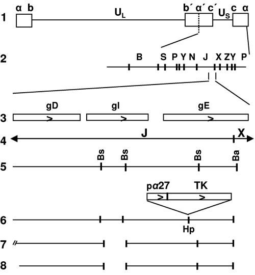
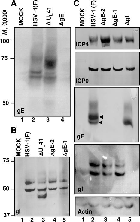
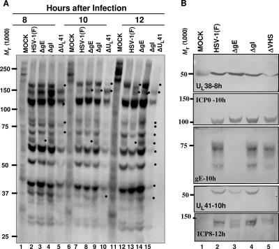
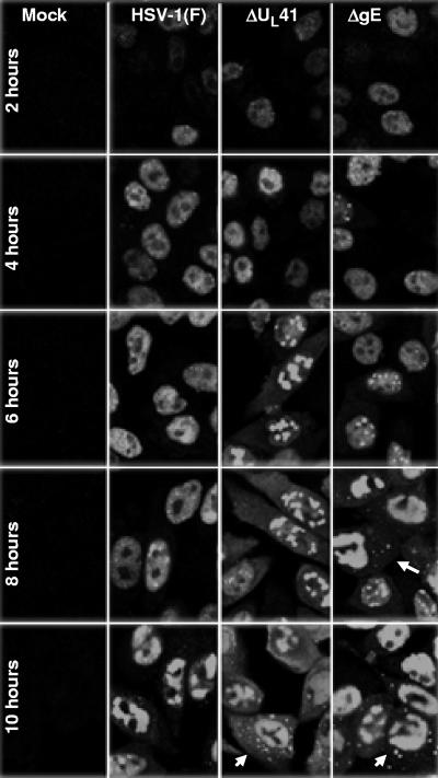
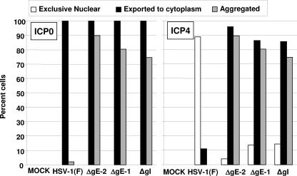
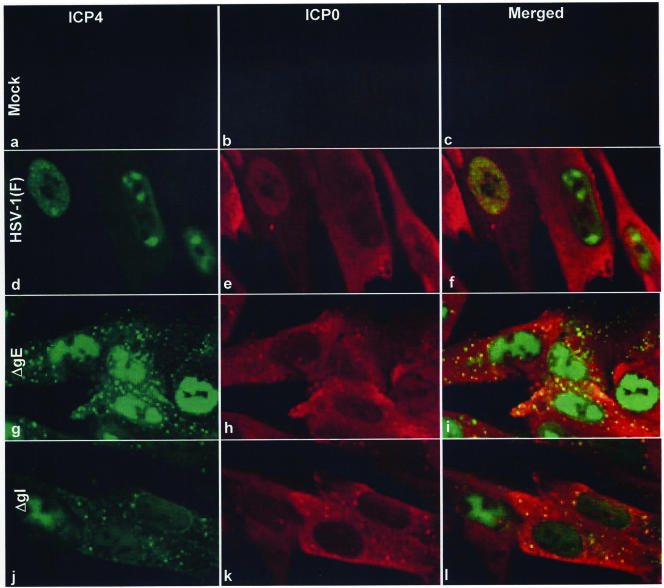
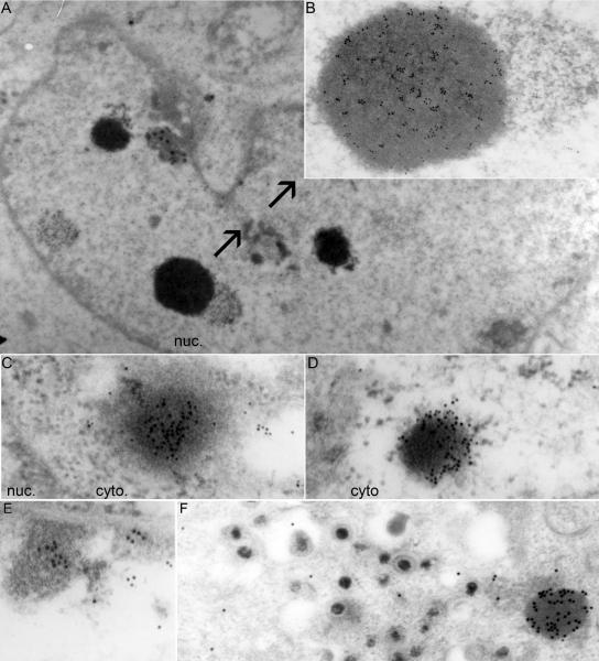
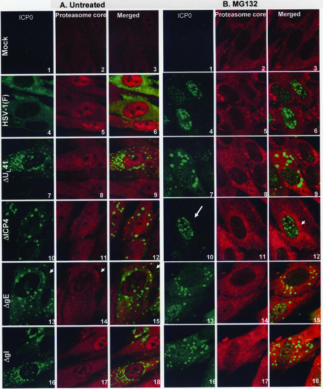
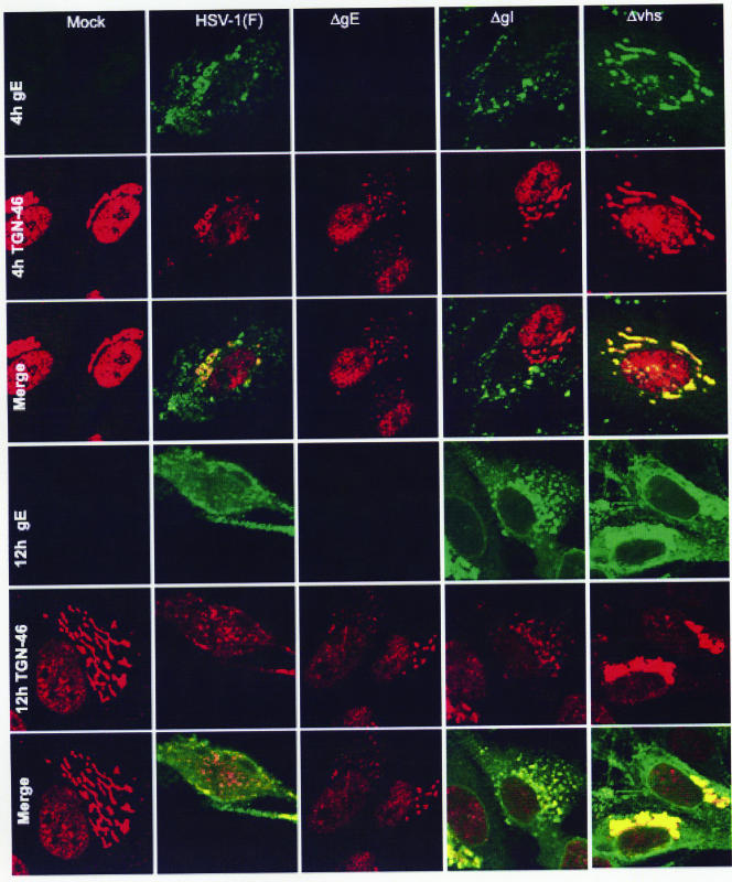
In wild-type herpes simplex virus 1-infected cells, the major regulatory protein ICP4 resides in the nucleus whereas ICP0 becomes dynamically associated with proteasomes and late in infection is translocated and dispersed in the cytoplasm. Inhibition of proteasomal function results in retention or transport of ICP0 to the nucleus. We report that in cells infected with mutants lacking glycoprotein E (gE), glycoprotein I (gI), or the product of the U(L)41 gene, both ICP4 and ICP0 are translocated to the cytoplasm and coaggregate in small dense structures that, in the presence of proteasomal inhibitor MG132, also contain proteasomal components. Gold particle-conjugated antibody to ICP0 reacted in thin sections with dense protein aggregates in the cytoplasm of mutant virus-infected cells. Similar aggregates were present in the nuclei but not in the cytoplasm of wild-type virus-infected cells. Exposure of cells early in infection to MG132 does not result in retention of ICP0 as in wild-type virus-infected cells. The results suggest that the retention of ICP4 and ICP0 in the nucleus is a dynamic process that involves the function of other viral proteins that may include the Fc receptor formed by the gE/gI complex and is not merely the consequence of expression of a nuclear localization signal. It is noteworthy that in DeltaU(L)41-infected cells gE is retained in the trans-Golgi network and is not widely dispersed in cellular membranes.


Figures

FIG. 1.
FIG. 1.
Construction of the ΔgI mutant virus. Schematic representations of the DNA sequence arrangements in HSV-1(F) and of the viral recombinants and plasmids used in these studies. Line 1, sequence arrangement of the HSV-1(F) genome. Boxes represent the terminal sequences repeated internally in inverted orientation and dividing the genome into the long (UL) and short (US) components. Line 2, BamHI restriction endonuclease map of HSV-1(F) DNA in the prototype (P) arrangement. Line 3, coding domains of gD, gI, and gE genes. Line 5, restriction endonuclease maps for the BamHI J and X DNA fragments shown in line 4 (Bs, BstEII; Ba, BamHI). Line 6, unique HpaI (Hp) site into which the α27-TK gene fusion was inserted, interrupting the coding domain of the gE gene. Line 7, sequence arrangement of the pΔgI plasmid, from which the 531-bp BstEII subfragment of the gI coding sequence has been deleted. Line 8, DNA sequence arrangement of the progeny ΔgI virus, R7081, with a deletion of 531 bp in the gI coding sequence.
FIG. 2.
FIG. 2.
Properties of the ΔgE, ΔgI, and ΔUL41 viruses. (A and B) Expression patterns of gE and gI in ΔUL41 virus-infected cells. HEp-2 cells were either mock infected or exposed (10 PFU/cell) to HSV-1(F), ΔUL41, ΔgE-2 (R7032), or ΔgE-1 (R7030) mutant viruses. The cells were harvested at 18 h after infection and lysed, and equal amounts of proteins were electrophoretically separated on 7% (A) or 10% (B) denaturing polyacrylamide gels, transferred to nitrocellulose sheets, and immunoblotted with either the gE (clone 3114) (A) or the gI (B) mouse monoclonal antibody, as detailed in Materials and Methods. (C) Characterization of the ΔgI mutant virus. HEL cells were either mock infected or exposed (10 PFU/cell) to HSV-1(F), ΔgE-2 (R7032), ΔgE-1 (R7030), or ΔgI mutant viruses. Cells were harvested at 18 h after infection and lysed, and equal amounts of protein were separated on replicate 7% or 10% denaturing polyacrylamide gels. The electrophoretically separated proteins were transferred to nitrocellulose sheets and reacted with the gE (clone 3114), gI, ICP4, or ICP0 mouse monoclonal antibody, as detailed in Materials and Methods. The reaction with the β-actin antibody served as a loading control.
FIG. 3.
FIG. 3.
Viral protein synthesis in ΔUL41-, ΔgE-, or ΔgI-infected cells. (A) HEL cells grown in 25-cm2 flasks were either mock infected or exposed (10 PFU/cell) to HSV-1(F), ΔgE, ΔgI, or ΔUL41 mutant viruses. At 7, 9, or 11 h after infection, the cells were rinsed extensively with l-methionine-free, serum-free 199 medium and overlaid with the same medium supplemented with 100 μCi [35S]methionine for 1 h. The cells were harvested at 8, 10, and 12 h postinfection and solubilized, and the proteins were electrophoretically separated on an 8% denaturing polyacrylamide gel, transferred to nitrocellulose, and subjected to autoradiography, as detailed in Materials and Methods. Dots shows viral proteins synthesized with different kinetics in the viruses tested. (B) After autoradiography, the nitrocellulose membrane shown in panel A was cut and immunoblotted for the UL38, ICP0, gE, UL41, or ICP8 protein.
FIG. 4.
FIG. 4.
Exportation of ICP4 from the nucleus and aggregation in the cytoplasm of the ΔgE and ΔUL41 virus-infected cells. Hep-2 cells, seeded in four-well slides, were either mock infected or exposed (10 PFU/cell) to HSV-1(F), ΔgE, or ΔUL41 mutant viruses. At 2, 4, 6, 8, or 10 h after infection, the cells were fixed in 4% paraformaldehyde and reacted with the mouse monoclonal antibody to ICP4, followed by reaction with the goat-anti mouse antibody conjugated to Alexa Fluor 488, as detailed in Materials and Methods. The images were captured with the same settings of a Zeiss confocal microscope with the aid of software provided by the manufacturer.
FIG. 5.
FIG. 5.
Quantification of infected cells with different forms of ICP4 and ICP0. HEL cells were either mock infected or exposed (10 PFU/cell) to HSV-1(F), the ΔgE mutants (ΔgE-2 or ΔgE-1), or the ΔgI virus. The cells were fixed in 4% paraformaldehyde at 9 h after infection and doubly stained with the mouse monoclonal antibody to ICP4 and the rabbit polyclonal antibody to ICP0 exon II, followed by reactions with the goat anti-mouse Alexa Fluor 488- and the goat anti-rabbit Alexa Fluor 594-conjugated antibodies, respectively, as detailed in Materials and Methods. Approximately 200 cells from sequential fields were counted to determine the percentages of cells with exclusive nuclear ICP4 and ICP0, ICP4 and ICP0 exported to the cytoplasm, or aggregated ICP4 and ICP0. The results for each protein are presented in different plots.
FIG. 6.
FIG. 6.
Colocalization and coaggregation of ICP4 and ICP0 in the cytoplasm of ΔgE or ΔgI mutant virus-infected cells. HEL cells seeded in four-well slides were either mock infected (a, b, and c) or exposed (10 PFU/cell) to HSV-1(F) (d, e, and f), ΔgE (g, h, and i), or ΔgI (j, k, and l) mutant virus. At 9 h after infection, the cells were fixed in 4% paraformaldehyde and doubly stained with the mouse monoclonal antibody to ICP4 and the rabbit polyclonal antibody to ICP0 exon II, followed by reactions with the goat anti-mouse antibody conjugated to Alexa Fluor 488 (green fluorescence) and the goat anti-rabbit antibody conjugated to Alexa Fluor 594 (red fluorescence), respectively, as detailed in Materials and Methods. The images were captured as described in the legend to Fig. 4.
FIG. 7.
FIG. 7.
Electron photomicrographs of ICP0-containing structures in HSV-1(F)- and ΔgE-infected cells. HEp-2 cells were harvested at 18 h after infection with 10 PFU/cell of HSV-1(F) or the ΔgE mutant and processed for immunoelectron microscopy, as described in Materials and Methods. Thin sections of cells were labeled with the ICP0 exon II rabbit polyclonal antibody, followed by reaction with a goat anti-rabbit IgG antibody conjugated with 10-nm colloidal gold (Ted Pella). (A) Low magnification of an HSV-1(F)-infected cell, showing a small number of electron-dense structures in the nucleus (nuc.) that contain multiple 10-nm gold particles. (B) Higher magnification of an electron-dense, gold particle-positive nuclear structure from panel A is shown with arrows. (C and D) Electron-dense structures in the cytosol of the ΔgE mutant-infected cells containing multiple 10-nm gold particles. (E) Gold particles present in association with marginated chromatin in nuclei of wild-type virus-infected cells. The density of the gold particles in the marginated chromatin was lower than that in electron-dense structures observed in the cytoplasm (cyto) of mutant virus-infected cells. (F) No gold particles were found in association with virions. In the same field, numerous gold particles were associated with electron-dense aggregates in the cytoplasm of cells infected with ΔgE virus. Control experiments involving secondary antibody conjugated to gold particles were negative (data not shown).
FIG. 8.
FIG. 8.
Coaggregation of the ICP4/ICP0 structures with proteasomal proteins in the ΔgE, ΔgI, or ΔUL41 mutant-infected cells. HEL cells, seeded in four-well slides, were either mock infected or exposed (10 PFU/cell) to HSV-1 (F) (A and B, images 4 to 6), ΔUL41 (A and B, images 7 to 9), ΔICP4 (A and B, images 10 to 12), ΔgE (A and B, images 13 to 15), or ΔgI (A and B, images 16 to 18) mutant virus. At 2 h after infection, the cells were either mock treated (A, images 1 to 18) or treated with 5 μM MG132 (B, images 1 to 18). At 9 h after infection, the cells were fixed in 4% paraformaldehyde and doubly stained with the mouse monoclonal antibody to ICP0 and the rabbit polyclonal antibody to the 20S proteasome core subunits, followed by reactions with the goat anti-mouse Alexa Fluor 488- and the goat anti-rabbit Alexa Fluor 594-conjugated antibodies, as detailed in Materials and Methods. All images were captured as described in the legend to Fig. 4. Arrows (A, images 13 to 15) show the halo effect caused by reaction of the antibody to the core components with the surface of the aggregates. The arrows in panel B show the nuclear localization of ICP0 upon MG132 treatment (image 10) and aggregated proteasomal proteins (image 12).
FIG. 9.
FIG. 9.
Subcellular distribution of gE in the ΔUL41 and ΔgI mutants. HEL cells, seeded in four-well slides, were exposed to 10 PFU of HSV-1(F), ΔUL41, ΔgE, or ΔgI mutant virus per cell. At 4 or 12 h after infection, the cells were fixed in 4% paraformaldehyde and reacted with the gE mouse monoclonal antibody (clone 1108) and the TGN46 rabbit polyclonal antibody, followed by reactions with the goat anti-mouse Alexa Fluor 488 (green fluorescence)- and the goat anti-rabbit Alexa Fluor 594 (red fluorescence)-conjugated antibodies, as detailed in Materials and Methods. The images were captured as described in the legend to Fig. 4.

Similar articles

Cited by

References

    1. Alconada, A., U. Bauer, and B. Hoflack. 1996. A tyrosine-based motif and a casein kinase II phosphorylation site regulate the intracellular trafficking of the varicella-zoster virus glycoprotein I, a protein localized in the trans-Golgi network. EMBO J. 156096-6110. - PMC - PubMed
    1. Basu, S., G. Dubin, M. Basu, V. Nguyen, and H. M. Friedman. 1995. Characterization of regions of herpes simplex virus type 1 glycoprotein E involved in binding the Fc domain of monomeric IgG and in forming a complex with glycoprotein I. J. Immunol. 154260-267. - PubMed
    1. Basu, S., G. Dubin, T. Nagashunmugam, M. Basu, L. T. Goldstein, L. Wang, B. Weeks, and H. M. Friedman. 1997. Mapping regions of herpes simplex virus type 1 glycoprotein I required for formation of the viral Fc receptor for monomeric IgG. J. Immunol. 158209-215. - PubMed
    1. Brideau, A. D., L. W. Enquist, and R. S. Tirabassi. 2000. The role of virion membrane protein endocytosis in the herpesvirus life cycle. J. Clin. Virol. 1769-82. - PubMed
    1. DeLuca, N. A., M. A. Courtney, and P. A. Schaffer. 1984. Temperature-sensitive mutants in herpes simplex virus type 1 ICP4 permissive for early gene expression. J. Virol. 52767-776. - PMC - PubMed

Publication types

MeSH terms

Substances

LinkOut - more resources