. 2008;25(2):109-18.
doi: 10.1007/s10585-007-9126-2. Epub 2007 Dec 5.

Analyses of the role of endogenous SPARC in mouse models of prostate and breast cancer

Affiliations

Analyses of the role of endogenous SPARC in mouse models of prostate and breast cancer

Sunny Y Wong et al. Clin Exp Metastasis. 2008.

Abstract

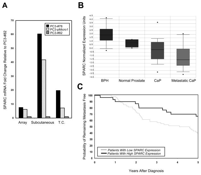
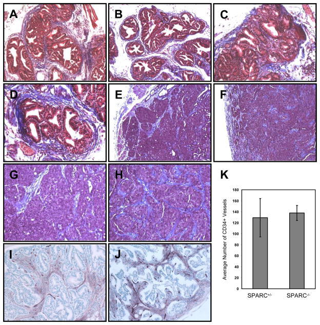
Secreted protein, acidic and rich in cysteine (SPARC, also known as osteonectin or BM-40) is a glycoprotein component of the extracellular matrix that has been reported to be involved with a variety of cellular processes. Although SPARC expression levels are frequently altered in a variety of tumor types, the exact implications of deregulated SPARC expression--whether it promotes, inhibits or has no effect on tumor progression--have remained unclear. Our recent gene expression analyses have shown that SPARC is significantly downregulated in highly metastatic human prostate cancer cells. To test the role of endogenous SPARC in tumorigenesis directly, we examined cancer progression and metastasis in SPARC(+/-) and SPARC(-/-) mice using two separate transgenic mouse tumor models: transgenic adenocarcinoma of the mouse prostate (TRAMP) and murine mammary tumor virus-polyoma middle T (MMTV-PyMT). Surprisingly, in both instances, we found that loss of SPARC had no significant effects on tumor initiation, progression or metastasis. Tumor angiogenesis and collagen deposition were also largely unaffected. Our results indicate that, although differential SPARC expression may be a useful marker of aggressive, metastasis-prone tumors, loss of SPARC is not sufficient either to promote or to inhibit cancer progression in two spontaneous mouse tumor models.


Figures

Figure 1
Figure 1. SPARC expression is downregulated in aggressive prostate and breast tumors
(A) SPARC expression levels, as assessed by gene expression analyses on tumors (“Array”), and by quantitative PCR on tumors (“Subcutaneous”) and on tissue culture cells (“Tissue Culture”), are shown for poorly metastatic PC3-#78 cells (black bars), moderately metastatic PC3-pMicro1 cells (dotted bars) and highly metastatic PC3-#82 cells (gray bars). Within each set of comparisons, the fold change in SPARC expression was expressed relative to SPARC expression in PC3-#82 cells, which was set to 1. (B) Analysis of data originally obtained by Dhanasekaran et al. [44], revealed that SPARC is highly expressed in benign prostate hyperplasia (BPH) and normal prostate tissue, and significantly downregulated in both prostate cancer (CaP) and metastatic CaP (p < 0.01 for CaP or metastatic CaP versus BPH or normal prostate tissue). (C) Analysis of data obtained by van’t Veer et al. [42], revealed that breast cancer patients with low SPARC expression exhibited increased likelihood of developing metastases within 5 years of diagnosis, relative to patients with higher SPARC expression (p = 0.026 by log rank test).
Figure 2
Figure 2. SPARC protein expression in spontaneous prostate and mammary tumors derived from SPARC−/− and SPARC+/− animals
Total lysates from TRAMP prostate and MMTV-PyMT mammary tumors were probed for SPARC using two different antibodies. Top and middle panels, SPARC is present as a ~45 kDa protein in SPARC+/−, but not SPARC−/−, tissues. The lower bands in the top panel for the TRAMP samples are non-specific bands detected by the ab14071 SPARC antibody but not by the AF942 SPARC antibody (middle). Bottom, GAPDH loading controls are shown for all samples.
Figure 3
Figure 3. Absence of SPARC does not affect prostate cancer progression or metastasis
(A) Average urogenital masses of TRAMP (left, p = 0.44) or TRAMP+ animals (right, p = 0.75) did not differ between genotypes. (B) The incidence of palpable prostate carcinomas and macroscopic para-aortic/lumbar lymph node metastases did not differ between the two cohorts. (C) Disease progression also did not differ between SPARC+/− and SPARC−/− tumors, as assessed by histological grading of the individual dorsolateral (DL) and ventral (VN) prostate lobes. (D) When an overall highest grade was assigned to each prostate sample (identical to the highest grade given to any constituent lobe), no significant differences were observed between genotypes in the distribution of grades awarded.
Figure 4
Figure 4. Absence of SPARC does not affect breast cancer initiation, progression or metastasis
(A) The time to tumor initiation for MMTV-PyMT tumors did not differ between genotypes. (B) Average total tumor burden, ~4 weeks after the initial date of tumor initiation, also did not vary between the two cohorts (p = 0.2). (C) When tumors were sub-divided by quadrant (UL, upper left; UR, upper right; LL, lower left; LR, lower right), average masses did not differ between SPARC+/− and SPARC−/− tumors. (D) The average number of lung metastases observed per animal was not significantly changed between genotypes (p = 0.17).
Figure 5
Figure 5. Collagen deposition and angiogenesis were largely unaffected by loss of SPARC
Masson’s trichrome staining was performed on TRAMP prostate cancer lesions (A–D) and on MMTV-PyMT mammary carcinomas (E–H). 10X magnification of images is shown in A, B, E and F; 20X magnification of images is shown in C, D, G and H. CD34 staining of blood vessels at 20X magnification is shown for prostate lesions from SPARC+/− (I) and SPARC−/− (J) animals, and the results are quantitated in (K).

Similar articles

Cited by

References

    1. Brekken RA, Sage EH. SPARC, a matricellular protein: at the crossroads of cell-matrix communication. Matrix Biol. 2001;19:816–827. - PubMed
    1. Framson PE, Sage EH. SPARC and tumor growth: where the seed meets the soil? J Cell Biochem. 2004;92:679–690. - PubMed
    1. Bradshaw AD, Sage EH. SPARC, a matricellular protein that functions in cellular differentiation and tissue response to injury. J Clin Invest. 2001;107:1049–1054. - PMC - PubMed
    1. Bradshaw AD, Reed MJ, Sage EH. SPARC-null mice exhibit accelerated cutaneous wound closure. J Histochem Cytochem. 2002;50:1–10. - PubMed
    1. Murphy-Ullrich JE. The de-adhesive activity of matricellular proteins: is intermediate cell adhesion an adaptive state? J Clin Invest. 2001;107:785–790. - PMC - PubMed

Publication types

MeSH terms