Review
. 2007 Sep;8(2-3):121-40.
doi: 10.1007/s10969-007-9036-1. Epub 2007 Dec 5.

Structural genomics of protein phosphatases

Affiliations
Review

Structural genomics of protein phosphatases

Steven C Almo et al. J Struct Funct Genomics. 2007 Sep.

Abstract

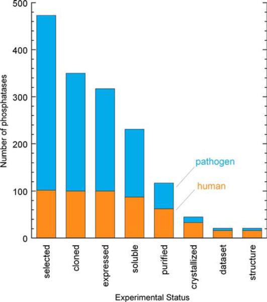
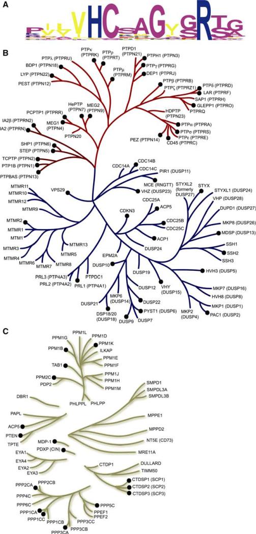
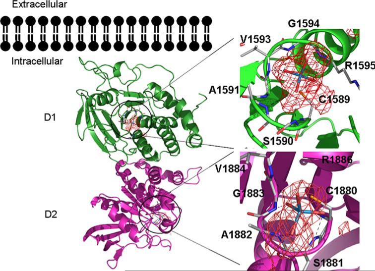
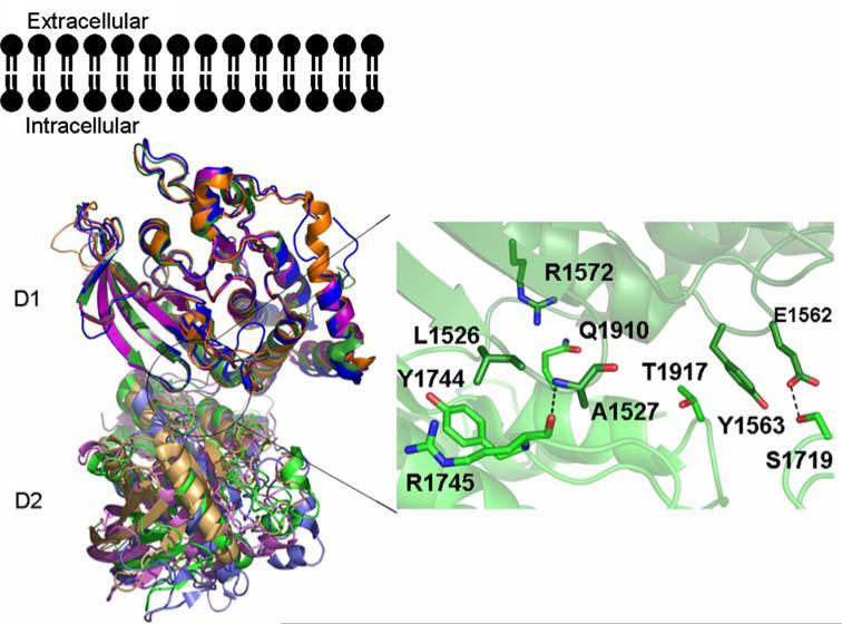
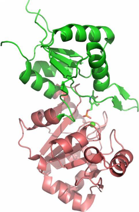
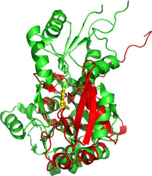
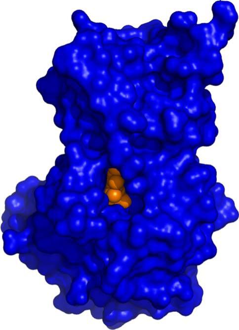
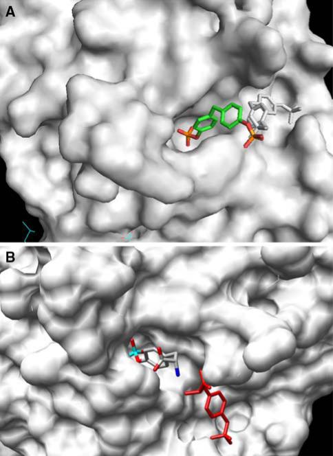
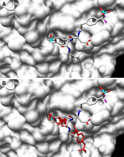
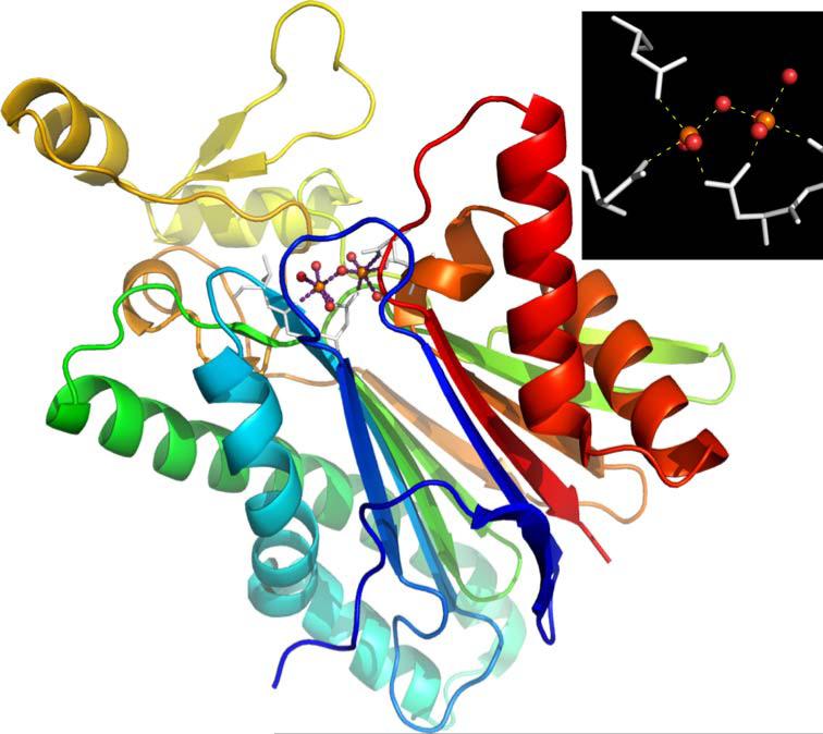
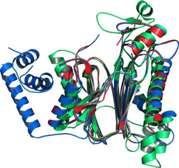
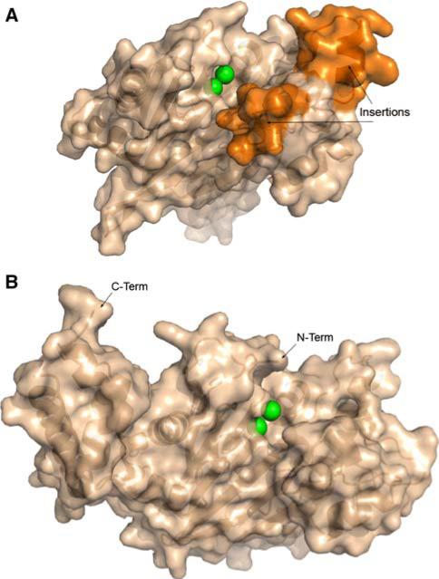
The New York SGX Research Center for Structural Genomics (NYSGXRC) of the NIGMS Protein Structure Initiative (PSI) has applied its high-throughput X-ray crystallographic structure determination platform to systematic studies of all human protein phosphatases and protein phosphatases from biomedically-relevant pathogens. To date, the NYSGXRC has determined structures of 21 distinct protein phosphatases: 14 from human, 2 from mouse, 2 from the pathogen Toxoplasma gondii, 1 from Trypanosoma brucei, the parasite responsible for African sleeping sickness, and 2 from the principal mosquito vector of malaria in Africa, Anopheles gambiae. These structures provide insights into both normal and pathophysiologic processes, including transcriptional regulation, regulation of major signaling pathways, neural development, and type 1 diabetes. In conjunction with the contributions of other international structural genomics consortia, these efforts promise to provide an unprecedented database and materials repository for structure-guided experimental and computational discovery of inhibitors for all classes of protein phosphatases.


Figures

Fig. 1
Fig. 1
Progress on protein phosphatase structural studies. Number of protein phosphatase targets at each experimental stage of the NYSGXRC structural genomics pipeline. Human phosphatases (or mammalian orthologs) are shown in orange and pathogen phosphatases are shown in blue
Fig. 2
Fig. 2
Human phosphatome phylogenetic tree. (a) Sequence logo [2] depicting the conservation of active site residues in the protein tyrosine and dual-specificity phosphatases. (b) Dendrogram of protein tyrosine (red branch) and dual-specificity (blue branch) phosphatases based on variation in the active site motif. (c) Dendrogram of all other human protein phosphatases based on alignment of the entire catalytic domain, including the metal dependent phosphatases (e.g. PPMs and PPPs) and the members of the haloacid dehalogenase (HAD) superfamily (e.g. CTDSPs, EYAs, MDP-1 and PDXP). Phosphatases with structures in the PDB are indicated by black circles
Fig. 3
Fig. 3
Structure of the human PTPσ tandem phosphatase domains. The structure of the PTPσ tandem phosphatase domains D1 and D2 is shown as a ribbon diagram with bound tungstate ions as stick and overlapping anomalous difference electron density in red. Domain D1 is shown in dark green and D2 in magenta. Interactions with the tungstate ion in the D1 and D2 active sites are magnified with hydrogen bonds represented as black dashes
Fig. 4
Fig. 4
Structural comparison of tandem phosphatase domains of RPTPs. Superposition of the structures of the tandem phosphatase domains of PTPσ (green), LAR (purple), CD45 (blue), and PTPγ (orange). Amino acids involved in interdomain interactions for PTPσ are shown to the right of the D1–D2 structures. For all structures the D1 and D2 domains are shown in dark and light shades of color, respectively
Fig. 5
Fig. 5
Comparison of the PTPσ D1 and D2 domain active sites. Superposition of the PTPσ D1 (green) and D2 (magenta) domain active sites. Active site residues and residues making up the WPD and KNRY loops are shown as stick figures. Root-mean-square deviation of D1/D2 superposition for 254 structurally equivalent Cα atoms is ~1.0Å
Fig. 6
Fig. 6
Comparison of the structures of IA-2 and PTP1B. Superposition of the structures of IA2 (green) and PTP1B (cyan), with active site residues shown as stick figures. Active site residues of IA2 and PTP1B bound to phosphotyrosine are magnified, highlighting differences responsible for the lack of catalytic activity of IA-2
Fig. 7
Fig. 7
Structure of SCP3. (a) Ribbon diagram of SCP3 showing the DXDX catalytic loop (yellow) and the catalytic Mg2+ ion modeled from SCP1 (magenta). (b) Atomic details of the SCP3 catalytic site, again with the Mg2+ ion modeled from SCP1
Fig. 8
Fig. 8
Structural comparisons of SCP3. (a) Superposition of SCP3 (green) with Methanococcus jannaschii phosphoserine phosphatase (red, PDB ID: 1F5S). The SCP3 catalytic site is freely accessible to solvent, whereas the alpha-helical capping domain in phosphoserine phosphatase shields its active site. (b) Superposition of SCP3 (green) with a dimer of the tetrameric Haemophilus influenzae deoxy-d-mannose-oculosonate 8-phosphatase (red and grey, PDB ID: 1K1E). Mg2+ ions are shown as pink spheres, and conserved phosphate-binding loops are shown in yellow. The capping domain of 1F5S occludes the active site entrance. In 1K1E, the second subunit of the dimer plays a similar role
Fig. 9
Fig. 9
Cofilin-mediated F-actin severing. F-actin severing activity of cofilin is regulated by a phosphorylation cycle involving the LIM kinase and the slingshot and chronophin phosphatases. Actin monomers are represented by blue ellipses
Fig. 10
Fig. 10
Chronophin structure. Cartoon representation of chronophin/ PLP phosphorylase with bound PLP and Ca2+. The core domain is colored raspberry, the capping domain green, PLP is shown with stick representation and the Ca2+ is shown as a green sphere. The catalytic site lies at the interface between the core and capping domains
Fig. 11
Fig. 11
Chronophin catalytic site. The active site of chronophin with its ligand PLP and inhibitory Ca2+. The Ca2+ (green sphere) is hepta-coordinated and participates in a bidentate interaction with the active site nucleophile Asp-25
Fig. 12
Fig. 12
Chronophin capping domain. Superposition of chronophin (green) and SCP (CTD phosphatase) (red; PDB ID: 2HHL), which lacks a capping domain. The core domains share 11% sequence identity and superimpose with a DALI Z-score of 6.6 and an RMSD of ~2.9Å for 116 structurally equivalent Cα atoms
Fig. 13
Fig. 13
Inaccessibility of PLP in the Chronophin Catalytic Site. Surface representation of chronophin (blue) with bound ligand PLP (orange) in the same orientation as shown in Fig. 12. The PLP is viewed on the edge and the phosphoryl group is completely buried from solvent
Fig. 14
Fig. 14
Compounds that Bind PTP1B. The low Km, low kcat substrate, compound 1, compound 2; and a highly-selective, nonhydrolyzable bidentate inhibitor, compound 3
Fig. 15
Fig. 15
Surface representation of the PTP1B active site. (a) Compound 1 binds to a catalytically incompetent form of PTP1B via one of two mutually incompatible binding modes, which encompass either the active site (multicolor) or a secondary peripheral site (black and white). (b) Phosphotyrosine simultaneously binds to the active site (multicolor) and a secondary peripheral site (red)
Fig. 16
Fig. 16
Structure of the PTP1B/Compound 3 Complex. (a) The bidentate ligand, compound 3, is bound to the active site and a secondary peripheral site different from that observed with compound 1 and phosphotyrosine. (b) Overlay of the double binding mode of phosphotyrosine (red) and the bidentate ligand compound 3 (multicolor)
Fig. 17
Fig. 17
X-ray structure of T. gondii PP2C (PDB ID: 2i44). Inset: two calcium ions supported by conserved aspartate residues and coordinated waters
Fig. 18
Fig. 18
Structural alignment and overlay of PP2Cs. PP2Ctg (green), PP2Cα (blue), and PP2Cβ (red)
Fig. 19
Fig. 19
Surface representations of PP2Cs. (a) PP2Ctg: Ca2+ shown as green spheres; surface corresponding to amino acid insertions 207– 213 and 218–232 colored orange. (b) PP2Cα in similar orientation as PP2Ctg: Mn2+ shown as green spheres

Similar articles

Cited by

References

    1. Tonks NK. Nat Rev Mol Cell Biol. 2006;7(11):833–846. - PubMed
    1. Schneider TD, Stephens RM. Nucl Acids Res. 1990;18:6097–6100. - PMC - PubMed
    1. McCluskey A, Sim AT, Sakoff JA. J Med Chem. 2002;45(6):1151–1175. - PubMed
    1. Peisach E, Selengut JD, Dunaway-Mariano D, Allen KN. Biochemistry. 2004;43(40):12770–12779. - PubMed
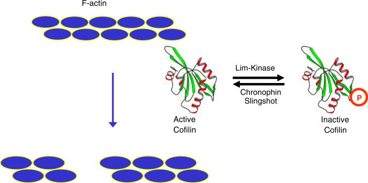
    1. Wiggan O, Bernstein BW, Bamburg JR. Nat Cell Biol. 2005;7(1):8–9. - PubMed

Publication types

Substances