Comparative Study
. 2008 May 15;71(3):1360-72.
doi: 10.1002/prot.21824.

Effect of oxidatively damaged DNA on the active site preorganization during nucleotide incorporation in a high fidelity polymerase from Bacillus stearothermophilus

Affiliations
Comparative Study

Effect of oxidatively damaged DNA on the active site preorganization during nucleotide incorporation in a high fidelity polymerase from Bacillus stearothermophilus

Ravindra Venkatramani et al. Proteins. .

Abstract

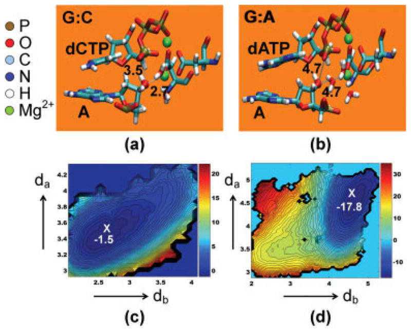
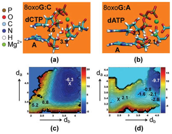
We study the effect of the oxidative lesion 8-oxoguanine (8oxoG) on the preorganization of the active site for DNA replication in the closed (active) state of the Bacillus fragment (BF), a Klenow analog from Bacillus stearothermophilus. Our molecular dynamics and free energy simulations of explicitly solvated model ternary complexes of BF bound to correct dCTP/incorrect dATP opposite guanine (G) and 8oxoG bases in DNA suggest that the lesion introduces structural and energetic changes at the catalytic site to favor dATP insertion. Despite the formation of a stable Watson-Crick pairing in the 8oxoG:dCTP system, the catalytic geometry is severely distorted to possibly slow down catalysis. Indeed, our calculated free energy landscapes associated with active site preorganization suggest additional barriers to assemble an efficient catalytic site, which need to be overcome during dCTP incorporation opposite 8oxoG relative to that opposite undamaged G. In contrast, the catalytic geometry for the Hoogsteen pairing in the 8oxoG:dATP system is highly organized and poised for efficient nucleotide incorporation via the "two-metal-ion" catalyzed phosphoryl transfer mechanism. However, the free energy calculations suggest that the catalytic geometry during dATP incorporation opposite 8oxoG is considerably less plastic than that during dCTP incorporation opposite G despite a very similar, well organized catalytic site for both systems. A correlation analysis of the dynamics trajectories suggests the presence of significant coupling between motions of the polymerase fingers and the primary distance for nucleophilic attack (i.e., between the terminal primer O3' and the dNTP P(alpha.) atoms) during correct dCTP incorporation opposite undamaged G. This coupling is shown to be disrupted during nucleotide incorporation by the polymerase with oxidatively damaged DNA/dNTP substrates. We also suggest that the lesion affects DNA interactions with key polymerase residues, thereby affecting the enzymes ability to discriminate against non-complementary DNA/dNTP substrates. Taken together, our results provide a unified structural, energetic, and dynamic platform to rationalize experimentally observed relative nucleotide incorporation rates for correct dCTP/incorrect dATP insertion opposite an undamaged/oxidatively damaged template G by BF.


Figures

Figure 1
Figure 1
(a) The active site region for the BF-DNA-dNTP ternary complex used in our correlation analysis. The circled region depicts the catalytic site. (b) The fully solvated and neutralized ternary complex used in our MD simulations. The circled region depicts the location of the active site fragment. (c) The catalytic site, depicting the key components required for a two-metal-ion assisted phosphoryl transfer reaction.
Figure 2
Figure 2
Average values (solid-lines) and standard deviations of metal-ligand and nucleophilic attack distances for the four model systems (G:C, G:A, 8oxoG:C and 8oxoG:A) obtained from 5 ns classical simulations. Also shown are values of metal ligand distances expected for an ideal two-metal-ion geometry (solid bars) taken from Ref. .
Figure 3
Figure 3
The average catalytic site geometry obtained from 5 ns classical simulations for the G:C (a) and G:A (b) systems. The primary reactive distances for catalysis, O3′-Pα (da) and O3′-MG2 (db) are shown. Corresponding free energy landscapes are shown in Figures (c) and (d), respectively with distances da and db given in Å. The location of the average simulation geometry is marked by X in these plots. The scale for energies (shown on the right for each free energy plot) is in units of kBT ~ 0.6 kcal/mol (for a T value of 300 K). The errors in free energies are estimated to be approximately ± 0.9 kBT (0.54 kcal/mol). [Color figure can be viewed in the online issue, which is available at www.interscience.wiley.com.]
Figure 4
Figure 4
Values of various metal–ligand distances for the four model systems (G:C, G:A, 8oxoG:C and 8oxoG:A) obtained during our umbrella sampling simulations. The nucleophilic attack distance dNTP:PαA:O3′ and the MG2—A:O3′ distance were constrained to a range of values between 5.0 and 2.0 Å (see Methods). For all systems except the control G:C (which shows two water molecules at the catalytic site) the catalytic Mg2+ is liganded to three water molecules for structures where da and db values are large and far from those for an ideal two-metal-ion geometry (from Ref. 58) represented by the solid bars. The extra water is expelled as da and db are constrained to approach their ideal two-metal-ion values.
Figure 5
Figure 5
The average catalytic site geometry obtained from 5 ns classical simulations for the 8oxoG:C (a) and 8oxoG:A (b) systems. The primary reactive distances for catalysis, O3′-Pα (da) and O3′-MG2 (db) are shown. Corresponding free energy landscapes are shown in Figures (c) and (d), respectively with distances da and db given in Å. The location of the average simulation geometry is marked by X in these plots. The scale for energies (shown on the right for each free energy plot) is in units of kBT ~0.6 kcal/mol (for a T value of 300 K). The errors in free energies are estimated to be approximately ± 0.9 kBT (0.54 kcal/mol). [Color figure can be viewed in the online issue, which is available at www.interscience.wiley.com.]

Similar articles

Cited by

References

    1. Kunkel TA. DNA replication fidelity. J Biol Chem. 2004;279:16895–16898. - PubMed
    1. Lindahl T. Instability and decay of the primary structure of DNA. Nature. 1993;362:709–715. - PubMed
    1. Friedberg EC. DNA damage and repair. Nature. 2003;421:436–439. - PubMed
    1. Hollstein M, Shomer B, Greenblatt M, Soussi T, Hovig E, Montesano R, Harris CC. Somatic point mutations in the p53 gene of human tumors and cell lines: updated compilation. Nucleic Acids Res. 1996;24:141–146. - PMC - PubMed
    1. Brieba LG, Eichman BF, Kokoska RJ, Doublie S, Kunkel TA, Ellenberger T. Structural basis for the dual coding potential of 8-oxoguanosine by a high-fidelity DNA polymerase. Embo J. 2004;23:3452–3461. - PMC - PubMed

Publication types

MeSH terms

LinkOut - more resources