

Expression of ERK signaling inhibitors Dusp6, Dusp7, and Dusp9 during mouse ear development
- PMID: 18058922
- PMCID: PMC2377012
- DOI: 10.1002/dvdy.21380
Expression of ERK signaling inhibitors Dusp6, Dusp7, and Dusp9 during mouse ear development
Abstract
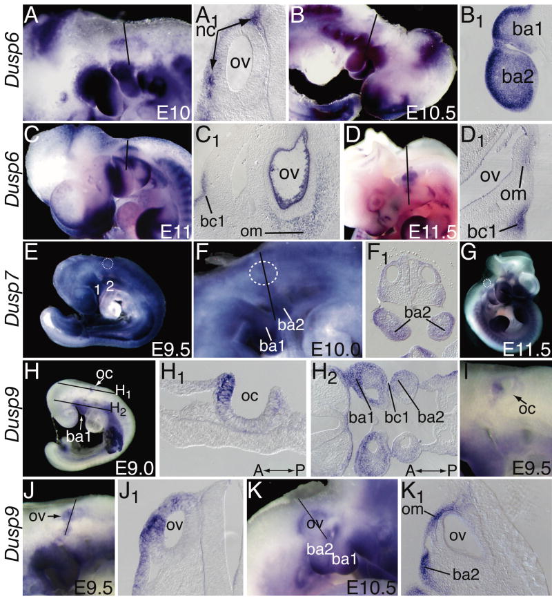
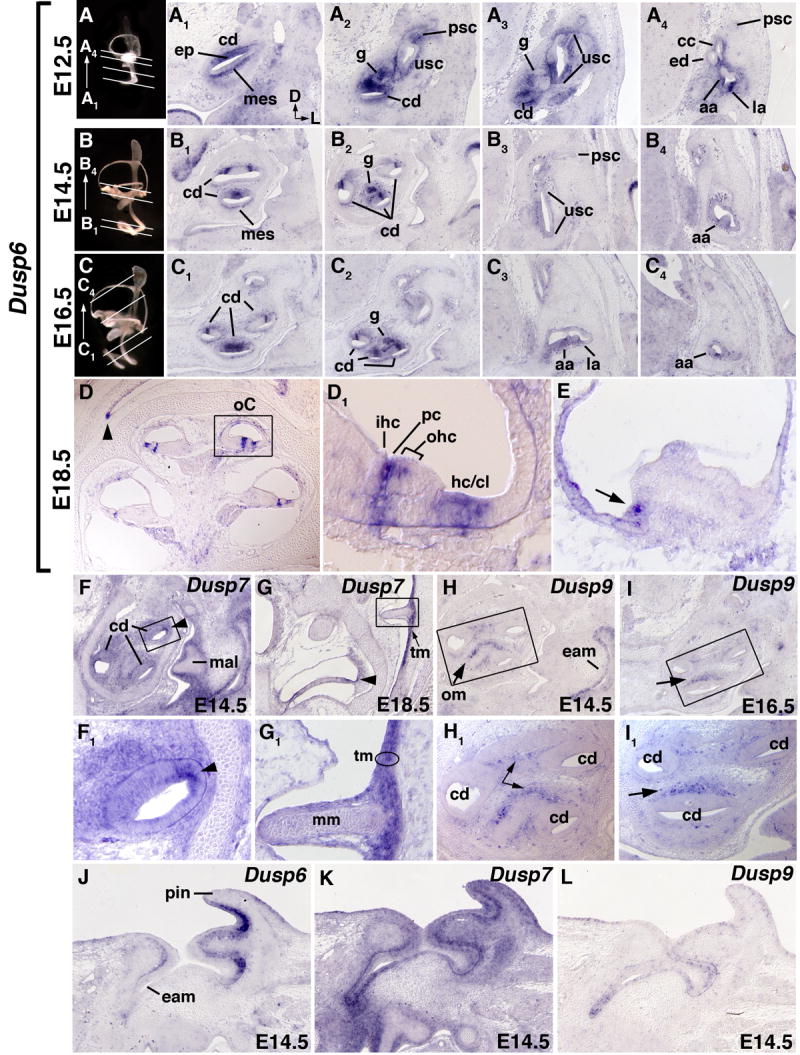
The levels of fibroblast growth factor (FGF) signaling play important roles in coordinating development of the mouse inner, middle, and outer ears. Extracellular signal-regulated kinases (ERKs) are among the effectors that transduce the FGF signal to the nucleus and other cellular compartments. Attenuation of ERK activity by dephosphorylation is necessary to modulate the magnitude and duration of the FGF signal. Recently, we showed that inactivation of the ERK phosphatase, dual specificity phosphatase 6 (DUSP6), causes partially penetrant postnatal lethality, hearing loss and skeletal malformations. To determine whether other Dusps may function redundantly with Dusp6 during otic development, we surveyed the expression domains of the three ERK-specific DUSP transcripts, Dusp6, Dusp7, and Dusp9, in the embryonic mouse ear. We show that each is expressed in partially overlapping patterns that correspond to regions of active FGF signaling, suggesting combinatorial roles in negative regulation of this pathway during ear development.
Figures
Similar articles
-
Dusp6 (Mkp3) is a negative feedback regulator of FGF-stimulated ERK signaling during mouse development.Development. 2007 Jan;134(1):167-76. doi: 10.1242/dev.02701. Development. 2007. PMID: 17164422 Free PMC article.
-
Differential expression profiles and roles of inducible DUSPs and ERK1/2-specific constitutive DUSP6 and DUSP7 in microglia.Biochem Biophys Res Commun. 2015 Nov 13;467(2):254-60. doi: 10.1016/j.bbrc.2015.09.180. Epub 2015 Oct 3. Biochem Biophys Res Commun. 2015. PMID: 26435497
-
DUSP5 and DUSP6, two ERK specific phosphatases, are markers of a higher MAPK signaling activation in BRAF mutated thyroid cancers.PLoS One. 2017 Sep 14;12(9):e0184861. doi: 10.1371/journal.pone.0184861. eCollection 2017. PLoS One. 2017. PMID: 28910386 Free PMC article.
-
Regulation of Dual-Specificity Phosphatase (DUSP) Ubiquitination and Protein Stability.Int J Mol Sci. 2019 May 30;20(11):2668. doi: 10.3390/ijms20112668. Int J Mol Sci. 2019. PMID: 31151270 Free PMC article. Review.
-
FGF signaling in ear development and innervation.Curr Top Dev Biol. 2003;57:225-59. doi: 10.1016/s0070-2153(03)57008-9. Curr Top Dev Biol. 2003. PMID: 14674483 Review. No abstract available.
Cited by
-
Anthocyanins from Hibiscus syriacus L. Inhibit Melanogenesis by Activating the ERK Signaling Pathway.Biomolecules. 2019 Oct 24;9(11):645. doi: 10.3390/biom9110645. Biomolecules. 2019. PMID: 31653006 Free PMC article.
-
Characterization of the Transcriptomes of Lgr5+ Hair Cell Progenitors and Lgr5- Supporting Cells in the Mouse Cochlea.Front Mol Neurosci. 2017 Apr 26;10:122. doi: 10.3389/fnmol.2017.00122. eCollection 2017. Front Mol Neurosci. 2017. PMID: 28491023 Free PMC article.
-
Retinoid signaling in inner ear development: A "Goldilocks" phenomenon.Am J Med Genet A. 2010 Dec;152A(12):2947-61. doi: 10.1002/ajmg.a.33670. Am J Med Genet A. 2010. PMID: 21108385 Free PMC article. Review.
-
Genetic rescue of Muenke syndrome model hearing loss reveals prolonged FGF-dependent plasticity in cochlear supporting cell fates.Genes Dev. 2013 Nov 1;27(21):2320-31. doi: 10.1101/gad.228957.113. Epub 2013 Oct 21. Genes Dev. 2013. PMID: 24145799 Free PMC article.
-
The role of foxi family transcription factors in the development of the ear and jaw.Curr Top Dev Biol. 2015;111:461-95. doi: 10.1016/bs.ctdb.2014.11.014. Epub 2015 Jan 21. Curr Top Dev Biol. 2015. PMID: 25662269 Free PMC article. Review.
References
-
- Abu-Issa R, Smyth G, Smoak I, Yamamura K, Meyers EN. Fgf8 is required for pharyngeal arch and cardiovascular development in the mouse. Development. 2002;129:4613–4625. - PubMed
-
- Alonso A, Sasin J, Bottini N, Friedberg I, Friedberg I, Osterman A, Godzik A, Hunter T, Dixon J, Mustelin T. Protein tyrosine phosphatases in the human genome. Cell. 2004;117:699–711. - PubMed
-
- Alvarez Y, Alonso MT, Vendrell V, Zelarayan LC, Chamero P, Theil T, Bosl MR, Kato S, Maconochie M, Riethmacher D, Schimmang T. Requirements for FGF3 and FGF10 during inner ear formation. Development. 2003;130:6329–6338. - PubMed
-
- Barald KF, Kelley MW. From placode to polarization: new tunes in inner ear development. Development. 2004;131:4119–4130. - PubMed
-
- Boulet AM, Moon AM, Arenkiel BR, Capecchi MR. The roles of Fgf4 and Fgf8 in limb bud initiation and outgrowth. Dev Biol. 2004;273:361–372. - PubMed
Publication types
MeSH terms
Substances
Grants and funding
LinkOut - more resources
Full Text Sources
Other Literature Sources
Molecular Biology Databases
Miscellaneous
