. 2008 May 1;86(6):1267-76.
doi: 10.1002/jnr.21584.

Postnatal developmental regulation of Bcl-2 family proteins in brain mitochondria

Affiliations

Postnatal developmental regulation of Bcl-2 family proteins in brain mitochondria

Lucian Soane et al. J Neurosci Res. .

Abstract

Although it has been long recognized that the relative balance of pro- and antiapoptotic Bcl-2 proteins is critical in determining the susceptibility to apoptotic death, only a few studies have examined the level of these proteins specifically at mitochondria during postnatal brain development. In this study, we examined the age-dependent regulation of Bcl-2 family proteins using rat brain mitochondria isolated at various postnatal ages and from the adult. The results indicate that a general down-regulation of most of the proapoptotic Bcl-2 proteins present in mitochondria occurs during postnatal brain development. The multidomain proapoptotic Bax, Bak, and Bok are all expressed at high levels in mitochondria early postnatally but decline in the adult. Multiple BH3-only proteins, including direct activators (Bid, Bim, and Puma) and the derepressor BH3-only protein Bad, are also present in immature brain mitochondria and are down-regulated in the adult brain. Antiapoptotic Bcl-2 family members are differentially regulated, with a shift from high Bcl-2 expression in immature mitochondria to predominant Bcl-x(L) expression in the adult. These results support the concept that developmental differences in upstream regulators of the mitochondrial apoptotic pathway are responsible for the increased susceptibility of cells in the immature brain to apoptosis following injury.


Figures

Fig. 1
Fig. 1
Expression of mitochondrial OXPHOS complexes and AIF during postnatal brain development. A: Expression of several subunits of the mitochondrial OXPHOS complexes I–V was examined in isolated rat brain mitochondria at the indicated postnatal day (PND) and in adult brain mitochondria using the OXPHOS Western blotting kit (Mitosciences). The expression of VDAC was examined on the same membranes to control for equal loading of mitochondrial protein. A representative of three different immunoblots is shown. B: Developmental regulation of AIF was examined as described above at the indicated ages in isolated mitochondria. A representative of four different immunoblots is shown. C: Densitometric analysis of the level of subunits of OXPHOS complexes II–V (n = 3), VDAC (n = 6), and AIF (n = 4). Data are expressed as mean ± SEM. *P < 0.05 vs. P3, **P < 0.05 vs. P7.
Fig. 2
Fig. 2
Expression of antiapoptotic Bcl-2 and Bcl-xL in mitochondria during postnatal brain development. A: Level of antiapoptotic Bcl-2 in rat brain mitochondria was examined by immunoblotting using equal amounts of mitochondria (50 μg/lane) isolated from rat brain at various ages postnatal (P3–P31) and from the adult rat brain. VDAC immunoblotting was performed on the same membranes. The relative levels of Bcl-2 were normalized to VDAC, and the results are expressed as percentage of the P3 level. Data are expressed as mean ± SEM (n = 5–6). *P < 0.05 vs. P3. **P < 0.05 vs. P7. B: Expression of Bcl-xL in mitochondria was examined similarly by immunoblotting, using VDAC as control (n = 4). The relative Bcl-xL level after normalization to VDAC was expressed as percentage of the P3 level.
Fig. 3
Fig. 3
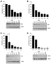
Developmental regulation of multidomain proapoptotic Bcl-2 proteins in brain mitochondria. A: Expression of proapoptotic Bax was examined by immunoblotting at the indicated ages in isolated brain mitochondria. The relative levels of Bax are expressed as percentage of the P3 level. Data are expressed as mean ± SEM (n = 6–7). *P ≤ 0.002 vs. P3 and P7, **P < 0.05 vs. P14. B: Immunoblot analysis of Bak expression in mitochondria during postnatal development and scan densitometric analysis of the relative Bak/VDAC levels; n = 4–5. *P < 0.05 vs. P3, P7, P14, P21, and P31. C: Expression of the multidomain proapoptotic protein Bok in mitochondria during postnatal development. A representative immunoblot is shown, and the relative Bok/VDAC levels during postnatal brain development are expressed as percentage of the P3 level (n = 4). *P < 0.005 vs. P3, P < 0.05 vs. P7.
Fig. 4
Fig. 4
Developmental expression of mitochondrial BH3-only proteins during postnatal brain development. The presence of several BH3-only proapoptotic proteins in isolated brain mitochondria was analyzed at the indicated age by immunoblotting using VDAC as control. Representative immunoblots demonstrating that significant levels of Bad, Bim, Bid, and Puma are present in the immature brain mitochondria. The relative level of the proteins is also expressed as percentage of the P3 value. Data are expressed as mean ± SEM (n = 4). A: *P < 0.01 vs. P3, P < 0.05 vs. P7. B: *P < 0.001 vs. P3, **P < 0.01 vs. P3. C: *P = < 0.001 vs. P3, **P < 0.001 vs. P3, P < 0.005 vs. P7. D: *P < 0.001 vs. P3 and P7.
Fig. 5
Fig. 5
Relative mitochondrial vs. total cellular level of proapoptotic Bcl-2 family proteins during postnatal brain development. A: Protein fraction present in mitochondria (mito) from the total cellular level (brain) was examined in immature (P7) and adult rat brain mitochondria for several proapoptotic Bcl-2 family proteins by loading lysates of isolated mitochondria and total brain homogenates on the same gels and immunoblotting with the indicated antibodies for multidomain (Bax and Bak) and BH3-only proteins (Bad, Bid, Bim). The F1α subunit of the F1Fo ATPase localized predominantly in mitochondria was used to verify protein loading and to normalize the relative levels of proapoptotic proteins. B: Results of quantification analysis following normalization to F1α were expressed as ratio of mitochondria to total protein levels present in brain homogenates for each of the proteins in A at P7 and in the adult. Data are expressed as mean ± SEM (n = 4). *P < 0.05 vs. adult.

References

    1. Adams JM, Cory S. The Bcl-2 protein family: arbiters of cell survival. Science. 1998;281:1322–1326. - PubMed
    1. Becker EB, Bonni A. Pin1 mediates neural-specific activation of the mitochondrial apoptotic machinery. Neuron. 2006;49:655–662. - PubMed
    1. Bittigau P, Sifringer M, Pohl D, Stadthaus D, Ishimaru M, Shimizu H, Ikeda M, Lang D, Speer A, Olney JW, Ikonomidou C. Apoptotic neurodegeneration following trauma is markedly enhanced in the immature brain. Ann Neurol. 1999;45:724–735. - PubMed
    1. Blomgren K, Hagberg H. Free radicals, mitochondria, and hypoxia-ischemia in the developing brain. Free Rad Biol Med. 2006;40:388–397. - PubMed
    1. Chen L, Willis SN, Wei A, Smith BJ, Fletcher JI, Hinds MG, Colman PM, Day CL, Adams JM, Huang DC. Differential targeting of prosurvival Bcl-2 proteins by their BH3-only ligands allows complementary apoptotic function. Mol Cell. 2005;17:393–403. - PubMed

Publication types

Substances