. 2007 Dec;25(12):1483-7.
doi: 10.1038/nbt1355. Epub 2007 Dec 2.

Redirecting lipoic acid ligase for cell surface protein labeling with small-molecule probes

Affiliations

Redirecting lipoic acid ligase for cell surface protein labeling with small-molecule probes

Marta Fernández-Suárez et al. Nat Biotechnol. 2007 Dec.

Abstract

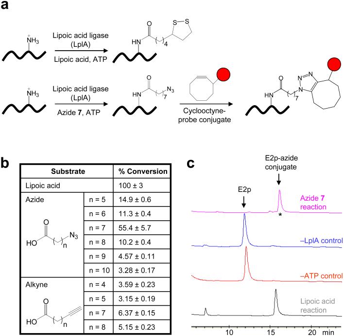
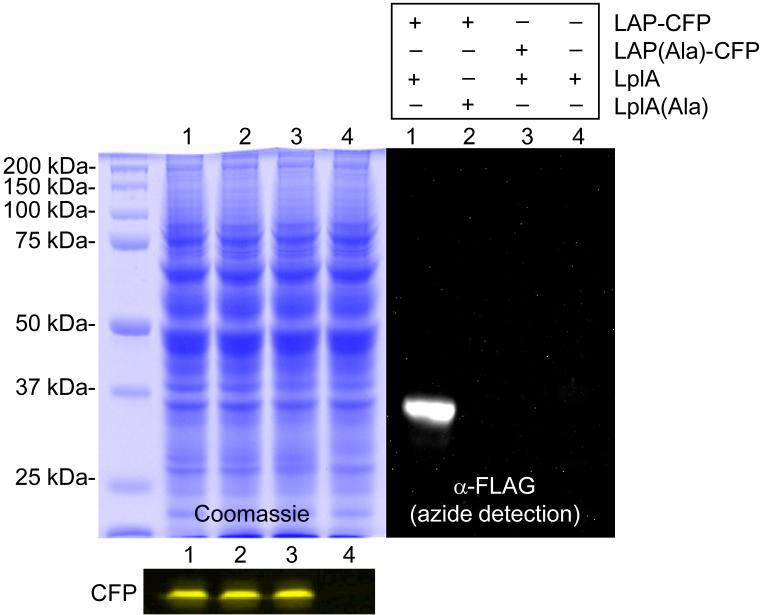
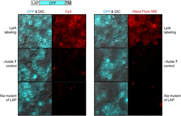
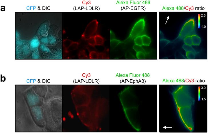
Live cell imaging is a powerful method to study protein dynamics at the cell surface, but conventional imaging probes are bulky, or interfere with protein function, or dissociate from proteins after internalization. Here, we report technology for covalent, specific tagging of cellular proteins with chemical probes. Through rational design, we redirected a microbial lipoic acid ligase (LplA) to specifically attach an alkyl azide onto an engineered LplA acceptor peptide (LAP). The alkyl azide was then selectively derivatized with cyclo-octyne conjugates to various probes. We labeled LAP fusion proteins expressed in living mammalian cells with Cy3, Alexa Fluor 568 and biotin. We also combined LplA labeling with our previous biotin ligase labeling, to simultaneously image the dynamics of two different receptors, coexpressed in the same cell. Our methodology should provide general access to biochemical and imaging studies of cell surface proteins, using small fluorophores introduced via a short peptide tag.


Figures

Figure 1
Figure 1. Re-directing LplA for site-specific protein labeling with fluorescent probes
(a) Natural reaction catalyzed by LplA (top), and scheme for LplA-catalyzed fluorescent tagging in cells (bottom). Instead of lipoic acid, LplA ligates an alkyl azide to a lysine sidechain within a peptide recognition sequence. The azide is then selectively functionalized with a cyclooctyne-probe conjugate (red circle), to give a triazole adduct. (b) Comparison of alkyl azide and alkyne substrates of LplA. Conversions are given relative to lipoic acid, which is normalized to 100%. (c) HPLC assay showing the ligation of the azide 7 substrate to E2p protein. The starred peak was analyzed by mass-spectrometry in Supplementary Figure 2 online.
Figure 2
Figure 2. LplA labels the LAP peptide without modifying endogenous mammalian proteins
Lysates from HEK cells expressing a LAP fusion to CFP were labeled in vitro with LplA and azide 7. The azide was derivatized with phosphine-FLAG via the Staudinger ligation, and the FLAG epitope was detected by blotting with an anti-FLAG antibody. Controls are shown with LAP-CFP replaced by its alanine point mutant (lane 3), or with LplA replaced by its catalytically inactive Lys133Ala mutant (lane 2). Coomassie staining demonstrates equal loading in all lanes. Fluorescence visualization of CFP demonstrates equal expression levels of the LAP fusion in lanes 1-3.
Figure 3
Figure 3. Site-specific labeling of LAP fusion proteins with fluorophores
A LAP-CFP fusion was targeted to the cell surface using a transmembrane (TM) domain. Cell-surface LAP was first labeled with azide 7 by LplA, and the introduced azide was then labeled with a cyclooctyne probe conjugated to Cy3 (left) or Alexa Fluor 568 (right). Live cell images of the introduced fluorophores are shown to the right of the merged CFP and DIC images, which highlight the transfected cells. Negative controls with azide 7 omitted from the labeling reaction, or with the LAP-CFP-TM replaced by its alanine point mutant are shown.
Figure 4
Figure 4. Simultaneous labeling and imaging of two receptors in polarized cells in a wound healing assay
HEK cells co-expressing a LAP-LDLR fusion and either AP-EGFR (a) or AP-EphA3 (b) were labeled during wound healing by first treating with LplA, BirA, azide 7, and biotin, followed by OCT-Cy3 to derivatize the azide, followed by monovalent streptavidin-Alexa Fluor 488 to detect the biotin. The Cy3 images show the non-polarized distribution of surface LAP-LDLR. The Alexa Fluor 488 images show the polarized distribution of AP-EGFR (a) and AP-EphA3 (b) at the wound edge. CFP is a transfection marker. The images on the far right depict the intensity ratios of Alexa Fluor 488 and Cy3. The white arrows point toward the wound.

References

    1. Debant A, Ponzio G, Clauser E, Contreres JO, Rossi B. Receptor cross-linking restores an insulin metabolic effect altered by mutation on tyrosine 1162 and tyrosine 1163. Biochemistry. 1989;28:14–17. - PubMed
    1. Weiss A, Littman DR. Signal transduction by lymphocyte antigen receptors. Cell. 1994;76:263–274. - PubMed
    1. Anderson RG, Brown MS, Beisiegel U, Goldstein JL. Surface distribution and recycling of the low density lipoprotein receptor as visualized with antireceptor antibodies. J. Cell Biol. 1982;93:523–531. - PMC - PubMed
    1. Barak LS, Webb WW. Fluorescent low density lipoprotein for observation of dynamics of individual receptor complexes on cultured human fibroblasts. J. Cell Biol. 1981;90:595–604. - PMC - PubMed
    1. Green DE, Morris TW, Green J, Cronan JE, Jr., Guest JR. Purification and properties of the lipoate protein ligase of Escherichia coli. Biochem. J. 1995;309:853–862. - PMC - PubMed

Publication types