. 2008 Jan;9(1):89-96.
doi: 10.1038/ni1550. Epub 2007 Dec 2.

Defective mast cell effector functions in mice lacking the CRACM1 pore subunit of store-operated calcium release-activated calcium channels

Affiliations

Defective mast cell effector functions in mice lacking the CRACM1 pore subunit of store-operated calcium release-activated calcium channels

Monika Vig et al. Nat Immunol. 2008 Jan.

Abstract

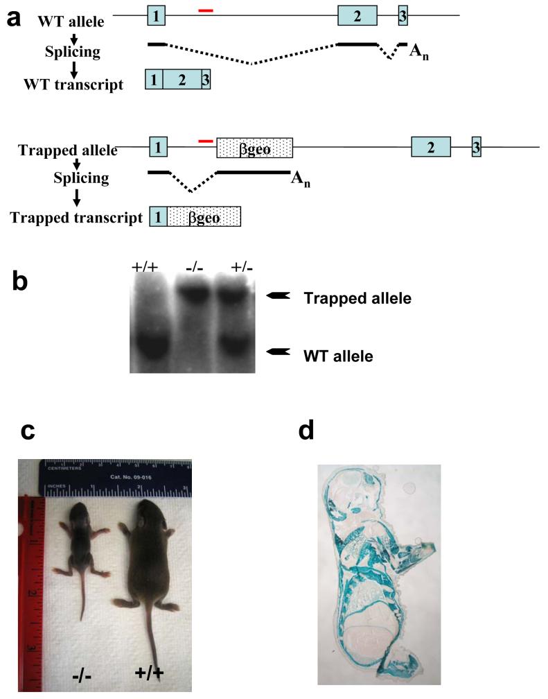
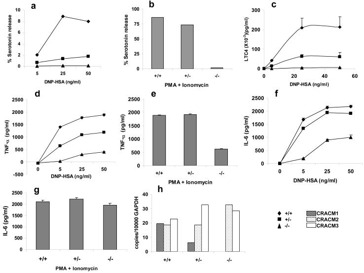
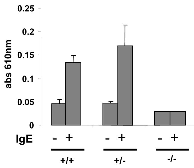
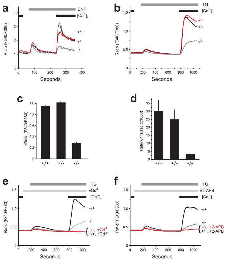
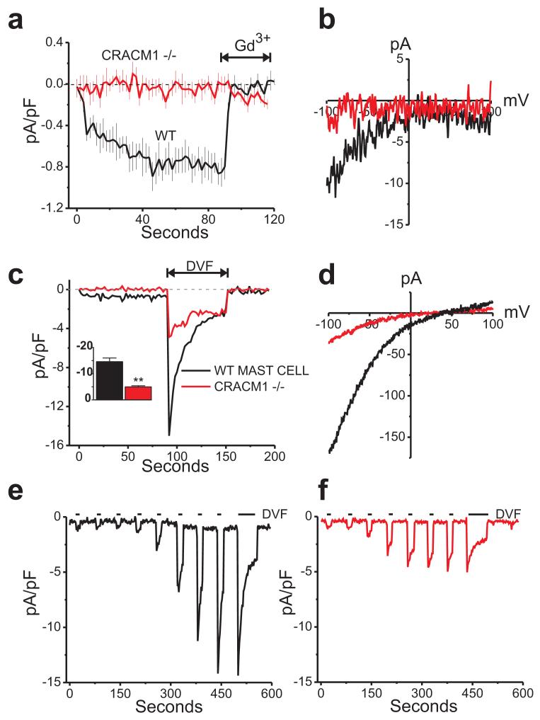
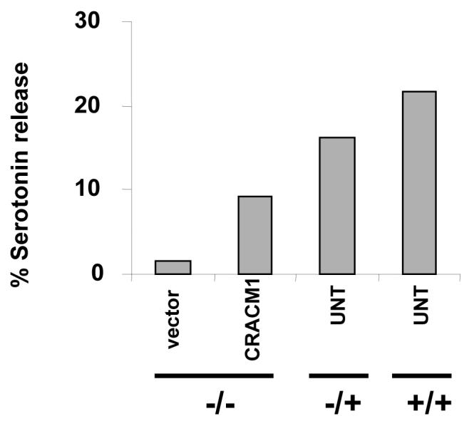
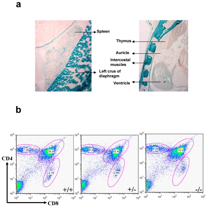
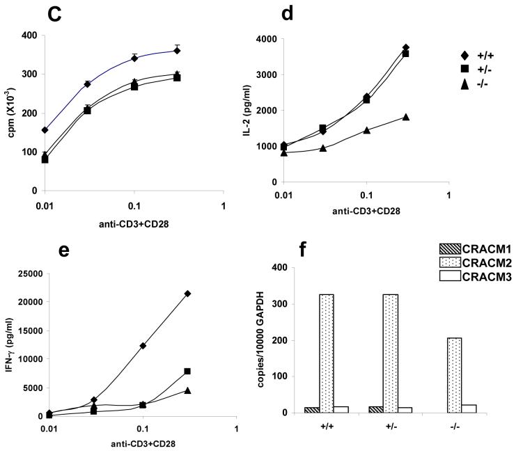
CRACM1 (also called Orai1) constitutes the pore subunit of store-operated calcium release-activated calcium channels. A point mutation in the gene encoding CRACM1 is associated with severe combined immunodeficiency disease in humans. Here we generated CRACM1-deficient mice in which beta-galactosidase activity 'reported' CRACM1 expression. CRACM1-deficient mice were smaller in size. Mast cells derived from CRACM1-deficient mice showed grossly defective degranulation and cytokine secretion, and the allergic reactions elicited in vivo were inhibited in CRACM1-deficient mice. We detected robust CRACM1 expression in skeletal muscles and some regions of the brain, heart and kidney but not in the lymphoid regions of thymus and spleen. In contrast, we found CRACM2 expression to be much higher in mouse T cells. In agreement with those findings, the store-operated calcium influx and development and proliferation of CRACM1-deficient T cells was unaffected. Thus, CRACM1 is crucial in mouse mast cell effector function, but mouse T cell calcium release-activated calcium channels are functional in the absence of CRACM1.


Figures

Figure 1
Figure 1. Generation of CRACM1 Mutant Mice Using Gene Trap
(a) Schematic representation of trapped CRACM1 gene in the ES cells. Blue boxes represent exons. Dotted box labelled ‘βgeo’ shows the site of gene trap vector insertion. The trapped transcript would result in the generation of truncated CRACM1-LacZ fusion protein. (b) Southern blot showing screening of CRACM1-/- mice using a gene specific probe. Red line depicts the site of probe hybridization. The size of WT allele is 11kb and that of trapped allele is 19.5kb. (c) Picture showing 10 day old CRACM1-/- mouse (left) with a WT littermate (right). (d) LacZ staining to detect the tissue distribution of CRACM1 protein. Neonatal (1 Day old) CRACM1-/- pups were cryo-sectioned and stained O/N with X-Gal.
Figure 2
Figure 2. CRACM1 deletion in mast cells strongly suppresses degranulation and LTC4 synthesis but affects cytokine secretion differentially
Serotonin release from CRACM1 +/+ (diamonds), +/- (squares) and -/- (triangles) mast cells loaded with anti- DNP IgE (0.5μg/ml) and stimulated either with 50, 25, 5 and 0 ng/ml of DNP-HSA to induce FcεRI cross-linking (a) or PMA (1.5μg/ml) plus Ionomycin (2μM) (b) for 15min. Results are expressed as percentage of total serotonin uptake (values represent pooled triplicates from 1 experiment, representative of n=3). (c) LTC4 secretion induced by FcεRI aggregation as in panel a (results are mean ± SEM of triplicates, from 1 experiment, representative of n=2). TNF (d, e) and IL-6 (f, g) secretion by cells loaded with anti-DNP IgE (0.5μg/ml) and stimulated either with 50, 25, 5 and 0 ng/ml of DNP-HSA (d, f) or PMA (1.5μg/ml) plus Ionomycin (2μM) (e, g) for 6 hrs. (h) Real time PCR showing CRACM1, CRACM2 and CRACM3 mRNA levels in mast cells derived from CRACM1 +/+ (hashed bars), +/- (dotted bars) and -/- (open bars) littermates.
Figure 3
Figure 3. CRACM1 deletion suppresses passive cutaneous anaphylaxis (PCA) reaction in mice
Mice were sensitized intra-dermally with 25 ng anti-DNP IgE in the left ears and with saline in the right ears. 24 h later PCA was induced by i.v. injection of DNP-HSA along with 2% Evan’s Blue dye. 60 min post injection, mice were sacrificed and ears were excised. Evan’s Blue was extracted and quantified by absorbance at 610 nm. The results are expressed as mean ± STD.
Figure 4
Figure 4. Store-operated Ca2+ influx is significantly reduced but not entirely abolished in CRACM1-/- mast cells
(a) Calcium influx measured in response to FcεRI aggregation with 25ng/ml of DNP-HSA in cells pre-loaded with anti-DNP IgE (0.5μg/ml) (representative of 3 experiments). Mast cells derived from CRACM1 +/+ (thick black), +/- (thick grey), -/- (thin black) were loaded with Fura-2AM, re-suspended in a zero calcium buffer ([Ca2+]o) and stimulated as shown. (b) Fura-5F-loaded mast cells were treated with 2 μm thapsigargin (TG) under nominally-Ca2+-free conditions for 10 minutes after which extracellular Ca2+ ([Ca2+]o) was restored to 1mM to observe Ca2+-entry (mean of 3 experiments). The peak Ca2+-entry response (ΔRatio =peak ratio-basal ratio) and the maximal rate of Ca2+ entry (Ratio units/sec) for each mast cell population were calculated and presented in (c) and (d) respectively (mean ± SEM of 3 experiments). (e) Using the same protocol as in (b), the effects of 1 μm Gd3+ (e; mean of 4 experiments) and 30 μm 2-APB (f; mean of 3 experiments) on thapsigargin-activated Ca2+ influx were observed on wild type +/+ and CRACM1 -/- primary mast cells.
Figure 5
Figure 5. CRACM1-/- mast cells show small store-operated CRAC currents
(a) Mean (± SE) time courses of store-operated current development in WT (black trace) and CRACM1 knockout mast cells (red trace). Whole cell currents were recorded from 0 to - 100 mV voltage steps and were activated with BAPTA and 20 μM IP3 in the pipet. 3 μM Gd3+ was added as indicated. (b) Current-voltage relationship recorded from wildtype (black trace) and CRACM1 -/- (red trace) mast cells under 20 mM Ca2+ conditions identical to the recordings shown in panel (a). (c) Small but detectable store-operated currents in CRACM1 knockout mast cells revealed by switching to divalent free solutions. Inset bar graph shows a significant reduction in the peak Na+ CRAC currents seen in the knockout cells as compared to the wildtype mast cells (unpaired t test, n=10 for each condition). (d) Current-voltage relationship of the traces shown in panel (c) taken at the peaks of the Na+ currents and showing the inward rectification of the store-operated currents in both the wildtype and CRACM1 -/- mast cells. (e, f) Typical passive depletion experiments confirming both the wildtype (black trace) and CRACM1 -/- (red trace) currents were indeed store-operated.
Figure 6
Figure 6. Reconstitution of CRACM1-/- mast cells with WT-CRACM1 reverses the defect in degranulation
Serotonin release from CRACM1-/- mast cells reconstituted with WT-CRACM1 expressing retrovirus (CRACM1-MIGW) or empty vector (MIGW). CRACM1+/+, +/- and the reconstituted -/- mast cells were stimulated with PMA (1.5μg/ml) plus Ionomycin (2μM) for 20 min. Results are expressed as percentage of total serotonin uptake.
Figure 7
Figure 7. CRACM1-/- mice have normal T cell development and proliferation but cytokine secretion is inhibited
(a) High resolution images of LacZ stained thymus and spleen of 1 day old CRACM1-/- neonates showing absence of staining in thymic lymphoid areas. Staining was done as described in Figure 1d. (b) Thymus staining for CD4 and CD8 antigens. Thymocytes harvested from 5 week old CRACM1 +/+ (left), +/- (middle) and -/- (right) littermates were stained with CD4-PE-Cy5 and CD8-FITC and analyzed using flowcytometry (c) Proliferation of CRACM1 +/+ (diamonds), +/- (squares) and -/- (triangles) splenocytes. Splenocytes were stimulated with different doses of anti-CD3 + anti-CD28 antibodies for 48 hrs and pulsed with 3H-thymidine for the last 6 hrs. Results are mean ± SEM of triplicates, representative of n=4. (d) IL-2 and (e) IFN-γ secretion by CRACM1 +/+ (diamonds), +/- (squares) and -/- (triangles) splenocytes stimulated as described in Fig. 2c. 48 hrs post-stimulation supernatants were collected and cytokine levels estimated. Results are from pooled triplicates, representative of n=4. (f) Real time PCR for CRACM1, CRACM2 and CRACM3 mRNA in thymocytes isolated from CRACM1 +/+ (hashed bars), +/- (dotted bars) and -/- (open bars) littermates.

References

    1. Vig M, et al. CRACM1 is a plasma membrane protein essential for storeoperated Ca2+ entry. Science. 2006;312:1220–1223. - PMC - PubMed
    1. Feske S, et al. A mutation in Orai1 causes immune deficiency by abrogating CRAC channel function. Nature. 2006;441:179–185. - PubMed
    1. Zhang SL, et al. Genome-wide RNAi screen of Ca(2+) influx identifies genes that regulate Ca(2+) release-activated Ca(2+) channel activity. Proc Natl Acad Sci U S A. 2006;103:9357–9362. - PMC - PubMed
    1. Vig M, et al. CRACM1 Multimers Form the Ion-Selective Pore of the CRAC Channel. Curr Biol. 2006 - PMC - PubMed
    1. Yeromin AV, et al. Molecular identification of the CRAC channel by altered ion selectivity in a mutant of Orai. Nature. 2006;443:226–229. - PMC - PubMed

Publication types