

Conformational changes in G-protein-coupled receptors-the quest for functionally selective conformations is open
- PMID: 18059316
- PMCID: PMC2268050
- DOI: 10.1038/sj.bjp.0707615
Conformational changes in G-protein-coupled receptors-the quest for functionally selective conformations is open
Abstract
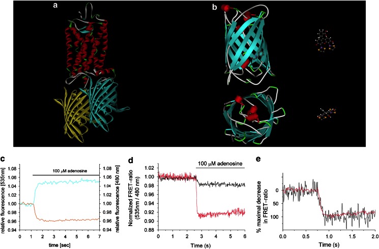
The G-protein-coupled receptors (GPCRs) represent one the largest families of drug targets. Upon agonist binding a receptor undergoes conformational rearrangements that lead to a novel protein conformation which in turn can interact with effector proteins. During the last decade significant progress has been made to prove that different conformational changes occur. Today it is mostly accepted that individual ligands can induce different receptor conformations. However, the nature or molecular identity of the different conformations is still ill-known. Knowledge of the potential functionally selective conformations will help to develop drugs that select specific conformations of a given GPCR which couple to specific signalling pathways and may, ultimately, lead to reduced side effects. In this review we will summarize recent progress in biophysical approaches that have led to the current understanding of conformational changes that occur during GPCR activation.
Figures

References
-
- Adams SR, Campbell RE, Gross LA, Martin BR, Walkup GK, Yao Y, et al. New biarsenical ligands and tetracysteine motifs for protein labeling in vitro and in vivo: synthesis and biological applications. J Am Chem Soc. 2002;124:6063–6076. - PubMed
-
- Adams SR, Harootunian AT, Buechler YJ, Taylor SS, Tsien RY. Fluorescence ratio imaging of cyclic AMP in single cells. Nature. 1991;349:694–697. - PubMed
-
- Barak LS, Ferguson SS, Zhang J, Caron MG. A beta-arrestin/green fluorescent protein biosensor for detecting G-protein-coupled receptor activation. J Biol Chem. 1997;272:27497–27500. - PubMed
-
- Bartl FJ, Fritze O, Ritter E, Herrmann R, Kuksa V, Palczewski K, et al. Partial agonism in a G-protein-coupled receptor: role of the retinal ring structure in rhodopsin activation. J Biol Chem. 2005;280:34259–34267. - PubMed
-
- Berg KA, Maayani S, Goldfarb J, Scaramellini C, Leff P, Clarke WP. Effector pathway-dependent relative efficacy at serotonin type 2A and 2C receptors: evidence for agonist-directed trafficking of receptor stimulus. Mol Pharmacol. 1998;54:94–104. - PubMed
Publication types
MeSH terms
Substances
LinkOut - more resources
Full Text Sources
Other Literature Sources
