. 2008 May;28(5):961-72.
doi: 10.1038/sj.jcbfm.9600590. Epub 2007 Dec 5.

Functional reactivity of cerebral capillaries

Affiliations

Functional reactivity of cerebral capillaries

Bojana Stefanovic et al. J Cereb Blood Flow Metab. 2008 May.

Abstract

The spatiotemporal evolution of cerebral microcirculatory adjustments to functional brain stimulation is the fundamental determinant of the functional specificity of hemodynamically weighted neuroimaging signals. Very little data, however, exist on the functional reactivity of capillaries, the vessels most proximal to the activated neuronal population. Here, we used two-photon laser scanning microscopy, in combination with intracranial electrophysiology and intravital video microscopy, to explore the changes in cortical hemodynamics, at the level of individual capillaries, in response to steady-state forepaw stimulation in an anesthetized rodent model. Overall, the microcirculatory response to functional stimulation was characterized by a pronounced decrease in vascular transit times (20%+/-8%), a dilatation of the capillary bed (10.9%+/-1.2%), and significant increases in red blood cell speed (33.0%+/-7.7%) and flux (19.5%+/-6.2%). Capillaries dilated more than the medium-caliber vessels, indicating a decreased heterogeneity in vessel volumes and increased blood flow-carrying capacity during neuronal activation relative to baseline. Capillary dilatation accounted for an estimated approximately 18% of the total change in the focal cerebral blood volume. In support of a capacity for focal redistribution of microvascular flow and volume, significant, though less frequent, local stimulation-induced decreases in capillary volume and erythrocyte speed and flux also occurred. The present findings provide further evidence of a strong functional reactivity of cerebral capillaries and underscore the importance of changes in the capillary geometry in the hemodynamic response to neuronal activation.


Figures
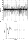
Figure 1
Figure 1
SEP recording over the latter 30-s period of the 60-s stimulation interval (containing 90 333-μs pulses) in a typical subject (a). The corresponding average SEP trace (b).
Figure 2
Figure 2
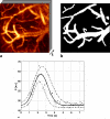
A 1024×1024-μm2 frame in the bolus tracking series of a typical subject at rest (a). The corresponding coarse intravascular map (b). (Note that the absence of smaller vessels from this map does not affect the estimate of the parameter of interest, namely the overall shortening in the transit time across the imaged vasculature.) Gamma variate fit (solid curve with standard error of the fit shown by dashed dotted lines) to the normalized time course data (shown as ‘x’) of a sample voxel in this data set (c).
Figure 3
Figure 3
Maps of transit time estimates (given by time-to-peak of bolus passage normalized to bolus arrival time), overlaid on a single frame of the bolus tracking time series in the subject of Figure 1 at baseline (a) and during activation (b). Note the dramatic drop in transit time at stimulation (b), relative to baseline (a). The corresponding histograms of intravascular voxel-wise transit times at rest (c) and during activation (d). The mean across-voxel transit time for this subject dropped from 1.34 ± 0.04 s at rest to 0.78 ± 0.02 s during activation.
Figure 4
Figure 4
Results of semi-automatic segmentation of vessels strictly included in the imaged stack (i.e., those found to both originate and terminate within the imaged volume) overlaid on the maximum intensity projection image (along axial direction) in a sample subject.
Figure 5
Figure 5
The vessel-wise blood volume change as a function of a point estimate of the resting vessel diameter (a) or vessel blood volume (b) (N=120 vessels; with 14-35 vessels/subject). The small vessels (green) are defined as having a resting diameter below 10μm; and medium vessels (red), a resting diameter above 10μm. Histograms of stimulation-induced volume changes of small vessels (N=87), (c) and medium vessels (N=33), (d). The mean CBV increase was 10.9 ± 1.2 % in small vessels and 6.4 ± 1.0 % in the medium caliber vessels. Data-driven clustering of the ΔCBV vs. resting CBV data produces an effective border between small and medium vessel clusters at about 2×105 μm3 (dashed line shown in (b)), resulting in very similar average small vs. medium vessel ΔCBV estimates (see text).
Figure 6
Figure 6
The histogram of RBC speed during baseline (a) and during activation (b) across all capillaries. Sample 1.5-s segments of line scans during rest and activation in a capillary (showing average vRBC increase of 17.0 ± 3.9 % upon activation) are shown in (c). The average stimulation-induced change in the RBC speed as a function of average baseline RBC speed in the responding vessels (d). The mean activation-induced change in RBC speed across all imaged capillaries (N=25) was 12.3 ± 7.2 % (p<10-3). The average change observed in the responding capillaries (N=16) was 16.3 ± 10.1 %, with 12 capillaries showing increases (33.0 ± 7.7 %) and 4 capillaries exhibiting RBC speed decreases (-33.8 ± 16.8 %) during forepaw stimulation.
Figure 7
Figure 7
The histogram of normalized fluorescent RBC flux during baseline (a) and during activation (b). The stimulation-induced change in the average FRBC flux as a function of average baseline fluorescent RBC flux (c). The mean activation-induced change in FRBC flux was 19.5 ± 6.2 % (p<10-6).

References

    1. Ances BM, Greenberg JH, Detre JA. The effects of graded hypercapnia on the activation flow coupling response due to forepaw stimulation in alpha-chloralose anesthetized rats. Brain Res. 2001;911:82–88. - PubMed
    1. Armstrong-James M, Fox K, Das-Gupta A. Flow of excitation within rat barrel cortex on striking a single vibrissa. J Neurophysiol. 1992;68:1345–1358. - PubMed
    1. Atkinson JL, Anderson RE, Sundt TM., Jr The effect of carbon dioxide on the diameter of brain capillaries. Brain Res. 1990;517:333–340. - PubMed
    1. Attwell D, Iadecola C. The neural basis of functional brain imaging signals. Trends Neurosci. 2002;25:621–625. - PubMed
    1. Berwick J, Johnston D, Jones M, Martindale J, Redgrave P, McLoughlin N, Schiessl I, Mayhew JE. Neurovascular coupling investigated with two-dimensional optical imaging spectroscopy in rat whisker barrel cortex. Eur J Neurosci. 2005;22:1655–1666. - PubMed

Publication types

LinkOut - more resources