. 2008 Aug;33(9):2093-107.
doi: 10.1038/sj.npp.1301621. Epub 2007 Dec 5.

Neural pathways underlying lactate-induced panic

Affiliations

Neural pathways underlying lactate-induced panic

Philip L Johnson et al. Neuropsychopharmacology. 2008 Aug.

Abstract

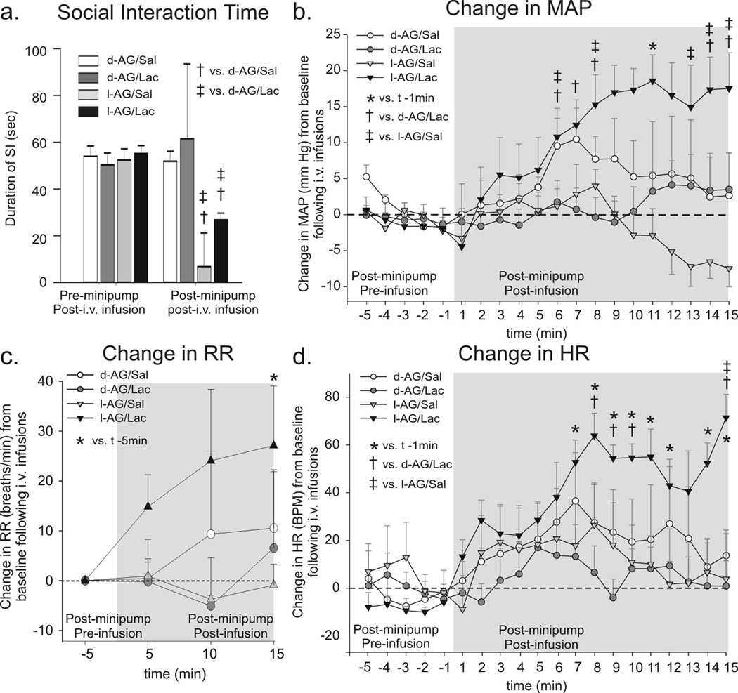
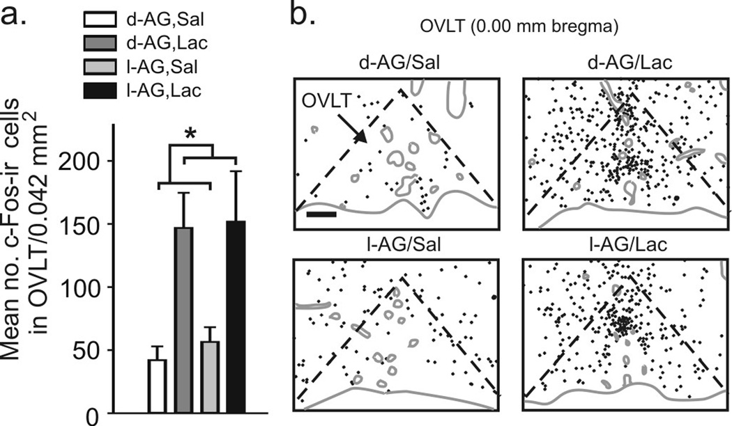
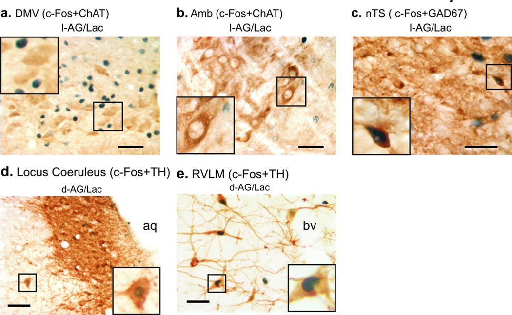
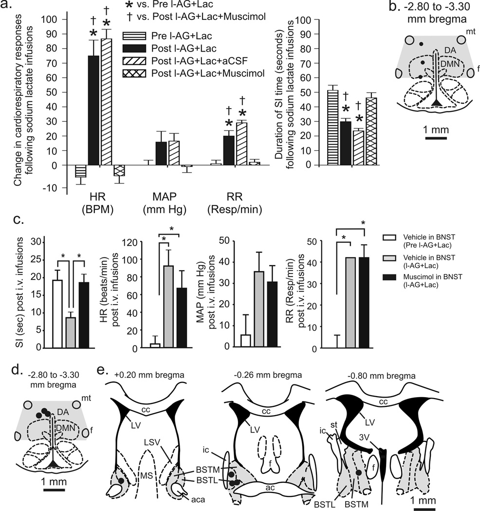
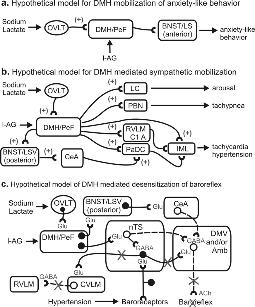
Panic disorder is a severe anxiety disorder characterized by susceptibility to induction of panic attacks by subthreshold interoceptive stimuli such as 0.5 M sodium lactate infusions. Although studied for four decades, the mechanism of lactate sensitivity in panic disorder has not been understood. The dorsomedial hypothalamus/perifornical region (DMH/PeF) coordinates rapid mobilization of behavioral, autonomic, respiratory and endocrine responses to stress, and rats with disrupted GABA inhibition in the DMH/PeF exhibit panic-like responses to lactate, similar to panic disorder patients. Utilizing a variety of anatomical and pharmacological methods, we provide evidence that lactate, via osmosensitive periventricular pathways, activates neurons in the compromised DMH/PeF, which relays this signal to forebrain limbic structures such as the bed nucleus of the stria terminalis to mediate anxiety responses, and specific brainstem sympathetic and parasympathetic pathways to mediate the respiratory and cardiovascular components of the panic-like response. Acutely restoring local GABAergic tone in the DMH/PeF blocked lactate-induced panic-like responses. Autonomic panic-like responses appear to be a result of DMH/PeF-mediated mobilization of sympathetic responses (verified with atenolol) and resetting of the parasympathetically mediated baroreflex. Based on our findings, DMH/PeF efferent targets such as the C1 adrenergic neurons, paraventricular hypothalamus, and the central amygdala are implicated in sympathetic mobilization; the nucleus of the solitary tract is implicated in baroreflex resetting; and the parabrachial nucleus is implicated in respiratory responses. These results elucidate neural circuits underlying lactate-induced panic-like responses and the involvement of both sympathetic and parasympathetic systems.


Figures

Figure 1
Figure 1
The effect of GABA synthesis inhibition in the DMH region and iv. lactate infusions on anxiety-like behavioral and cardiorespiratory responses from experiment 1. a) Social interaction (SI) time pre and post l-AG (GABA synthesis inhibitor)- or d-AG (control isomer) treatment in rats also receiving i.v. infusions of saline (Sal) or lactate (Lac). Change in b) mean arterial blood pressure (MAP), c) respiration rate (RR), d) and heart rate (HR) from 5 min baseline post-i.v. infusion. The RR data are presented as change from baseline for consistency, but the * symbol indicates a significant difference in actual RR over time. Bars or lines represent the mean + S.E.M. No differences were noted between baseline levels of MAP, RR and HR for each group (data not shown).
Figure 2
Figure 2
Graphs and illustrations of c-Fos-ir cells in the organum vasculosum lamina terminalis (OVLT) from experiment 1. a) Graph illustrates mean (± SEM) numbers of c-Fos-ir cells in the OVLT following i.v. infusions of saline (Sal) or lactate (Lac) in rats previously treated with d- or l-AG infusions into the DMH/PeF region (* P < 0.05). b) Illustrations of c-Fos-ir cells (each black dot = 1 c-Fos-ir cell) in the OVLT of a rat in each treatment group. Gray solid lines indicate the outline of the brain section and blood vessels. Black dashed lines indicate the OVLT. Scale bar: 136 µm.
Figure 3
Figure 3
Photomicrographs of cellular responses [i.e., c-Fos induction; blue/black immunostaining of nuclei] in neurochemically identified cell groups studied in experiment 1 represented brown cytoplasmic immunostaining. Photographs of c-Fos and choline acetyltransferase (ChAT) immunostaining in the a) dorsal motor nucleus of the vagus (DMV) and b) ambiguus nucleus (Amb) in lactate (Lac) + l-AG-treated rats. Photographs of c) c-Fos and glutamic acid decarboxylase (GAD67) immunostaining in the nucleus of the solitary tract (nTS) of a lactate+l-AG treated rat. Photomicrographs illustrating c-Fos and tyrosine hydroxylase (TH) immunostaining of d) pontine A6 noradrenergic cell group in the locus coeruleus (LC, −10.04 mm bregma) and e) rostroventrolateral medulla (RVLM) C1 adrenergic cell group (−11.96 mm bregma) in saline- (Sal, left) and Lac (right) infused rats with prior infusions of d-allylglycine (d-AG) in the DMH/PeF. Insets at the bottom of low magnification photographs contain higher magnification photographs. Scale bar: 50 µm; lower left insert; a–b) 25 µm, c) 22 µm, d–e) 20 µm.
Figure 4
Figure 4
Effects of restoring GABAergic tone in the DMH (experiment 2) or bed nucleus of the stria terminalis (BNST, experiment 3) on l-AG+lactate (Lac)-induced behavioral and/or cardiorespiratory responses. a) Graph illustrates the effects of injecting muscimol or artificial cerebrospinal fluid (aCSF) into the DMH region of l-AG+Lac treated rats. Bars represent the mean ± S.E.M. [* (versus Pre l-AG+Lac) P < 0.001, † (versus Post l-AG+Lac+Muscimol) P < 0.01)]. b) Illustration of cannula placements in the DMH region for experiment 2. Black circles represent cannula placements verified histologically (n=3) and gray shading indicates cannula placements from tissue that was micropunched for future neurochemical analyses (n=3). c) First graph, SI duration prior to and after l-AG infusions with aCSF or muscimol injections targeting the BNST followed by i.v. sodium lactate infusions. The following graphs illustrate the changes in the MAP, HR and RR in response to lactate prior to and after l-AG infusions in the DMH and with aCSF or muscimol injections targeting the BNST in experiment 3; (* P<0.05). d) Illustration of cannula placements in the DMH region for experiment 3. Black circle represents one histological placement (n=3) and gray shading represents distribution of cannula placements from tissue micropunched for neurochemical analysis (n=2). e) Illustration of the distribution injection sites in the BNST in experiment 3. Gray shading represents the BNST and black circles indicate aCSF or muscimol injection sites (n=5) verified with histology. See methods section for full list of abbreviations.
Figure 5
Figure 5
Hypothetical models of sodium lactate induced increases in panic-like behavioral and cardiorespiratory responses in panic-prone (i.e., l-AG treated) rats. a) Hypothetical model illustrating the neural pathways of lactate + l-AG-induced anxiety where plus signs indicate an excitatory pathway. b) Illustration of a hypothetical model of sodium lactate + l-AG mobilization of sympathetic nervous system activity. c) Hypothetical model illustrating sodium lactate + l-AG-induced desensitization of the vagally-mediated baroreflex, where solid lines with solid circles indicate excitatory neurons and dashed lines with open circles indicate inhibitory neurons and an “X” over a line indicates paths that are inhibited by the DMH/PeF. Abbreviations: bv, blood vessel; GABA, GABAergic path; Glu, glutamatergic path; IML, intermediolateral cell column; See methods for additional abbreviations.

Similar articles

Cited by

References

    1. Abshire VM, Hankins KD, Roehr KE, DiMicco JA. Injection of L-allylglycine into the posterior hypothalamus in rats causes decreases in local GABA which correlate with increases in heart rate. Neuropharmacology. 1988;27:1171–1177. - PubMed
    1. Catelli JM, Sved AF. Enhanced pressor response to GABA in the nucleus tractus solitarii of the spontaneously hypertensive rat. Eur J Pharmacol. 1988;151:243–248. - PubMed
    1. Chamberlin NL, Saper CB. Topographic organization of respiratory responses to glutamate microstimulation of the parabrachial nucleus in the rat. J Neurosci. 1994;14:6500–6510. - PMC - PubMed
    1. Chan RK, Sawchenko PE. Organization and transmitter specificity of medullary neurons activated by sustained hypertension: implications for understanding baroreceptor reflex circuitry. J Neurosci. 1998;18:371–387. - PMC - PubMed
    1. Chen T, Hui R, Dong YX, Li YQ, Mizuno N. Endomorphin 1- and endomorphin 2-like immunoreactive neurons in the hypothalamus send axons to the parabrachial nucleus in the rat. Neurosci Lett. 2004;357:139–142. - PubMed

Publication types

MeSH terms