. 2008 Jan 9;27(1):132-42.
doi: 10.1038/sj.emboj.7601949. Epub 2007 Dec 6.

Cdc2p controls the forkhead transcription factor Fkh2p by phosphorylation during sexual differentiation in fission yeast

Affiliations

Cdc2p controls the forkhead transcription factor Fkh2p by phosphorylation during sexual differentiation in fission yeast

Midori Shimada et al. EMBO J. .

Abstract

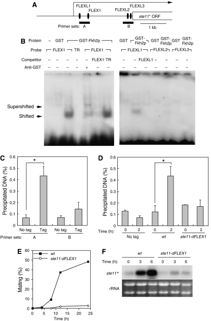
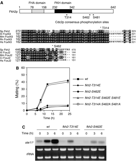
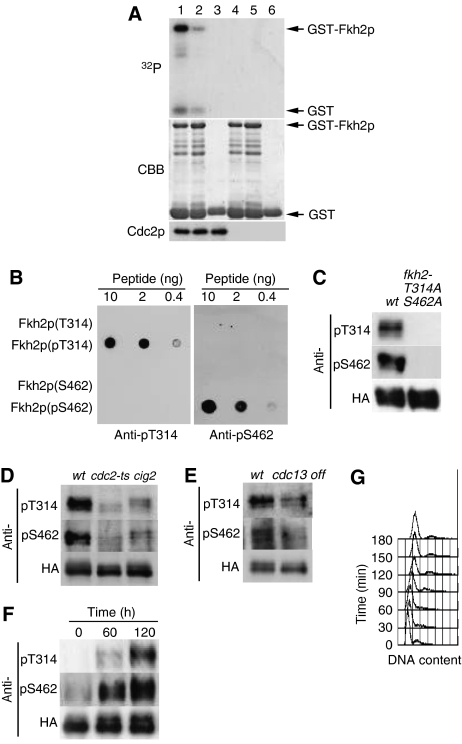
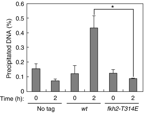
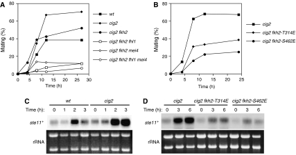
In most eukaryotes, cyclin-dependent kinases (Cdks) play a central role in control of cell-cycle progression. Cdks are inactivated from the end of mitosis to the start of the next cell cycle as well as during sexual differentiation. The forkhead-type transcription factor Fkh2p is required for the periodic expression of many genes and for efficient mating in the fission yeast Schizosaccharomyces pombe. However, the mechanism responsible for coordination of cell-cycle progression with sexual differentiation is still unknown. We now show that Fkh2p is phosphorylated by Cdc2p (Cdk1) and that phosphorylation of Fkh2p on T314 or S462 by this Cdk blocks mating in S. pombe by preventing the induction of ste11+ transcription, which is required for the onset of sexual development. We propose that functional interaction between Cdks and forkhead transcription factors may link the mitotic cell cycle and sexual differentiation.


Figures

Figure 1
Figure 1
Forkhead transcription factors are required for the induction of ste11+ mRNA and efficient mating. (A) wt (HM6), fkh2 (HM5657), fhl1 (HM4837), mei4 (HM50), fkh2 fhl1 (HM4887), fkh2 mei4 (HM5515), or fkh2 fhl1 mei4 (HM5544) cells were grown in EMM2 medium to a density of 1 × 10⁷ cells/ml, washed, and resuspended at a density of 2 × 10⁷ cells/ml in EMM2 medium lacking nitrogen. They were then cultured at 30°C and samples were collected at the indicated times for determination of mating frequency. Data are from representative experiments. (B) Total RNA was extracted from cells treated as in (A), and the abundance of ste11+ mRNA was examined by northern blot analysis. Ethidium bromide staining of rRNA is shown as a loading control. The ratios of intensities of ste11+ to rRNA signals were used to calculate the relative fold enrichment, shown below the rRNA. The samples from wt to fkh2, from fhl1 to mei4, from fkh2 mei4 to fkh2 fhl1 mei4 were from the same gel. All the samples were treated equally and the exposure time was the same. (C) Cells were transformed with pcL-ste11+ (ste11+), pAL-fkh2+ (fkh2+), or the empty vector pcL-X (Vec) and were cultured as in (A) for the determination of mating efficiency at 24 h after transfer to EMM2 medium without nitrogen. Data are from representative experiments.
Figure 2
Figure 2
Fkh2 binds to FLEX and FLEXL sequences in the putative promoter region of ste11+ and thereby induces ste11+ mRNA. (A) Schematic representation of the region upstream of the open reading frame (ORF) of ste11+ showing FLEX and FLEXL sequences. The major transcription initiation site of ste11+ is indicated by the arrow, and the regions targeted by primer sets in ChIP analysis are also shown. (B) An EMSA was performed with recombinant GST–Fkh2p(216–330) (or GST alone) and with FLEX1, FLEXL1, FLEXL2, FLEXL3, or TR (negative control) probes labeled with 32P. Competition was evaluated with excess amounts of unlabeled FLEX1, TR, or FLEXL1 oligonucleotides, and supershift analysis was performed with antibodies to GST, as indicated. The positions of shifted and supershifted bands are shown. (C) No tagged cells (no tag, HM6) and cells expressing GFP-tagged Fkh2p (Tag, HM5719) were grown to late log-phase, washed, and resuspended in medium without nitrogen. After incubation for 2 h at 30°C, cells were collected and analyzed by ChIP with antibodies to GFP and with the primer sets indicated in (A). Data are means±s.e. *P<0.006 (Student's t-test). (D) No tagged cells (no tag, HM6) and cells expressing GFP-tagged Fkh2p (wt; HM5719 and ste11-dFLEX1: HM6124) were treated and analyzed as in (C) with primer set A. Samples were collected at 0 and 2 h after nitrogen withdrawal. Data are means±s.e. of values from three separate experiments. *P<0.007 (Student's t-test). (E) wt (HM6) or ste11-dFLEX1 (HM5832) cells were treated and analyzed for mating efficiency as in Figure 1A. (F) Total RNA was extracted from cells treated as in (E) and was subjected to northern blot analysis of ste11+ mRNA.
Figure 3
Figure 3
Phosphorylation of Fkh2p by Cdc2p negatively regulates mating. (A) A schematic representation of Fkh2p indicating consensus phosphorylation sites (T314, S462, S481) for Cdc2p as well as the FHA and FKH domains is shown in the upper panel. Multiple alignment of Fkh2, FoxN3, and FoxJ2 proteins of S. pombe (Sp), Xenopus laevis (Xl), Mus musculus (Mm), Rattus norvegicus (Rn), Homo sapiens (Hs), and Ciona intestinalis (Ci) is shown in the lower panels. Identical (shaded black) and similar (shaded gray) amino acids as well as two Cdc2p consensus phosphorylation sites (T314 and S462) of Fkh2p are indicated. Dashes represent gaps introduced to optimize alignment. (B) Cells expressing wild type (wt, HM5145) or T314E (fkh2-T314E, HM5910), S462E (fkh2-S462E, HM5911), T314E S462E S481E (fkh2-T314E S462E S481E, HM5827) or T314A S462A S481A (fkh2-T314A S462A S481A, HM5722) mutant forms of Fkh2p were treated and analyzed for mating efficiency as in Figure 1A. (C) Total RNA was extracted from cells treated as in (B) and was subjected to northern blot analysis of ste11+ mRNA.
Figure 4
Figure 4
Phosphorylation of Fkh2p on T314 and S462 by Cdc2p in vitro and in vivo. (A) Kinase assays were performed with Cdc2p precipitates prepared from protein extracts of exponentially growing cells expressing hemagglutinin epitope (HA)-tagged forms of Cdc2p (HM6118; lanes 1–3) or not expressing HA (HM6; lanes 4–6) with anti-HA antibody. Substrates (lanes 1–3, respectively) included GST–Fkh2p(305–492), GST–Fkh2p(305–492) containing T314A, S462A, and S481A mutations, or GST alone. Reaction mixtures were separated by SDS–polyacrylamide gel electrophoresis, and proteins were detected by staining with Coomassie brilliant blue (CBB) and autoradiography (32P). Arrows indicate GST and the GST–Fkh2p fusion proteins. The Cdc2p input into each reaction mixture was also examined separately by Western blotting. (B) Various amounts (10, 2, or 0.4 ng) of Fkh2p peptides containing phosphorylated or nonphosphorylated T314 or S462 were spotted onto a nitrocellulose membrane and subjected to immunodetection with affinity-purified antibodies (anti-pT314 and anti-pS462) generated in response to the corresponding phosphorylated peptides. (C) Cells expressing HA-tagged forms of wild-type Fkh2p (HM5145) or the Fkh2p(T314A,S462A) mutant (HM5722) were grown to mid-log phase at 30°C. Cell lysates were then subjected to immunoprecipitation with antibodies to HA, and the resulting precipitates were subjected to immunoblot analysis with anti-pT314, anti-pS462, and anti-HA, as indicated. (D) Cells expressing HA-tagged Fkh2p were either grown to mid-log phase in EMM2 at 24°C and then incubated at 36.5°C for 7 h (wt, HM5145; cdc2-ts, HM5444) or grown as in (C) (cig2, HM5530). Cell lysates were subjected to immunoprecipitation and immunoblot analysis as in (C). (E) Cells expressing HA-tagged Fkh2p (wt, HM5146; cdc13 off, HM5554) were grown to mid-log phase in EMM2 at 30°C, after which thiamine was added to the culture medium to switch off cdc13+ expression and the cells were incubated for an additional 5 h. Cell lysates were subjected to immunoprecipitation and immunoblot analysis as in (C). (F) Cells expressing HA-tagged Fkh2p (HM6107) were synchronized in G1 by transient temperature arrest and samples taken every 1 h upon release to the permissive temperature. Cell lysates were subjected to immunoprecipitation and immunoblot analysis as in (C). (G) DNA content of the cells in (F) was determined by flow cytometric analysis.
Figure 5
Figure 5
Phosphorylation of Fkh2p on T314 reduces its binding to the FLEX1 sequence upstream of ste11+. No tagged cells (No tag, HM6) and cells expressing GFP-tagged wild-type (wt, HM5719) or T314E (fkh2-T314E, HM5912) mutant form of Fkh2p were treated and subjected to ChIP analysis with antibodies to GFP and the primer set A as in Figure 2D. Data are means±s.e. of values from three independent experiments. *P<0.013 (Student's t-test).
Figure 6
Figure 6
The mating efficiency of cig2 cells is reduced by mutation of forkhead transcription factors. (A) wt (HM6), cig2 (HM5555), cig2 fkh2 (HM5701), cig2 fkh2 fhl1 (HM5702), cig2 fkh2 mei4 (HM5703), or cig2 fkh2 fhl1 mei4 (HM5704) cells were assayed for mating efficiency as in Figure 1. (B) cig2 (HM5530), cig2 fkh2-T314E (HM5924), or cig2 fkh2-S462E (HM5925) cells were assayed for mating efficiency as in Figure 1. (C, D) Total RNA was extracted from cells treated as in (A) or (B) and was subjected to northern blot analysis of ste11+ mRNA.

Similar articles

Cited by

References

    1. Bahler J (2005) Cell-cycle control of gene expression in budding and fission yeast. Annu Rev Genet 39: 69–94 - PubMed
    1. Bahler J, Wu JQ, Longtine MS, Shah NG, McKenzie A III, Steever AB, Wach A, Philippsen P, Pringle JR (1998) Heterologous modules for efficient and versatile PCR-based gene targeting in Schizosaccharomyces pombe. Yeast 14: 943–951 - PubMed
    1. Buck V, Ng SS, Ruiz-Garcia AB, Papadopoulou K, Bhatti S, Samuel JM, Anderson M, Millar JB, McInerny CJ (2004) Fkh2p and Sep1p regulate mitotic gene transcription in fission yeast. J Cell Sci 117: 5623–5632 - PubMed
    1. Bulmer R, Pic-Taylor A, Whitehall SK, Martin KA, Millar JB, Quinn J, Morgan BA (2004) The forkhead transcription factor Fkh2 regulates the cell division cycle of Schizosaccharomyces pombe. Eukaryot Cell 3: 944–954 - PMC - PubMed
    1. Costa RH (2005) FoxM1 dances with mitosis. Nat Cell Biol 7: 108–110 - PubMed

Publication types

MeSH terms